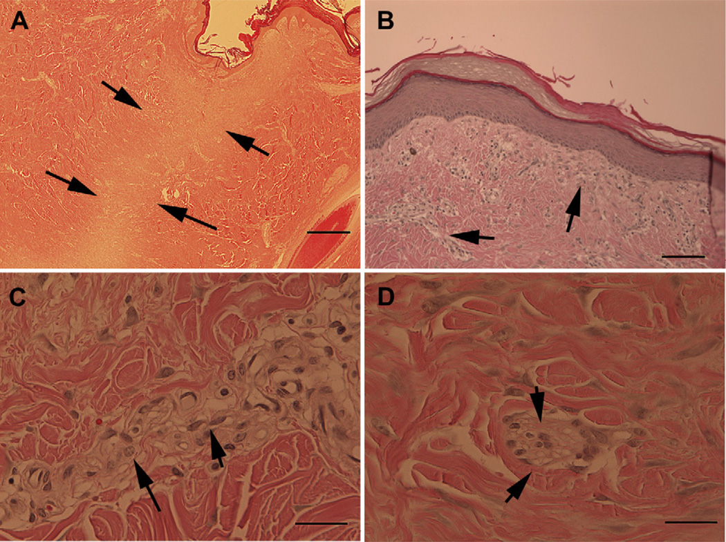
Fig. 4.
Dermal lesions heal normally with minor residual fibrosis evident at six months. Skin re-epithelialized and hair follicles had reformed by the conclusion of the six month study (Panel A) although a band of disorganization could still be discerned where the lesion was created. Higher magnification revealed bands of fibrotic tissue that could still be detected underlying the epidermis and in other areas of the regenerated dermis (panel B, arrows). The cells comprising the fibrotic tissue were mainly fibroblasts (Panel C, short arrows) with some macrophages (Panel C, long arrow), lymphocytes and rare giant multi-nuclear cells (Panel D, arrows). Scale bars = 1000 um (Panel A), 200 um (Panel B), and 50 um (Panels C and D).

