Abstract
The interrelationship between genetic susceptibility and carcinogenic exposure is important in cancer development. Polymorphisms in detoxification enzymes of the glutathione-S-transferases (GST) family are associated with an increased incidence of lymphoma. Here we investigated the molecular connection of the genetic polymorphism of GSTT1 to the response of lymphocytes to polycyclic aromatic hydrocarbons (PAH). In neoplastic situation, GSTT1 deletions were more frequently observed in lymphoma patients (54.9%) than in normal controls (42.0%, P = 0.009), resulting in an increased risk for lymphoma in individuals with GSTT1-null genotype (Odds ratio = 1.698, 95% confidence interval = 1.145–2.518). GSTT1 gene and protein expression were accordingly decreased in GSTT1-deleting patients, consistent with activated profile of cell cycle regulation genes. Mimicking environmental exposure using long-term repeat culture with low-dose PAH metabolite Hydroquinone, malignant B- and T-lymphocytes presented increased DNA damage, pCHK1/MYC expression and cell proliferation, which were counteracted by ectopic expression of GSTT1. Moreover, GSTT1 expression retarded xenograft tumor formation of Hydroquinone-treated lymphoma cells in nude mice. In non-neoplastic situation, when zebrafish was exposed to PAH Benzo(a)pyrene, molecular silencing of gstt1 enhanced the proliferation of normal lymphocytes and upregulated myca expression. Collectively, these findings suggested that GSTT1 deletion is related to genetic predisposition to lymphoma, particularly interacting with environmental pollutants containing PAH.
Introduction
During the past decades, the incidence of lymphoma has been significantly increased, ranging it among the ten most frequent cancers [1]. The etiologies of lymphoma remain largely undetermined. However, epidemiological studies revealed that exposure to environmental pollutants is a susceptibility factor [2]. Polycyclic aromatic hydrocarbons (PAH) represent the main components of environmental pollutants that have genotoxic and carcinogenic properties.
Genetic polymorphisms in detoxification enzymes are important determinants of individual variation in cancer risk. Glutathione S-transferases (GST) are the major detoxification enzymes in humans. As phase II biotransformation enzymes, GST catalyze the conjugation of reduced glutathione to electrophilic centres on a wide range of substrates, including activated exogenous molecules like PAH.
Several GST polymorphisms commonly occurring in humans are associated with an increased susceptibility to cancers, when combined with environmental factors. Recently, the role of GST genotypes in the pathogenesis of lymphoma has been addressed [3]. GSTT1 is an important member of GST family and involved in the detoxification of various carcinogens, particularly PAH. Evidence of an elevated risk for lymphoma in individuals with GSTT1-null homozygotes has been reported [4], [5], [6]. Proposed reasons could include an impaired neutralization of reactive oxygen species or reduced deactivation of carcinogenic intermediates of PAH. However, the exact molecular connection between GSTT1 deletions and lymphoma development remained to be investigated.
In the present study, we examined the genetic polymorphisms of GSTT1 in Chinese patients with lymphoma in comparison with a health control cohort, correlating the GSTT1-null genotype with the progression of lymphoma cells and the proliferative behavior of normal lymphocytes under the exposure of PAH. Our results showed that GSTT1 deletion could be a potential risk factor of lymphomagenesis. Genetic susceptibility may interact with the genotoxic effect of environmental carcinogens to eventually predispose to lymphoma.
Patients and Methods
Ethics Statement
Written informed consent was obtained from all the patients (from the next of kin, caretakers, or guardians on the behalf of the minors/children patients) in accordance with the Declaration of Helsinki. The study was approved by the Shanghai Rui Jin Hospital Review Boards. Animals were used according to the protocols approved by the Shanghai Rui Jin Hospital Animal Care and Use Committee.
Patients
Two hundred and four patients with lymphoma [103 diffuse large B-cell lymphoma (DLBCL) and 101 T-cell lymphoma (TCL) cases], 127 men and 77 women aged 14 to 82 years were included in this study. Histological diagnoses were established according to the World Health Organization classifications. Frozen tumor specimen was available in 114 lymphoma patients and 40 age- and sex-matched reactive hyperplasia cases.
Cell Lines and Reagents
The B-lymphoma cell line Namalwa and T-lymphoma cell line Jurkat were obtained from American Type Culture Collection. Hydroquinone (Sigma-Aldrich) was dissolved in normal saline before use. Benzo(a)pyrene (BaP, Sigma-Aldrich) was dissolved in dimethyl sulfoxide (DMSO) as a stock solution of 400 mg/ml.
Cell Proliferation and Flow Cytometric Assay
Cell growth was measured by MTT assay and cell proliferation was determined by 5-ethynyl-2′-deoxyuridine (EdU) incorporation assay using Cell-Light EdU kit (Ribobio Co., Ltd., China) at 48 h. Cell cycle and cell apoptosis were assessed at 48 h as previous reported [7].
Genome-wide Copy Number Variation (CNV) Analysis
Genomic DNA was extracted using Wizard Genomic DNA Purification Kit (Promega). Genome-wide CNV genotyping was performed on frozen tumor samples of 25 DLBCL, 20 TCL and 8 reactive hyperplasia cases, using Human 610-Quad_v1 (610 k SNP probes) or 660 W-Quad_v1 (660 k SNP probes) DNA Analysis BeadChips. Regions were determined based on the Log R Ratio (LRR) of the signal intensity and B allele frequency (BAF) of genotyping call from the sample using platform of GenomeStudio V2011.1 with CnvPartition 3.1.6 (Illumina). All the data is available on NCBI (Accession number GSE47357).
GSTT1 Genotyping and Expression
GSTT1 deletion was detected on frozen tumor (114 cases) and peripheral blood (the rest 90 cases without frozen tumor specimen) of lymphoma patients by multiplex polymerase chain reaction (PCR) method, using albumin gene as an internal positive control, as previously reported [8]. The normal control group comprised 205 unrelated healthy volunteers. Blood samples were collected and leukocytes were isolated after hypotonic lysis of erythrocytes. Genomic DNA was extracted and amplified using the primers: GSTT1∶5′-TTCCTTACTGGTCCTCACATCTC-3′ and 5′-TCACCGGATCATGGCCAGCA-3′, and albumin:5′-GCCCTCTGCTAACAAGTCCTAC-3′ and 5′-GCCCTAAAAAGAAAATCGCCAATC-3′.
Total RNA was extracted using Trizol reagent and reverse-transcribed using PrimeScript RT reagent Kit with gDNA Eraser (TaKaRa). Real-time PCR was performed on frozen samples of lymphoma and reactive hyperplasia patients, using ABI PRISM 7900HT and specific probes for GSTT1 (Assay ID: Hs01091675_g1) and GAPDH (Life Technologies). A relative quantification was calculated using the 2−ΔΔCT method.
Gene Network and Pathway Analysis
Human Genome U133 Plus 2.0 Array GeneChip microarray (Affymetrix) was performed on tumor samples of 8 DLBCL patients and analyzed by Expression Console software (Partek GS 6.5, Affymetrix). The data is available on NCBI (Accession number GES47355). Human LncRNA Microarray V2.0 (Arraystar Inc.) was performed on tumor samples of 8 TCL cases and analyzed by Agilent Feature Extraction Software (Agilent Technologies).
Genes were subsequently filtered by comparing their expression levels between the GSTT1-deleting and non-deleting patients. Statistical differences were calculated and the genes with P<0.05 were analyzed for enrichment of KEGG pathways using Database for Annotation, Visualization and Integrated Discovery (DAVID v6.7, http://david.abcc.ncifcrf.gov) for network composition analyses. Genes of pathway(s) significantly involved in both DLBCL and TCL were hierarchical clustered using MeV v4.8.1 (Dana-Farber Cancer Institute).
GSTT1 Transfection
GSTT1 expression vector (GSTT1, NM_000853.2) and the negative control vector (FU, pReciever-M46) were obtained from GeneCopoeia. The recombinant lentivirus vector PGC-GSTT1-IRES-GFP-LV and PGC-FU-GFP-LV were produced and packaged by co-transfecting 293T cells with the package vector. The supernatant of 293T cell culture were condensed and the virus titers were approximately 3×10⁹ TU/ml. To transfect Namalwa and Jurkat cells, the multiplicities of infection were 50 and 10, respectively.
Comet Assay
DNA damage was determined by the comet assay with Reagent Kit for Single Cell Gel Electrophoresis Assay (Trevigen, Inc.). In addition to frank DNA strand breaks, oxidised bases were measured by conversion to breaks using endonuclease III (recognizing oxidised pyrimidines) or formamidopyrimidine DNA glycosylase (FPG, specific for oxidised purines). Measurements of comet parameters % DNA in the tail, tail length and tail moment were obtained. Net enzyme-sensitive sites were calculated by subtracting the comet score after incubation with buffer alone from the score with enzyme.
Tissue Array
A human lymphoma tissue array (NHL482) was obtained from US Biomax, Inc. GSTT1 and p53-binding protein 53 BP1 expression were scored semi-quantitatively based on staining intensity and distribution using the immunoreactive score, as previously reported [7].
Immunohistochemistry and Immunofluorescence Assay
Immunohistochemical analyses were carried out on 5-µm-paraffin sections or acetone-fixed cells with an indirect immunoperoxidase method using the antibodies against GSTT1, 53 BP1 (Abcam), Ki67 (Dako) and MYC (Abcam). Immunofluorescence assay was performed on acetone-fixed cells using mouse anti-human-γH2AX antibody followed by donkey anti-mouse-IgG antibody, as well as rabbit anti-human-53 BP1 and rabbit anti-human-pCHK1 antibody followed by donkey anti-rabbit-IgG antibody (Abcam). Nuclei were counterstained with DAPI.
Western Blot
Western blot was performed as previously described [7]. Actin (Sigma) and LaminB (Abcam) were used to ensure equivalent loading of total and nuclear protein, respectively. Antibodies against GSTT1 and CHK1 were obtained from EPITOMICS. Anti-pCHK1, pCHK2 and CHK2 antibodies were from Cell Signaling. Anti-MYC and 53 BP1 antibodies were from Abcam. Horseradish peroxidase-conjugated goat anti-mouse-IgG and goat anti-rabbit-IgG were from Santa Cruz Biotechnology Inc.
Tumorigenicity Assay in Murine Models
Five-week-old female BALB/c nude mice were obtained from Shanghai Laboratory Animal Center. Mice were injected subcutaneously into the right flank with lymphoma cells. For each cell line, mice were divided into 3 subgroups. Namalwa cells were injected with 1×10⁷, 2×10⁶ and 5×10⁵, and Jurkat cells were injected with 4×10⁷, 1×10⁷ and 2×10⁶, respectively. The number of the tumors formed was determined until 4 weeks after injection. Then mice were sacrificed, with tumor tissue samples fixed in formaldehyde and further processed for paraffin embedding.
Cloning and Plasmid Construction in Zebrafish
Adult zebrafish (Danio rerio) were maintained following established guidelines [9] at 28°C on a 14 h:10 h light:dark cycle. Zebrafish gstt1a and gstt1b genes were identified based on homology to human GSTT1. The specific primers were designed according to genomic sequence in the UCSC data base (University of California, Santa Cruz) to amplify part of gstt1a and gstt1b genes: gstt1a:5′- CGGGATCCGGCGCCTCTCTTCTTTTCTT-3′ and 5′-CCCTCGAG CTGCATCATCTCCACAATGG-3′; gstt1b:5′-CGGGATCCTGATCCCAAAGGGTTGAGTC-3′ and 5′-CCCTCGAGGAACAAAAAGGTGCCATTCG-3′; gstt1a-EGFP reporter:5′- GATCCACTTACTTCCACAATGCCGCTGGAGCTGTATCTCGATCTGCATTCCCAGCCG-3′ and 5′- AATTCGGCTGGGAATGCAGATCGAGATACAGCTCCAGCGGCATTGTGGAAGTAAGTG-3′; gstt1b-EGFP reporter:5′- GATCCCTTTTTAAACATGACTCTGGAAATTTACTTGGACCTGTTTTCCCAGCCCTGG-3′ and 5′- AATTCCAGGGCTGGGAAAACAGGTCCAAGTAAATTTCCAGAGTCATGTTTAAAAAGG-3′; myca:5′-CGGGATCCTTTCCAAGAACTCCCACCCC-3′ and 5′-CCCTCGAGTTTAATCTAGGGCTGCGCAG-3′. The amplified PCR product was purified and subcloned into pCS2+ for subsequent in vitro synthesis of mRNA.
Whole-mount in situ Hybridization (WISH) in Zebrafish
To localize gstt1a and gstt1b in zebrafish, whole-mount in situ hybridization (WISH) was carried out. pCS2+ containing part of gstt1a, gstt1b, rag1 or myca (729 bp, 473 bp, 1000 bp and 231 bp, respectively) was utilized to generate antisense RNA probes for gstt1a, gstt1b, rag1 and myca using digoxigenin-11-uridine 5′-triphosphate (Roche Applied Science). Zebrafish embryos were fixed in 4% paraformaldehyde at the stages indicated and WISH was performed as previously described [10].
Microinjection of Zebrafish Embryos
The gstt1a and gstt1b morpholino oligonucleotides (MO) and its 5 bp-mismatch control were synthesized by Gene Tools LLC. The sequences were as follows: gstt1a:5′-CGAGATACAGCTCCAGCGGCATTGT-3′; 5 bp-mismatch of gstt1a: 5′-CGAcATAgAcCTCCAcCcGCATTGT-3′; gstt1b:5′- GGTCCAAGTAAATTTCCAGAGTCAT-3′; 5 bp-mismatch of gstt1b:5′- GGTCgAAcTAAATTTgCAcAcTCAT -3′. MOs were diluted to 1 mM stock solution in 1×Danieau buffer and were microinjected at a volume of 2 nl into one-cell stage embryos using an air pressure injector and glass capillaries. Injection experiments were performed in triplicate.
The vectors pCS2+-MO-gstt1a and pCS2+-MO-gstt1b were linearized with XhoI and transcribed in vitro with SP6 RNA polymerase in the presence of m7G (59)ppp(59)G (Ambion) to produce capped transcripts and were microinjected into one-cell stage embryos for MO efficiency evaluation.
Transmission Electron Microscopy
The embryos of zebrafish were fixed overnight with 2% glutaraldehyde/0.1 M phosphate-buffered saline (pH 7.3) at 4°C. The embryos were then post-fixed in 1% osmium tetroxide at 4°C for 1 h, rinsed thoroughly with distilled water, dehydrated by graded ethanol and freeze-dried. The samples were sputter-coated in Epon812 (TAAB Laboratories) and ultrathin sections were prepared, stained with uranyl acetate and lead citrate, and examined with a PhilipsCM120 transmission electron microscopy (Philips).
Statistic Analysis
Assays were set up in triplicate and the results were presented as Mean±S.E. Variance between the experimental groups was determined by two-tailed t-test or Mann-Whitney test. The logistic regression test was used to compare the differences in genotype frequencies between patients and controls adjusted by sex and age. Differences were considered significant when the 2-sided P<0.05.
Results
GSTT1-null Genotype is Frequently Observed in Lymphoma Patients
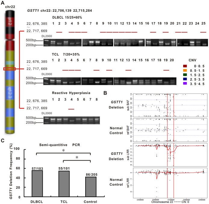
A homozygous loss in chr22q11.23 spanning GSTT1 gene (22,706,139 bp to 22,715,284 bp, NCBI Build 36) was detected in 15/25 (60%) of DLBCL and 7/20 (35%) of TCL patients, comparing with 1/8 (12.5%) of reactive hyperplasia cases (Figure 1A). Figure 1B depicted data for the region of 22,500,000 bp to 22,900,000 bp of chromosome 22. In the normal control, the LRR was distributed around zero corresponding to DNA copy number 2, whilst the BAFs were clustered around values of 0, 0.5 and 1 that correspond to the diploid genotypes AA, AB and BB. The GSTT1-null genotype presented a much more complex scenario with extensive genomic rearrangements leading to considerable variation in the SNP data.
Figure 1. GSTT1-null genotype is frequently observed in patients with lymphoma.
A: GSTT1 deletion detected by copy number variation (CNV) analysis (upper panel) and validated by semi-quantitative PCR (lower panel) in diffuse large B-cell lymphoma (DLBCL, 15/25, 60.0%), T-cell lymphoma (TCL, 7/20, 35.0%) and reactive hyperplasia (1/8, 12.5%). B: SNP data illustrating the distribution of B allele frequencies (BAF) and Log R Ratio (LRR) values across the region of chromosome 22 (22,676,385 bp to 22,717,669 bp) in the GSTT1-deleting cases and normal controls. C: Semi-quantitative PCR validation of GSTT1 deletion in expanded sample-set of DLBCL (57/103, 55.3%), TCL (55/101, 54.5%) and normal controls (86/205, 42.0%). *P<0.05 comparing with normal controls.
Using semi-quantitative PCR, GSTT1 genotypes were further investigated. GSTT1 gene deficiency was deduced from the absence of the specific 460 bp fragment. Presence of a 350 bp albumin fragment confirmed proper functioning of the method (Figure 1A). Of the 204 patients, 112 cases (54.9%) were GSTT1-null. Prevalence of the GSTT1 deletions was 55.3% (57/103) in DLBCL and 54.5% (55/101) in TCL, significantly higher than that in normal controls (86/205, 42.0%, P = 0.027 and P = 0.034, respectively, Figure 1C), adjusted by sex and age. This translated into an increased risk of lymphoma for individuals with the GSTT1-null genotype (Odds ratio = 1.698; 95% confidence interval = 1.145–2.518).
GSTT1 Deletion Results in Loss of Protein Expression
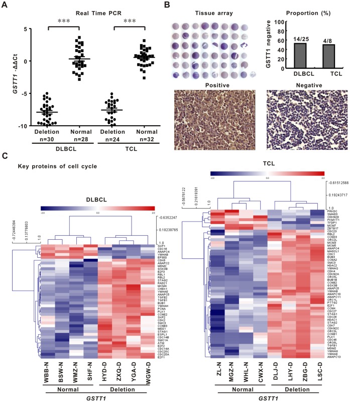
GSTT1 gene expression was measured by real-time quantitative PCR in 114 patients with frozen tissue specimen. GSTT1-deleting patients presented significant downregulation of GSTT1 gene expression in both DLBCL and TCL, comparing with those without deletion (P both <0.001, Figure 2A). GSTT1 protein was detected by tissue array. GSTT1 was negative in 56.0% (14/25) of DLBCL and in 50.0% (4/8) of TCL, respectively, in accordance with the percentage of GSTT1 deletion in the two subtypes (Figure 2B). Moreover, 53BP1 was more frequently expressed in GSTT1-negative cases than in GSTT1-positive cases [DLBCL, 85.7% (12/14) vs 45.5% (5/11), P = 0.032, T-NHL, 100% (4/4) vs 0% (0/4), P = 0.028].
Figure 2. GSTT1 deletion is related to decreased gene and protein expression and enhanced cell cycle progression.
A: GSTT1 gene expression assessed by quantitative real-time PCR in the GSTT1-deleting patients and normal controls. B: GSTT1 protein expression detected by tissue array. C: Geneset of cell cycle related proteins revealed by gene network and pathway analysis on microarray data of DLBCL and TCL.
Gene expression profile was assessed by microarray in frozen tissue sample of 8 DLBCL and 8 TCL patients with the history of PAH exposure (four each for GSTT1-deleting and non-deleting cases). The pathway identified with statistical significance by the differentially expressed GSTT1 revolved around key proteins of cell cycle progression (Figure 2C), indicating that GSTT1 deletion is biologically functional and relevant to lymphoma cell proliferation.
GSTT1 Protects Lymphoma Cells from PAH-induced DNA Damage
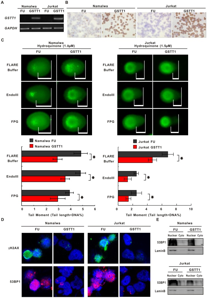
To further elucidate its biological function in lymphoma, GSTT1 gene was transfected to GSTT1-negative Namalwa and Jurkat cells. Comparing with the negative control (FU), lymphoma cells expressing GSTT1 (GSTT1) showed increased levels of gene and protein expression, as revealed by semi-quantitative PCR (Figure 3A) and by immunostaining assay (Figure 3B), respectively.
Figure 3. GSTT1 expression protects lymphoma cells from PAH-induced DNA damage.
A: GSTT1 gene expression assessed by semi-quantitative PCR in Namalwa and Jurkat cells transfected with GSTT1 (GSTT1) and the negative control vector (FU). B: Images represent results from three independent experiments. GSTT1 protein expression detected by immunohistochemistry assay. C: DNA damage measured by alkaline and modified comet assay in Namalwa and Jurkat cells treated with Hydroquinone (Upper panels). Mean tail moments were calculated in the same cells (Lower panels). Data represents Mean ± SE from at least 50 cells in each group. D: Immunofluorescence assay of γH2AX and 53BP1 in Hydroquinone-treated lymphoma cells. *P<0.05 comparing with the FU cells.
Hydroquinone, common metabolites of PAH [11], was used to treat lymphoma cells. Fifty percent of growth inhibition (IC50) was first measured in both cell lines at concentrations ranging from 1.25 to 40 µM. The IC50 value was 14.4 µM in Namalwa cells and 18.7 µM in Jurkat cells, respectively. To mimic the environmental exposure status, lymphoma cells were then cultured with low-dose Hydroquinone (at a concentration of approximately 10% of IC50 with minimal cytotoxicity, 1 µM for Namalwa GSTT1/FU cells and 1.5 µM for Jurkat GSTT1/FU cells, respectively) in a repeat manner [12]. Briefly, cells were treated once a week with Hydroquinone for 24 h and weekly for 4 times in total. Normal saline was referred as the solvent control.
DNA strand breaks were assessed using comet assay, and in addition DNA oxidation damage, namely oxidised pyrimidines and purines, using lesion specific enzymes Endo III and FPG. As manifested by increased parameters % DNA in the tail and prolonged tail length, DNA damage was less intensive in Hydroquinone-treated GSTT1-expressing lymphoma cells (GSTT1) than in the negative control cells (FU). Tail moment was further assessed by parameters % DNA×tail length. The results showed that tail moment, as well as EndoIII- and FPG-sensitive sites, were significantly reduced in the GSTT1 groups than in the FU groups (Namalwa, P = 0.037, P = 0.042 and P = 0.042, Jurkat, P = 0.041, P = 0.049 and P = 0.049, respectively, Figure 3C).
γH2AX and 53 BP1 are sensitive markers of DNA damage [13]. As detected by immunofluorescence assay, FU cells presented increased nuclear levels of γH2AX and 53 BP1, which were significantly prohibited in GSTT1 cells (Figure 3D). These data indicated that DNA damage is constitutively activated in response to PAH and could be protected by GSTT1 expression.
GSTT1 Inhibits PAH-mediated Lymphoma Cell Proliferation
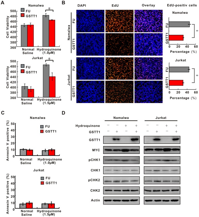
Upon treatment with normal saline, there is no difference of cell growth between the FU and GSTT1 groups. However, cell growth was enhanced in Hydroquinone-treated FU cells, which could be significantly reduced by ectopic expression of GSTT1 (Namalwa, P = 0.045 and Jurkat, P = 0.043, respectively, Figure 4A). Cell proliferation was further determined by EdU assay. Comparing with the FU groups, EdU-positive cells in the GSTT1 groups were accordingly reduced (Namalwa, P = 0.043 and Jurkat, P = 0.039, respectively, Figure 4B). Cell cycle analysis showed a lower percentage of S-phase cells in the GSTT1 groups than in the FU groups (Namalwa, 17.5±0.52% vs 14.3±0.55%, P = 0.013, and Jurkat, 14.1±0.44% vs 11.8±0.54%, P = 0.032, respectively). As for cell apoptosis, neither Namalwa nor Jurkat cells showed obvious change in the percentage of ANX-V-positive cells between the FU and GSTT1 groups when treated with Hydroquinone (Figure 4C).
Figure 4. GSTT1 expression inhibits PAH-mediated lymphoma cell proliferation.
A: Effect of GSTT1 expression on cell proliferation. 3×10⁵ cells treated with normal saline or Hydroquinone were seeded and cell number was counted at 48 h by typan blue. Data represent Mean±S.E. of densitometric values from three individual experiments. B: EdU assay of Namalwa and Jurkat cells treated with normal saline or Hydroquinone. C: Cell apoptosis analyzed by flow cytometry. Histography indicates Mean±S.E. from three individual experiments. D: Key proteins of DNA damage and cell cycle detected by Western blot in Namalwa and Jurkat cells with or without Hydroquione treatment. *P<0.05 comparing with the FU cells.
By Western blot, expressions of cell cycle regulation proteins were assessed with or without Hydroquinone treatment. In Hydroquinone-treated Namalwa and Jurkat cells, GSTT1 expression was consistent with downregulation of MYC, pCHK1 and nuclear 53 BP1, while pCHK2, total CHK1 and CHK2 levels remained constant (Figure 4D).
GSTT1 Retards Tumor Formation of PAH-treated Lymphoma Cells
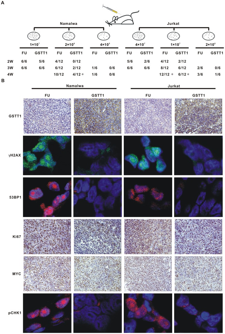
To assess the carcinogenic potential in vivo, the FU and GSTT1 cells were treated with Hydroquinone as mentioned above and injected subcutaneously in nude mice (Figure 5A). Expression of GSTT1 could significantly prolong the latency of tumor formation. Of note, 2×10⁶ Namalwa cells derived from the FU group could form tumors in 10 of 12 nude mice in 4 weeks. However, when 2×10⁶ cells from GSTT1 group were injected, only 4 tumors were found in 12 nude mice (P = 0.013). Similar results were obtained in Jurkat cells: tumor were formed in all mice at 4 weeks injected with 1×10⁷ FU cells, but only 6 of 12 mice in those treated with the same amount of GSTT1 cells (P = 0.014).
Figure 5. GSTT1 expression reduces tumorogeneity of PAH-treated lymphoma cells.
A: Tumor formation in nude mice. Indicated amount of Hydroquinone treated cells were injected subcutaneously and tumorigenicity was reported as numbers of tumors formed per numbers of mice injected. B: Expression of GSTT1, Ki67 and MYC detected in tumor tissues by immunohistochemistry assay, as well as pCHK1, γH2AX and 53 BP1 by immunofluorescence assay. *P<0.05 comparing with the FU cells.
To search for in situ evidence of DNA damage and tumor cell proliferation, immunoflurescence assay of γH2AX, 53 BP1 and pCHK1, as well as immunohistochemistry assay of Ki67 and MYC were performed on mice tumor sections. In parallel with in vitro results, all markers were increased in the FU groups, compared with the GSTT1 groups following Hydroquinone treatment (Figure 5B).
GSTT1 Knock-down Stimulates Lymphocyte Proliferation in PAH-treated Zebrafish
After searching the zebrafish genome data base (Zv9), two GSTT1 genes were identified, referred as gstt1a and gstt1b, sharing part of nucleotide acids identical to human GSTT1 gene. To confirm the evolutionary conservation of GSTT1, we investigated the distribution of genes located adjacent to the GSTT1 locus in zebrafish and in human genomes. As shown in Figure S1A, 9 genes, along with GSTT1, define an approximately 840-kb genomic region on human chromosome 22, which is syntenic to the zebrafish gstt1a genomic locus on linkage group 8 and zebrafish gstt1b on linkage group 21. Thus, zebrafish gstt1a and gstt1b are evolutionarily conserved orthologs of human GSTT1.
Then the temporal and spatial expression patterns of gstt1a and gstt1b were examined in zebrafish embryos from 0.25 h to 120 hpf by WISH using digoxigenin-labeled antisense RNA probe. Strong signals were detected in the blastomeres of the two-cell stage and the shield period (6 h, Figure S1B), indicating that both gstt1a and gstt1b transcripts were maternally derived and played a role in early embryonic development. Although both genes exhibited similar patterns of expression during the early stage, differential expression was observed after 30 hpf: high levels of gstt1a transcripts were mostly restricted to the liver (2d), while gstt1b transcripts gradually disappeared after 30 hpf and was no longer observed after 3d (Figure S1B).
To further determine the function of GSTT1 on normal lymphocytes, morpholino was applied to efficiently block gstt1a and gstt1b gene expression in zebrafish. Injection of MO of gstt1a at the dosage of 8 ng per embryo, but not mismatch oligo control, was able to completely suppress the EGFP expression of co-injected capped gstt1a-EGFP mRNAs reporter (Figure S1C, left panels), indicating that the antisense RNA that could effectively silence gene expression. Similar results were obtained in gstt1b (Figure S1C, right panels).
Exposure of BaP, a carcinogenic PAH, was conducted at the concentration of 10 µg/L, based on previous experiments investigating the teratogenicity of PAH on zebrafish [14], [15]. Zebrafish embryos were microinjected with gstt1a and gstt1b morpholinos or 5 bp mismatch oligo controls, and treated with BaP from 24 hpf to 4 d. DMSO was used as the solvent control at a final concentration of 0.1%. At least 100 embryos were included in each group.
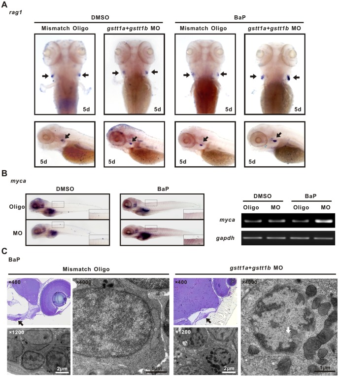
Comparing with those injected with mismatch oligos, gstt1a and gstt1b silenced embryos showed an increase of the rag1 signal in the thymus under exposure to BaP, as detected by WISH (Figure 6A, right panels). No significant difference of the rag1 signal was observed in embryos treated with DMSO (Figure 6A, left panels).
Figure 6. Knock-down of gstt1a and gstt1b promotes lymphocyte proliferation exposed to BaP.
A; WISH images showed the rag1 expression in the thymus (arrows) of differently treated 5 dpf embryos. B: In situ analysis of myca at 5 dpf. The morphants showed increased expression of myca in microinjected gstt1a and gstt1b morpholino exposed to BaP (Left panels), semi-quantitative PCR showed similar expression pattern in embyos (Right panels). C: Ultrastructure of thymic lymphocytes from 5 dpf larvae exposed to BaP. Images represent results from three independent experiments and each group contains 30 morphants.
To confirm that MYC activation is also involved in PAH-associated stimulation of lymphocyte proliferation in zebrafsh, the expression of myca, zebrafish homologue of human MYC, was assessed in BaP-treated embryos at 5 d. In accordance with rag1 expression, myca expression was significantly higher in embryo injected with gstt1a and gstt1b morpholinos than those injected with mismatched oligos by WISH (Figure 6B, left panels) and by semi-quantitative PCR (Figure 6B, right panels).
The structure of the thymus in 5 d-embryos was examined by transmission electron microscopy (Figure 6C). Ultrastructural analysis of both cells (microinjected with gstt1a and gstt1b morpholino or oligo control) revealed no sign of apoptosis. At a higher magnification, abundant euchromatin and robust mitochondrias were observed in BaP-treat gstt1-silenced lymphocytes, possibly related to a hyper-proliferative status of the cells.
Therefore, in addition to lymphoma cells, inactivation of GSTT1 was able to confer a proliferative advantage induced by PAH in their normal counterparts.
Discussion
Although environmental factors are proven relevant for tumorogenesis, few genetic variations with confirmed associations to date and the difficulty in accurately assessing exposures are main challenges to evaluate the interaction between gene and environment in cancer [16]. Our study provided evidence that genetic polymorphisms in GSTT1 gene, both in neoplastic and non-neoplastic situation, could modulate the response of the lymphocytes to the major component of environmental pollutants PAH and might link to lymphoma development.
The GSTT1-null genotype was more prevalent in lymphoma patients than in normal controls, conferring approximately a 1.7-fold increase in the risk of lymphoma. This coincides with epidemiological studies from Western [4], African [5], and other Asian countries [6], although the incidence of GSTT1 deletion varied among the geographical areas. The genetic polymorphism was associated with loss of gene and protein expression, indicating that GSTT1 is functionally impaired in lymphoma.
It is previously reported that deletions in the GSTT1 gene contribute to individual susceptibility to PAH-induced DNA damage and carcinogenesis [17], [18]. Our study further showed in lymphoma that introduction of GSTT1 to GSTT1-negative tumor cells is associated with increased DNA stability and repair of oxidative DNA damage in response to PAH. These circumstances fit to the idea that GSTT1 was involved in the susceptibility of individuals to PAH-induced DNA damage, loss of which favoring the accumulation of cytogenetic aberrations and thereby leading to the initiation of lymphoma. Therefore, genetic polymorphisms in detoxification enzymes might account for individual variation in lymphoma risk and should be considered in a more complex scenario involving the gene-environment interactions.
GSTT1 can modulate multiple cellular processes, including cell proliferation and cell death [19]. Among the lymphoma patients with the history of PAH exposure, instead of cell apoptosis, the GSTT1-deleting cases displayed a genomic profile of cell cycle progression, referring dysregulation of cell proliferation as the major target of GSTT1 deletions in lymphoma. Experimentally, in GSTT1-negative lymphoma cells, expression of GSTT1 significantly prohibited PAH to enhance tumor cell growth, with tumor aggressiveness accordingly decreased in murine models. These data thus suggested a potential role for GSTT1 in protecting against lymphoma cell proliferation provoked by PAH.
MYC is essential for cell proliferation [20] and is a proto-oncogene frequently upregulated in lymphoma [21]. More importantly, MYC itself can localize onto sites of active DNA replication and directly controls S-phase cell progression [22]. MYC was activated following PAH treatment in our study, particularly in GSTT1-negative lymphoma cells, corresponding to increased S-phase cells, enhanced cell proliferation and in vivo tumorigenicity, indicative the possible involvement of MYC on PAH-associated cell cycle dynamics and lymphoma progression in GSTT1-null status.
The MYC-induced DNA damage response acts as a double-edged sword in tumor progression. CHK1 and CHK2 are key factors involved in the replication stress response and controlled by MYC [23]. Indeed, activation of CHK1 is essential for tumor maintenance, while CHK2 activity constitutes a barrier to malignant transformation. As previously reported in Eμ-myc lymphoma models, tumor cells present increased levels of CHK1 phosphorylation, in turn limits MYC-induced apoptosis. In the clinical setting, lymphoma patients exhibit a striking correlation between high levels of MYC and CHK1 [24]. This was also proven by us in PAH-treated lymphoma cells, where MYC might ensure proliferative advantage through selectively activating CHK1. Recent reports have identified MYC-positive lymphoma as a subtype with poor disease prognosis [25], even resistant to high-dose chemotherapy [26] and newly developed bio-therapeutic agent [27]. Since MYC is difficult to be targeted directly, CHK1 inhibitors could thus become attractive candidates for therapeutic intervention on MYC-driven malignancies and warrant further investigation.
Genetic factors that impair DNA repair can increase the likelihood of pre-neoplastic changes [28]. This is particularly obvious when environmental factors were existed, as a previous report showing that the t(14;18)-positive clones are prominent in individuals exposed to pesticides and correlated with a higher risk of t(14;18) lymphoma [29]. In addition to lymphoma cells and murine xenograft models, we used zebrafish as an animal model to verify the cooperative effect of the genetic and environmental factor on their normal counterparts. In GSTT1-knock-down zebrafish, although may not initially be lethal, genomic lesions in lymphocytes could be modulated by PAH that promote lymphocyte proliferation and MYC upregulation, which could eventually link to malignant transformation of lymphoma.
Supporting Information
GSTT1 is evolutionarily conserved and expressed ubiquitously during embryonic development. A: Comparison of the syntenic relationship of the zebrafish gstt1 genes with the human orthologue. Orthologous gstt1 genes symbols were in bold. Other pairs of duplicated genes (e.g. mmp11a and mmp11b) on zebrafish and the human (e.g. MMP11) orthologue are underlined. Hs_Chr, Homo sapiens chromosome, Dr_LG, Danio rerio linkage group, Mb, megabase. B: Expression of zebrafish gstt1a and gstt1b in wild-type AB strain embryo during embryonic development. C: Efficiency validation of EGFP reporter expression by gstt1a morpholino and gstt1b morpholino. Images represent the typical outcome of three independent experiments and each group contains 30 morphants.
(TIF)
Funding Statement
The work was supported, in part, by the National Natural Science Foundation of China (81325003, 81172254 and 81101793), the Shanghai Commission of Science and Technology (11JC1407300), the Program of Shanghai Subject Chief Scientists (13XD1402700), and the “Shu Guang” project supported by Shanghai Municipal Education Commission and Shanghai Education Development Foundation. The funders had no role in study design, data collection and analysis, decision to publish, or preparation of the manuscript.
References
- 1. Siegel R, Naishadham D, Jemal A (2012) Cancer statistics, 2012. CA Cancer J Clin 62: 10–29. [DOI] [PubMed] [Google Scholar]
- 2.Clapp RW, Jacobs MM, Loechler EL (2008) Environmental and occupational causes of cancer: new evidence 2005–2007. Rev Environ Health 23: 1–37. PubMed: 18557596. [DOI] [PMC free article] [PubMed]
- 3. Skibola CF, Curry JD, Nieters A (2007) Genetic susceptibility to lymphoma. Haematologica 92: 960–969. [DOI] [PMC free article] [PubMed] [Google Scholar]
- 4.Yri OE, Ekstrom PO, Hilden V, Gaudernack G, Liestol K, et al.. (2013) The Influence Of Polymorphisms In Genes Encoding Immunoregulatory Proteins and Metabolizing Enzymes On Susceptibility And Outcome In Diffuse Large B-Cell Lymphoma Patients Treated With Rituximab. Leuk Lymphoma. PubMed: 23391141. [DOI] [PubMed]
- 5.Abdel Rahman HA, Khorshied MM, Elazzamy HH, Khorshid OM (2012) The link between genetic polymorphism of glutathione-S-transferases, GSTM1, and GSTT1 and diffuse large B-cell lymphoma in Egypt. J Cancer Res Clin Oncol 138: 1363–1368. PubMed: 22484853. [DOI] [PubMed]
- 6.Bin Q, Luo J (2013) Role of polymorphisms of GSTM1, GSTT1 and GSTP1 Ile105Val in Hodgkin and non-Hodgkin lymphoma risk: a Human Genome Epidemiology (HuGE) review. Leuk Lymphoma 54: 14–20. PubMed: 22734843. [DOI] [PubMed]
- 7.Shi WY, Xiao D, Wang L, Dong LH, Yan ZX, et al.. (2012) Therapeutic metformin/AMPK activation blocked lymphoma cell growth via inhibition of mTOR pathway and induction of autophagy. Cell Death Dis 3: e275. PubMed: 22378068. [DOI] [PMC free article] [PubMed]
- 8.Rimando MG, Chua MN, Yuson E, de Castro-Bernas G, Okamoto T (2008) Prevalence of GSTT1, GSTM1 and NQO1 (609C>T) in Filipino children with ALL (acute lymphoblastic leukaemia). Biosci Rep 28: 117–124. PubMed: 18444911. [DOI] [PubMed]
- 9.Westerfield O (1995) A prescription for hospital safety: treating workplace violence. Healthc Facil Manag Ser: 1–8. PubMed: 10156934. [PubMed]
- 10.Paffett-Lugassy NN, Zon LI (2005) Analysis of hematopoietic development in the zebrafish. Methods Mol Med 105: 171–198. PubMed: 15492396. [DOI] [PubMed]
- 11.Shimada T (2006) Xenobiotic-metabolizing enzymes involved in activation and detoxification of carcinogenic polycyclic aromatic hydrocarbons. Drug Metab Pharmacokinet 21: 257–276. PubMed: 16946553. [DOI] [PubMed]
- 12.Pang Y, Li W, Ma R, Ji W, Wang Q, et al.. (2008) Development of human cell models for assessing the carcinogenic potential of chemicals. Toxicol Appl Pharmacol 232: 478–486. PubMed: 18778725. [DOI] [PubMed]
- 13.Bonner WM, Redon CE, Dickey JS, Nakamura AJ, Sedelnikova OA, et al.. (2008) GammaH2AX and cancer. Nat Rev Cancer 8: 957–967. PubMed: 19005492. [DOI] [PMC free article] [PubMed]
- 14.Wassenberg DM, Di Giulio RT (2004) Synergistic embryotoxicity of polycyclic aromatic hydrocarbon aryl hydrocarbon receptor agonists with cytochrome P4501A inhibitors in Fundulus heteroclitus. Environ Health Perspect 112: 1658–1664. PubMed: 15579409. [DOI] [PMC free article] [PubMed]
- 15.Billiard SM, Timme-Laragy AR, Wassenberg DM, Cockman C, Di Giulio RT (2006) The role of the aryl hydrocarbon receptor pathway in mediating synergistic developmental toxicity of polycyclic aromatic hydrocarbons to zebrafish. Toxicol Sci 92: 526–536. PubMed: 16687390. [DOI] [PubMed]
- 16.Wang SS, Nieters A (2010) Unraveling the interactions between environmental factors and genetic polymorphisms in non-Hodgkin lymphoma risk. Expert Rev Anticancer Ther 10: 403–413. PubMed: 20214521. [DOI] [PubMed]
- 17.Xiong P, Bondy ML, Li D, Shen H, Wang LE, et al.. (2001) Sensitivity to benzo(a)pyrene diol-epoxide associated with risk of breast cancer in young women and modulation by glutathione S-transferase polymorphisms: a case-control study. Cancer Res 61: 8465–8469. PubMed: 11731429. [PubMed]
- 18.Koh WP, Nelson HH, Yuan JM, Van den Berg D, Jin A, et al.. (2011) Glutathione S-transferase (GST) gene polymorphisms, cigarette smoking and colorectal cancer risk among Chinese in Singapore. Carcinogenesis 32: 1507–1511. PubMed: 21803734. [DOI] [PMC free article] [PubMed]
- 19. Laborde E (2010) Glutathione transferases as mediators of signaling pathways involved in cell proliferation and cell death. Cell Death Differ 17: 1373–1380. [DOI] [PubMed] [Google Scholar]
- 20.Gustafson WC, Weiss WA (2010) Myc proteins as therapeutic targets. Oncogene 29: 1249–1259. PubMed: 20596078. [DOI] [PMC free article] [PubMed]
- 21.Nemajerova A, Petrenko O, Trumper L, Palacios G, Moll UM (2010) Loss of p73 promotes dissemination of Myc-induced B cell lymphomas in mice. J Clin Invest 120: 2070–2080. PubMed: 20484818. [DOI] [PMC free article] [PubMed]
- 22.Dominguez-Sola D, Ying CY, Grandori C, Ruggiero L, Chen B, et al.. (2007) Non-transcriptional control of DNA replication by c-Myc. Nature 448: 445–451. PubMed: 17597761. [DOI] [PubMed]
- 23.Campaner S, Amati B (2012) Two sides of the Myc-induced DNA damage response: from tumor suppression to tumor maintenance. Cell Div 7: 6. PubMed: 22373487. [DOI] [PMC free article] [PubMed]
- 24.Hoglund A, Nilsson LM, Muralidharan SV, Hasvold LA, Merta P, et al.. (2011) Therapeutic implications for the induced levels of Chk1 in Myc-expressing cancer cells. Clin Cancer Res 17: 7067–7079. PubMed: 21933891. [DOI] [PubMed]
- 25.Barrans S, Crouch S, Smith A, Turner K, Owen R, et al.. (2010) Rearrangement of MYC is associated with poor prognosis in patients with diffuse large B-cell lymphoma treated in the era of rituximab. J Clin Oncol 28: 3360–3365. PubMed: 20498406. [DOI] [PubMed]
- 26.Cuccuini W, Briere J, Mounier N, Voelker HU, Rosenwald A, et al.. (2012) MYC+ diffuse large B-cell lymphoma is not salvaged by classical R-ICE or R-DHAP followed by BEAM plus autologous stem cell transplantation. Blood 119: 4619–4624. PubMed: 22408263. [DOI] [PMC free article] [PubMed]
- 27.Gupta M, Maurer MJ, Wellik LE, Law ME, Han JJ, et al.. (2012) Expression of Myc, but not pSTAT3, is an adverse prognostic factor for diffuse large B-cell lymphoma treated with epratuzumab/R-CHOP. Blood 120: 4400–4406. PubMed: 23018644. [DOI] [PMC free article] [PubMed]
- 28.Duarte EC, Ribeiro DC, Gomez MV, Ramos-Jorge ML, Gomez RS (2008) Genetic polymorphisms of carcinogen metabolizing enzymes are associated with oral leukoplakia development and p53 overexpression. Anticancer Res 28: 1101–1106. PubMed: 18507060. [PubMed]
- 29.Agopian J, Navarro JM, Gac AC, Lecluse Y, Briand M, et al.. (2009) Agricultural pesticide exposure and the molecular connection to lymphomagenesis. J Exp Med 206: 1473–1483. PubMed: 19506050. [DOI] [PMC free article] [PubMed]
Associated Data
This section collects any data citations, data availability statements, or supplementary materials included in this article.
Supplementary Materials
GSTT1 is evolutionarily conserved and expressed ubiquitously during embryonic development. A: Comparison of the syntenic relationship of the zebrafish gstt1 genes with the human orthologue. Orthologous gstt1 genes symbols were in bold. Other pairs of duplicated genes (e.g. mmp11a and mmp11b) on zebrafish and the human (e.g. MMP11) orthologue are underlined. Hs_Chr, Homo sapiens chromosome, Dr_LG, Danio rerio linkage group, Mb, megabase. B: Expression of zebrafish gstt1a and gstt1b in wild-type AB strain embryo during embryonic development. C: Efficiency validation of EGFP reporter expression by gstt1a morpholino and gstt1b morpholino. Images represent the typical outcome of three independent experiments and each group contains 30 morphants.
(TIF)


