Abstract
Background:
Glenoid fossa fractures are rare injuries having a prevalence of 0.1%. These fractures may be managed operatively if substantially displaced. However, several fractures of glenoid fossa are managed nonoperatively, even if displaced, due to high incidence of associated injuries which may render patient unfit to undergo major orthopaedic surgery. There is a relative paucity of articles reporting on outcome of treatment of glenoid fossa fractures. We present our experience of treating these injuries over past decade with operative and nonoperative methods.
Materials and Methods:
21 patients of glenoid fossa fractures were included in this series with 14 males and 7 females. Patients with displacement of >5 mm who were fit to undergo surgery within 3 weeks of injury were operated using a posterior Judet's approach. Overall 8 patients with displaced fractures were operated (Group A) while 9 patients with displaced fractures (Group B) and 4 patients with undisplaced fractures (Group C) were managed nonoperatively.
Results:
The mean age and followup period in this series was 29 years and 7.3 years respectively. In group A, average constant score was 87.25. The least constant score was observed for group B (58.55) while group C had an average constant score of 86. Brachial plexus injury and fracture-dislocations had poorer outcome.
Conclusion:
Operative treatment for displaced glenoid fractures is a viable option at centers equipped to handle critically ill patients and subset of patients with fracture-dislocation as opposed to fracture alone should always be treated operatively due to persistent loss of function.
Keywords: Functional outcome, glenoid fracture, nonoperative, operative
INTRODUCTION
Scapular fractures are rare injuries and most often treated nonoperatively with acceptable results.1,2,3,4,5 Most scapular fractures are non or minimally displaced and do well with conservative treatment.1,6,7 This observation, however, has been based on the treatment of scapular fractures in general and its relevance is, therefore, very limited. A more differentiated approach is necessary as good results are not guaranteed with exclusively conservative treatment in all cases.8
As with any intra-articular fracture, displaced fractures of glenoid fossa may be managed operatively if substantially displaced.8,9,10,11,12,13,14,15,16,17,18 However, several fractures of glenoid fossa are managed nonoperatively, even if displaced, as high incidence of associated injuries may render the patient unfit to undergo major orthopedic surgery in view of more compelling urgencies.4,5,8,9,10,11,12,13,14 There is a relative paucity of articles reporting on the outcome of treatment of glenoid fossa fractures. We retrospectivly analysed the outcome in our patients of glenoid fossa fractures.
MATERIALS AND METHODS
On retrospective search of hospital records, we identified patients sustaining glenoid fossa fractures and admitted in our emergency department during the period ranging from 1998 to 2010. Fractures were classified according to the widely used Ideberg classification for glenoid fractures.5 We included only type II-V fractures in our analysis since these fractures have the distinction of being associated with other high energy injuries and are managed differently as compared to type I fractures which are generally associated with shoulder dislocations. We were able to identify 21 cases with glenoid fossa fracture who were available for assessment after followup periods ranging from 2 to 14 years. There were 6 type II, 7 type III, 1 type IV, 6 type V, and 1 type VI fractures [Table 1]. All subjects who were available for followup and gave informed consent for their inclusion in the present series were included.
Table 1.
Demographic and outcome details of patients included in the series
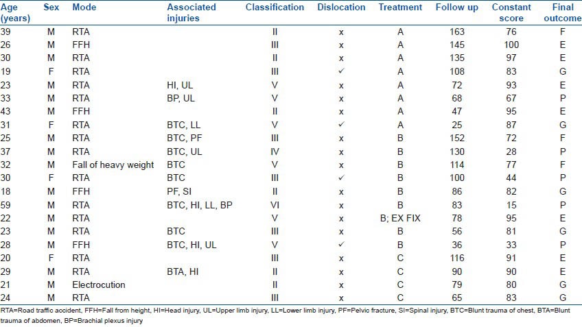
The mean age of patients at the time of trauma was 29 years (range 18-59) there were 17 males and 4 females. Road traffic accident was the most common mode of injury accounting for 15 cases, followed by fall from height (4), electrocution (1), and fall of heavy object (1). All except one case had closed injury. Associated injuries included brachial plexus injury (2), clavicle fracture (5), coracoids fracture (2), acromion fracture (2), scapular body fracture (3), ipsilateral upper limp fracture(s) (4), rib fracture(s) (9), spine injury (1), pelvic injury (2), lower limb fractures (2), head injury (4), blunt trauma chest (8), and blunt trauma abdomen (1). Overall, 12 patients had significant associated injury (excluding ipsilateral shoulder girdle fractures).
After initial resuscitation in the emergency department according to the protocol of advanced life trauma support, patients were assessed for musculoskeletal and associated injuries. Radiographic imaging for scapular fractures included standard scapular trauma series with computed tomography with 3D reconstruction for complex fractures and inability to assess fracture displacement on radiographs (10/21 cases). Further management was based on the amount of fracture displacement and general condition of the patient. There were 17 fractures which were displaced >5 mm, which was taken as the criterion for operative intervention, but only 8 were operated due to inability of the remaining 9 patients to undergo a major surgical procedure on account of poor general condition. We operated only on patients with displaced fractures who were able to undergo operative intervention within the first 3 weeks of injury [Figures 1 and 2]. All fractures were approached from the posterior side using the Judet's approach [Figure 1] and fixed with either plate (6), screws alone (1), or plate with additional screws outside plate (1), depending on fracture configuration. During this approach, we tried to access only the lateral border of scapula through the intermuscular interval between infraspinatus and teres minor and did not attempt to directly reduce or fix fractures extending to scapular body or vertebral border. In this regard, we agree with Bartoníček et al. that restoring the lateral border is of paramount importance.19 Although we did not use deltopectoral approach in any of the cases in the present series, we have used it in some recent cases where the main fragment was primarily anterior. External fixation was done for clavicle in one Gustilo Anderson type IIIa open displaced fracture of glenoid with ipsilateral clavicle fracture [Figure 3]. Remaining 13 patients, including 7 displaced fractures, were managed conservatively with a period of immobilization followed by early mobilization in a hope to achieve better clinical outcome [Figure 4]. Patients were thus divided into three groups [A: managed operatively (n = 8); B: displaced but managed nonoperatively (n = 9); and C: undisplaced fractures (n = 4)]. Disappearance of visible fracture lines on X-rays and pain on clinical examination were taken as indicators of union. At the final followup, patients were assessed for pain, function, range of movements, and strength using the Constant score20 and the final result was reported as excellent, good, fair, or poor, depending on the difference in scores of abnormal and normal shoulders (<11, excellent; 11-20, good; 21-30, fair; >30, poor). Complications during the perioperative period and at the latest followup were also recorded. The study was approved by our Institutional Ethics Committee.
Figure 1.
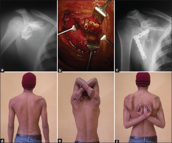
(a) Radiograph of right shoulder joint showing type V glenoid fracture in a 23 year old male (b) Peroperative photograph showing open reduction and internal fixation through posterior approach using two plates (c) followup radiograph at 6 years showing plates in situ and union (d,e,f) Clinical photographs showing excellent functional outcome with slight atrophy of infraspinatus muscle possibly due to surgical insult and the final Constant score was 93
Figure 2.
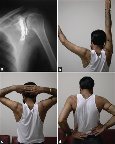
(a) Radiograph of left shoulder joint at followup of 13½ years in a case with open reduction shows some degree of degeneration (b,c,d) Clinical photographs showing restriction of abduction to 120° and restricted rotations. Final Constant score was 76
Figure 3.
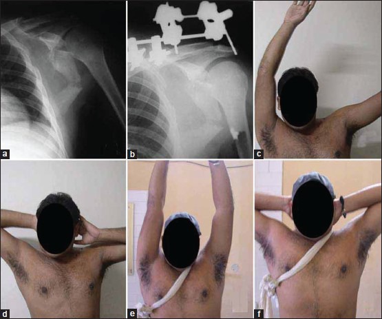
Preoperative (a) and postoperative (b) radiographs of a patient with open glenoid fossa fracture managed with external fixation for clavicle fracture. (c,d) Clinical photographs at 6 weeks followup showing the patient had restriction of abduction and external rotation. (e,f) Clinical photographs at 6 years followup showing abduction and external rotation. Final Constant score for this patient was 95
Figure 4.
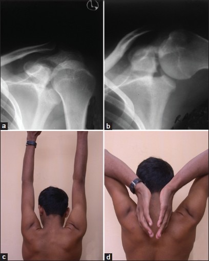
Neutral (a) and abduction (b) anteroposterior radiographs of shoulder showing a type III displaced fracture which was managed nonoperatively. Patient had good outcome with Constant score of 81 and unrestricted movements (c and d)
RESULTS
The incidence of associated injuries was 57.14% (12/21). The mean length of hospital stay was 15.2, 32.3, and 3.8 days in groups A, B, and C, respectively. Time for fracture union was the least in group C (5.5 weeks) followed by group A (6.7 weeks) and was the longest in group B (9.4 weeks), but union was achieved in all cases without further intervention, with overall mean time of 7.7 weeks for union in this series.
In group A, the average Constant score was 87.25 with four excellent, two good, one fair, and one poor result. Mean operative time was 105 min (range 45-150 min). The least Constant score amongst the three groups was observed for group B (58.55) with one excellent, two good, two fair, and four poor results. In group C, the average Constant score was 86 with two excellent and two good results [Table 1]. Amongst the different parameters of Constant score, pain and function were the least affected at the final followup, whereas range of movements followed by strength were the most severely affected.
Predictors of inferior outcome included brachial plexus injury [Figure 5] and fracture dislocation of glenoid. Four of five cases with poor result in this series had either brachial plexus palsy or fracture dislocation. Only one poor result in group B was not attributable to either of these two factors. Time taken till maximal improvement in shoulder Constant score was also compared amongst the three groups and yielded the least value for group A followed by groups C and B. There were two cases of superficial wound infection which resolved with prolonged course of antibiotic therapy for 6 weeks.
Figure 5.
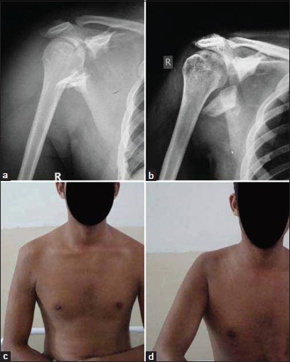
Radiographs (anteroposterior views) of shoulder at initial presentation (a) and final followup of a patient with type VI fracture with associated brachial plexus injury. Patient had visible atrophy of deltoid muscle (c) and no functional movements at shoulder joint (d). Patient had a final Constant score of 15
DISCUSSION
The relative infrequency (prevalence 1%) and “benign characteristics” of a scapular fracture probably explains the limited attention in the literature. Glenoid fossa fractures represent 10% of scapular fractures with overall prevalence of 0.1%.5,7,10,11 Majority of glenoid fossa fractures are undisplaced and can be managed nonoperatively. This is in contrast to the present series, where majority of fractures were displaced. This may be due to the referral system prevalent in our region whereby we receive higher percentage of patients with high-velocity trauma. Furthermore, inpatient records searched during this study did not include the records of patients with low-velocity trauma who are kept under observation for up to 24 h before being discharged.
The glenohumoral joint affords more degree of freedom of movement than any other joint and is therefore able to compensate for severe deformities and loss of movements. Although traditionally advocated treatment for scapular fractures has been nonoperative,21,22 recent authors have reported on favorable outcome after operative treatment for displaced glenoid fractures.4,8,11,12,13,14,15,16,17,18 Kavanagh et al.12 reported on nine patients treated surgically for intra-articular fractures with displacement >2 mm, and Mayo et al.16 reported good to excellent outcomes in 22 of 27 patients. Schandelmaier et al.,13 in a series of 22 operated cases, reported a median Constant score of 94% (mean 79%), while Adam14 reported excellent or good result in 8 out of 10 operated cases. Anavian et al.,18 in the largest published series of operated glenoid fossa fractures with 33 patients, reported that 27 of 30 patients available at final followup were able to resume previous level of activity and only four had mild pain while rest of the patients were completely pain free. We did not encounter any immediate complication related to the operative procedure, which is similar to the observation made in previously published reports, thus indicating the safety of the approach and feasibility of surgery. Nevertheless, postoperative infection remains a major cause of poor result.13,14
The most important predictor of poor outcome in the present series was nonoperative treatment in association with dislocation. Patients with persistent brachial plexus injuries also fared poorly, which has been universally accepted as an indicator of poor outcome in previously published series.13,14 Excluding these cases with dislocation (gross displacement) and brachial plexus palsy, only one patient of the remaining six in group B had poor result. Thus, a satisfactory result might still be achieved with nonoperative treatment of displaced fractures. Time taken to achieve maximal improvement in shoulder Constant score was the least in group A followed by groups C and B. This earlier recovery of shoulder function was perhaps in part due to shorter period of immobilization and earlier institution of physiotherapy in group A.
The most common mechanism of these injuries is a violent force applied laterally to the proximal part of the humerus, which is then driven into the glenoid cavity.10,14,19 A transverse fracture of the glenoid fossa occurs and then propagates in one of several directions, depending on the direction of the traumatic force.10,14,19 On account of the amount of force generally required to produce these fractures, the incidence of associated injuries is relatively high.1,7,10,14 Nearly half of these patients have a concomitant injury excluding the shoulder girdle.10,21 In the present series, 57% (12/21) cases had associated injuries, with rib fracture and blunt trauma of the chest being the most common injuries. The treatment of these associated injuries invariably assumes priority over scapular fracture on account of their severity and often precludes surgical treatment of displaced fractures during the initial period.
Goss was one of the first authors to recommend surgical treatment of glenoid fossa fractures.10 He emphasized on reduction of intra-articular step greater than 5 mm. While some authors have shown acceptable results in fractures displaced less than 5 mm, the advantage of achieving precise reduction in glenoid fossa fractures has not been proven objectively23 although it has been endorsed universally based on the treatment of intra-articular fractures elsewhere in the body.11,12,13,14,15,16,17 Instability of glenohumoral joint or of fracture fragments themselves is a more compelling indication for surgery, which can occur with fracture of more than one-fourth of the glenoid cavity.10,11,13,14 In a review of significant published series on operative treatment of scapular fractures, Lantry et al.24 found an intra-articular step of 5 mm as the most common indication used for glenoid fossa fractures, although most series, which included glenoid fossa fractures, universally accepted fracture dislocations as an indication of surgery. In view of lack of reports on the results of nonoperative treatment of glenoid fossa fractures, the amount of displacement necessitating operative treatment remains a matter of conjecture.25 Nevertheless, it seems reasonable to individualize treatment based on the associated injuries, feasibility of surgery and the risks involved, presence of instability between the fractured fragments or at the joint itself, presence of gross displacement of fragment, or a fracture involving >25% of glenoid cavity.
To conclude, due to rarity of these injuries, most reported series have included a relatively small number of patients treated operatively and even less often treated nonoperatively. Thus, endorsement of favorable results of these series might be an over simplification as the outcome of these fractures might be often dependent on factors other than the anatomy of the fracture alone. We believe that operative treatment for displaced glenoid fractures is a viable option at centers equipped to handle critically ill patients. However, lack of such treatment does not preclude a satisfactory outcome in all displaced fractures. A subset of patients with fracture dislocation as opposed to fracture alone should perhaps always be treated operatively due to persistent loss of function with nonoperative treatment, although the sample size is too small for deriving a meaningful conclusion.
Footnotes
Source of Support: Nil
Conflict of Interest: None
REFERENCES
- 1.Imatani RJ. Fractures of the scapula: A review of 53 cases. J Trauma. 1975;15:473–8. doi: 10.1097/00005373-197506000-00002. [DOI] [PubMed] [Google Scholar]
- 2.Lindholm A, Leven H. Prognosis in fractures of the body and neck of the scapula. Acta Chir Scand. 1974;140:33–6. [PubMed] [Google Scholar]
- 3.Zlowodzki M, Bhandari M, Zelle BA, Kregor PJ, Cole PA. Treatment of scapular fractures: Systematic review of 520 fractures in 22 case series. J Orthop Trauma. 2006;20:230–3. doi: 10.1097/00005131-200603000-00013. [DOI] [PubMed] [Google Scholar]
- 4.Aulicino PL, Reinert C, Kornberg M, Williamson S. Displaced intra-articular glenoid fractures treated by open reduction and internal fixation. J Trauma. 1986;26:1137–41. doi: 10.1097/00005373-198612000-00017. [DOI] [PubMed] [Google Scholar]
- 5.Ideberg R, Grevsten S, Larsson S. Epidemiology of scapular fractures: Incidence and classification of 338 fractures. Acta Orthop Scand. 1995;66:395–7. doi: 10.3109/17453679508995571. [DOI] [PubMed] [Google Scholar]
- 6.Wilber MC, Evans EB. Fractures of the scapula: An analysis of forty cases and review of the literature. J Bone Joint Surg Am. 1977;59:358–62. [PubMed] [Google Scholar]
- 7.McGahan JP, Rab GT, Dublin A. Fractures of the scapula. J Trauma. 1980;20:880–3. doi: 10.1097/00005373-198010000-00011. [DOI] [PubMed] [Google Scholar]
- 8.Bauer G, Fleischmann W, Dubler E. Displaced scapular fractures: Indications and long-term results of open reduction and internal fixation. Arch Orthop Trauma Surg. 1995;114:215–9. doi: 10.1007/BF00444266. [DOI] [PubMed] [Google Scholar]
- 9.Aston JW, Jr, Gregory CF. Dislocation of the shoulder with significant fracture of the glenoid. J Bone Joint Surg Am. 1973;55:1531–3. [PubMed] [Google Scholar]
- 10.Goss TP. Fractures of the glenoid cavity. J Bone Joint Surg Am. 1992;74:299–305. [PubMed] [Google Scholar]
- 11.Hardegger FH, Simpson LA, Weber BG. The operative treatment of scapular fractures. J Bone Joint Surg Br. 1984;66:725–31. doi: 10.1302/0301-620X.66B5.6501369. [DOI] [PubMed] [Google Scholar]
- 12.Kavanagh BF, Bradway JK, Cofield RH. Open reduction and internal fixation of displaced intra-articular fractures of the glenoid fossa. J Bone Joint Surg Am. 1993;75:479–84. doi: 10.2106/00004623-199304000-00001. [DOI] [PubMed] [Google Scholar]
- 13.Schandelmaier P, Blauth M, Schneider C, Krettek C. Fractures of the glenoid treated by operation. J Bone Joint Surg Br. 2002;84:173–7. doi: 10.1302/0301-620x.84b2.12357. [DOI] [PubMed] [Google Scholar]
- 14.Adam FF. Surgical treatment of displaced fractures of the glenoid cavity. Int Orthop. 2002;26:150–3. doi: 10.1007/s00264-002-0342-8. [DOI] [PMC free article] [PubMed] [Google Scholar]
- 15.Esenkaya I. Surgical treatment of scapular fractures. Acta Orthop Traumatol Turc. 2003;37:33–40. [PubMed] [Google Scholar]
- 16.Mayo KA, Benirschke SK, Mast JW. Displaced fractures of the glenoid fossa. Clin Orthop. 1998;347:122–30. [PubMed] [Google Scholar]
- 17.Leung KS, Lam TP, Poon KM. Operative treatment of displaced intra-articular glenoid fractures. Injury. 1993;24:324–8. doi: 10.1016/0020-1383(93)90056-c. [DOI] [PubMed] [Google Scholar]
- 18.Anavian J, Gauger EM, Schroder LK, Wijdicks CA, Cole PA. Surgical and functional outcomes after operative management of complex and displaced intra-articular glenoid fractures. J Bone Joint Surg Am. 2012;94:645–53. doi: 10.2106/JBJS.J.00896. [DOI] [PubMed] [Google Scholar]
- 19.Bartoníček J, Frič V. Scapular body fractures: Results of the operative treatment. Inter Orthop (SICOT) 2011;35:747–53. doi: 10.1007/s00264-010-1072-y. [DOI] [PMC free article] [PubMed] [Google Scholar]
- 20.Constant CR, Murley AH. A clinical method of functional assessment of the shoulder. Clin Orthop Relat Res. 1987;214:160–4. [PubMed] [Google Scholar]
- 21.Ideberg R. Unusual glenoid fractures: A report on 92 cases. Acta Orthop Scand. 1987;58:191–2. [Google Scholar]
- 22.Kligman M, Roffman M. Glenoid fracture: Conservative treatment versus surgical treatment. J South Orthop Assoc. 1998;7:1–5. [PubMed] [Google Scholar]
- 23.Zlowodzki M, Bhandari M, Zelle BA, et al. Treatment of scapular fractures: systematic review of 520 fractures in 22 case series. J Orthop Trauma. 2006;20:230–3. doi: 10.1097/00005131-200603000-00013. [DOI] [PubMed] [Google Scholar]
- 24.Lantry JM, Roberts CS, Giannoudis PV. Operative treatment of scapular fractures: A systematic review. Injury. 2008;39:271–83. doi: 10.1016/j.injury.2007.06.018. [DOI] [PubMed] [Google Scholar]
- 25.Nork SE, Barei DP, Gardner MJ, Schildhauer TA, Mayo KA, Benirschke SK. Surgical exposure and fixation of displaced type IV, V and VI glenoid fractures. J Orthop Trauma. 2008;22:487–93. doi: 10.1097/BOT.0b013e31817d5356. [DOI] [PubMed] [Google Scholar]


