

Introduction
The incidence of ectopic pregnancy is to be decreased from 1 in 100 deliveries to 1 in 250. Tubal ectopic often becomes symptomatic in first trimester by eroding the tubal wall and causing hemorrhage and shock. It is very rare for an ectopic to progress into second trimester and remain asymptomatic. Diagnosis of ectopic pregnancy in first trimester can avert rupture and potential mortality and morbidity. We are reporting a rare case of ampullary pregnancy which progressed unruptured until 16 weeks with live fetus in situ.
Case Report
Mrs. N, a 26-year-old G3P1L1A1 presented to the emergency department with amenorrhea of 16 week period. Ultrasound performed outside revealed a right-sided ectopic pregnancy. She had no complaints of bleed per vaginum, fainting attacks, or pain abdomen.
She had regular cycles with normal flow and had been married for 9 years. She had had one spontaneous abortion at 2 months of gestation and one full term vaginal delivery 6 years before. She had no risk factors for ectopic pregnancy.
On examination, the patient was hemodynamically stable. Abdominal examination revealed a mass in the right iliac region with its size being around 12 × 8 cm, nontender, and borders were not made out. Vaginal examination revealed a mass in the right fornix with a size of about 12 × 7 cm, felt separately from the uterus with restricted mobility. A bruit was felt in the right fornix.
On investigating, her hemoglobin was 12 g % and beta hcg was 1,18,500. Ultrasound showed uterus of size 8 × 5.1 × 5.1 with empty cavity and endometrial thickness of 19 mm. Gestational sac was seen in right adnexa with live fetus of gestational age 16 weeks' with fetal heart rate of 140 bpm. Bilateral ovaries were normal.
Patient was taken up for emergency laparotomy after duly obtaining informed consent. Laparotomy revealed a vascular cystic structure in the right tube at the ampullary region measuring about 10 × 7 × 7 cm. Right salpingectomy was done. Uterus was bulky; left fallopian tube and ovary were normal. The specimen revealed a live fetus with placenta attached to the anti-mesenteric part of the tube (Fig. 1). Histopathology of the specimen showed fallopian tube with ectopic gestation with fetus corresponding to 16 week's gestation. Post-operative course of the patient was uneventful.
Fig. 1.
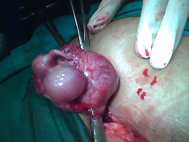
Reveals a live fetus with placenta attached
Discussion
Advanced tubal ectopic pregnancy is rare. Awareness of risk factors and improved technologies like the serum beta hcg doubling time and transvaginal ultrasonography allows for ectopic pregnancy to be identified early. In our patient case, the absence of transvaginal ultrasound done in the first trimester possibly led to the late diagnosis of the ectopic pregnancy. However, in spite of transvaginal ultrasound performed in the first trimester, ectopic pregnancy may be missed because of the inexperienced radiologists or the presence of an intrauterine gestation in women with heterotopic pregnancy [1]. Mert Gol et al. [2] have reported a case of 17-week ruptured tubal ectopic pregnancy. We believe that transvaginal ultrasound by experienced radiologist along with a serum beta hcg doubling time in first trimester is the best diagnostic modality for the early diagnosis of ectopic pregnancy.
References
- 1.Hasskios D, Bakas P, Pistofidis G, Creatsas G. Heterotopic pregnancy at 16 weeks of gestation after invitro fertilization and embryo transfer. Arch Gynecol Obstet. 2002;266:124–125. doi: 10.1007/s004040100202. [DOI] [PubMed] [Google Scholar]
- 2.Gol M, Aydin C, Guven C, et al. A 17 week tubal pregnancy. Türkiye Klinikleri Jinekoloji Obstetrik Dergisi. 2003;13:241–244. [Google Scholar]