

Abstract
Ophthalmomyiasis externa refers to the infestation of ocular surface by dipterous larvae. The term ophthalmomyiasis interna refers to the infestation of the anterior or posterior segment of the eyeball. Oestrus ovis is the most common aetiological agent for ophthalmomyiasis externa, and external disease is the most common form of ocular myiasis correspondingly. The larva is an obligate parasite of sheep. However, humans are accidentally infested. This article is related to the case series of three patients with the diagnosis of ophthalmomyiasis externa who were treated in Hakkari State Hospital in June 2013.
Background
Ophthalmomyiasis externa is the infestation of ocular surface and conjunctiva with the larval forms of flies from the phylogenic order Diptera. Ophthalmomyiasis interna results when the larva penetrates the globe of the eyes, whereas in orbital ophthalmomyiasis larva is in the orbital space. External disease is the most common form of the disease and orbital disease is the least one.1 Oestrus ovis is the main agent causing external ocular myiasis. Other species such as Rhinoestrus purpureus, Dermatobia hominis, Cochliomyia hominivorax, Chrysomyia bezziana, Wohlfartia magnifica, Oedemagena tarandi and Hypoderma bovis are implicated for ophthalmomyiasis interna. Infestations with these agents are in individuals with underlying illnesses and are frequently associated with ophthalmomyiasis interna.2
O ovis, larval form of the sheep nasal bot fly, is the main agent causing external ocular myiasis. It is an obligatory parasite of sheep and goats in their nasal and paranasal cavities. Humans are accidental hosts in its life cycle. The disease is common in sheep farming areas of the Mediterranean and Middle East countries during spring and summer.1
The infestation of conjunctiva by a fly larva is a rare event. However, I came across seven patients with ophthalmomyiasis externa in Hakkari from June to August 2013, which arouses the suspicion that this disease could be prevalent in this district. According to what the patients and their relatives said, I got the impression that the disease is rather common among those especially who deal with sheep growing. This paper issues three patients of ophthalmomyiasis externa who referred to Hakkari State Hospital Eye Clinics in June 2013.
Case presentation
Case 1: A 30-year-old man from Yukarı Akkaya farming field presented to the eye clinic, at Çukurca Town, on 5/06/2013 with a sensation of having a foreign body in his right eye as a result of being in close contact with his sheep a day ago. On ophthalmic examination, visual acuities were 20/20 in both eyes and intraocular pressures were normal bilaterally. Something translucent like a worm was moving in the precorneal tear meniscus with the 16–25 magnification of the biomicroscopy. The examination of anterior chamber and fundus images were normal bilaterally. Conjunctival hyperaemia and serous secretion were detected in the right eye. The worm in the conjunctiva was moving and it had two dark lines on its head, crawling over the bulbar conjunctiva and eating mucoid secretions. The larva was actively avoiding the light of the slit lamp and trying to burrow through the conjunctival fornices, as seen in the video 1.
Larva is seen on ocular surface.
Case 2: A 26-year-old woman from Bayköy Village presented to the eye clinic on 8/06/2013. She stated that a fly slipped into her eye a day ago before she came to the clinic. She also had a sensation of a foreign object moving in her eyes. Ophthalmic examination revealed that visual acuities were 20/20 bilaterally and intraocular pressures were normal as well. Eyelid cellulites and hyperaemic conjunctivas with mucopurulent secretion were seen in the right eye. Bilateral corneas, anterior chambers and fundus images were normal. On examination of fornix conjunctiva, a larva hiding in the secretion was seen (figure 1).
Figure 1.
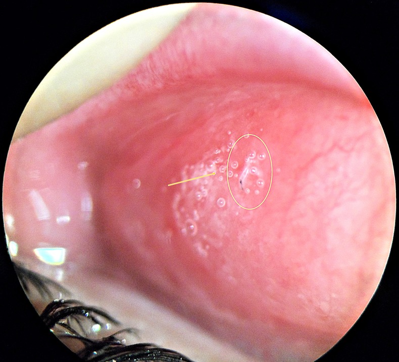
Larva buried itself in conjunctival fornix.
Case 3: A 10-year-old boy, from Yukarı Akkaya farming field, presented to my clinic on 10/6/2013 with his eyes red. Ophthalmic examination revealed that visual acuities in both eyes were 20/20 and intraocular pressures were normal bilaterally. Anterior and posterior chamber examination seemed normal as well. He said that a fly touched his eye and another fly slipped to his brothers’ throat. On slit lamp examination, a moving worm was detected in the tear meniscus of the eye (figure 2).
Figure 2.

Larva is seen in tear meniscus moving over conjunctiva.
Investigations
The larvae were taken off with a sterile cotton tip applicator and put into a container of fixator. The first-stage larval form of O ovis was detected which had a pair of sharp, dark brown oral hooks connected to the large internal cephalo-pharyngeal skeleton and numerous spines at the margins of each body segment, as seen in figure 3. The larva was translucent, small (0.8–1 mm) and avoiding the light with vermiform movements as seen in the video. The disease is a parasitic infestation of sheep whereas humans are accidental hosts. Patients were investigated for systemic illnesses and immune dysregulation, and all were found healthy.
Figure 3.
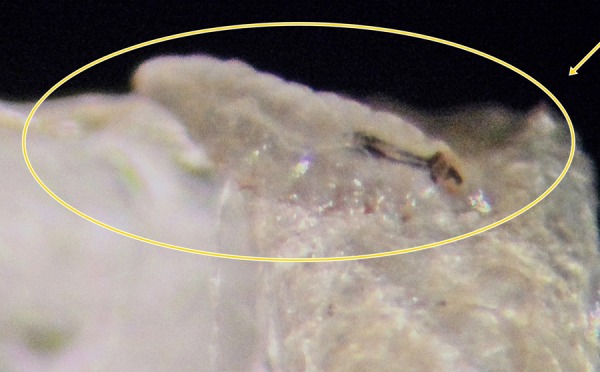
Larva has a pair of sharp, dark brown oral hooks which are connected to the large internal cephalopharyngeal skeleton and numerous spines at the margins of each body segment as seen.
Differential diagnosis
The symptoms of ophthalmomyiasis externa are very similar to symptoms of acute catarrhal conjunctivitis.3 Patients reported itching, burning, mobile foreign body sensation, photophobia, watery discharge and eyelid hyperaemia in their eyes. The symptoms begin after larval ovulation with acute onset of eye pain and inflammation unilaterally as a rule. Conjunctival pseudomembrane, follicular conjunctival reaction and punctate keratopathy may accompany the clinical picture. Viral, bacterial or foreign body conjunctivitis may cause the same symptoms. But visualisation of the larva concludes the diagnosis.
Treatment
The larvae were resisting to be taken off with a cotton tip applicator since they adhered to the conjunctiva with their oral hooks. After paralysing the larvae using topical proparacaine (% 0.5) drops, they were taken off with a sterile cotton tip applicator. We used topical ofloxacin (% 0.3) eye drops 4×1 and fluorometholone acetate (% 0.1) eye drops 4×1 in all patients to relieve inflammation and bacterial infection. Oral ampicillin sulbactam (1 g) was prescribed 1×1 for eyelid cellulites in the second patient. The clinical condition of all patients improved and conjunctival hyperaemia convalescenced in a few days with no clinical problem in the follow-up period.
Outcome and follow-up
The clinical condition of all patients improved and conjunctival hyperaemia convalescenced in a few days with no clinical problem in the follow-up period.
Discussion
Myiasis is the infestation of tissues or organs of living vertebrates by dipterous fly larvae. The most common site for infestation is skin, but eyes, noses, paranasal sinuses, throat and urogenital tract might also be infested.1 Keyt first described an ocular myiasis case in 1900; then Elliott notified ophthalmomyiasis from India in 1910.4 Ophthalmomyiasis due to O ovis was firstly described by James in 1947.5 O ovis is the most common agent for external ocular myiasis. The majority of the cases have been reported from the Mediterranean countries and Middle East region.1 O ovis, the sheep nasal bot fly, is an obligatory parasite in nasal and paranasal cavities of sheep and goats. Hakkari is a city in the south-east border of Turkey near Iran and Iraq and because sheep growing is commonly performed here, this region may be considered as an endemic site.
The sheep nasal bot fly, O ovis, is a large dark grey fly with dark spots on the dorsum of its thorax and abdomen and has light brown hair.6 Adult female fly deposits first-stage larvae into the host's external nares and the larvae make up their way into the nasal and paranasal passages where a 10-month maturation happens. When the larvae mature to pupa stage, they drop to the soil. After 3–6 weeks of next stage of maturation, adult fly emerges from the pupae and lives approximately up to a month. The adult female fly is capable of ejecting a jet of larva while in proximity to the eye.7
Patients with ophthalmomyiasis externa typically present a history of a close contact with sheep in early summer.8 Ocular involvement occurs less than 5% of all cases of human myiasis.9 Ophthalmomyiasis is classified as ophthalmomyiasis externa, if the larvae are present on the conjunctiva and ophthalmomyiasis interna, if the larvae penetrate the globe.9 The conjunctival ophthalmomyiasis may present with burning, itching, mobile foreign body sensation, photophobia, watery discharge and eyelid cellulites. External ophthalmomyiasis manifests as an acute foreign body sensation with eye pain and lacrimation unilaterally as a rule. Red eye, photophobia, conjunctival hyperaemia, lid oedema, subconjunctival haemorrhages, pseudomembrane formation and superficial punctate keratopathy may accompany the clinical picture. Differential diagnosis from preceptal cellulitis, keratitis and keratoconjunctivitis must be performed.
Ophthalmomyiasis interna is the term used when the larvae penetrate the globe. In this case, the larvae can be seen in the anterior segment, in the vitreous or in subretinal space. Anterior ophthalmomyiasis interna appears clinically as anterior uveitis, and posterior segment inflammation can accompany it. Posterior ophthalmomyiasis interna can be diagnosed by pigmented and atrophic retinal pigment epithelium changes in crisscrossing pattern, concomitant focal haemorrhages, multifocal scarring and retinal detachment.10 Red eye, vision loss, floaters, eye pain and scotomas are the symptoms that have been described for ophthalmomyiasis interna. In orbital ophthalmomyiasis, the larva is in the orbit. Eyelid tumours are the most common predisposing factor causing orbital myiasis.1
Cattle and sheep are natural hosts but larva of O ovis can also develop in human eye. The disease is self-limited and confined to the conjunctiva. Invasion of the disease due to O ovis is rarely notified.11 Treatment of ophthalmomyiasis externa is managed by removal of the larva. As the larva avoids from the slit lamp light and grips firmly to conjunctiva with its oral hooks, it could be difficult to remove easily. Topical anaesthetic drops may be used to paralyse the larva or liquid paraffin may be used to asphyxiate it. The erosion of conjunctiva by bristles of larvae may facilitate bacterial infections and thereby ocular inflammation.12 Topical corticosteroids relieve the inflammation and topical antibiotics prevent secondary bacterial superinfection.13–18 Ofloxacin and norfloxacin eye drops may be preferred for pathogenic bacteria and larvae in external disease.12 Ophthalmomyiasis is really a rare morbidity but it is common in sheep farming areas, so it could be assessed as an occupational disease. It can be suggested that injection of ivermectin, doramectin or moxidectin into the nasal passages of the sheep may be effective in preventing the disease.12
The infestation of human conjunctiva by an insect larva is a rare event but it is very common in countries where standards of hygiene are low. Most cases are seen during spring and summer seasons. Inspection of conjunctival fornix for foreign objects is essential in the diagnosis.
Learning points.
Parasitic infections of ocular surface may be seen in healthy individuals.
Sanitation and occupational knowledge is important in parasitic diseases.
Prompt diagnosis and treatment prevents serious complications.
Fornix examination is essential in the diagnosis of larva.
Ophthalmologists must take into consideration ophthalmomyiasis in the differential diagnosis of conjunctivitis.
Footnotes
Competing interests: None.
Patient consent: Obtained.
Provenance and peer review: Not commissioned; externally peer reviewed.
References
- 1.Francesconi F, Lupi O. Myiasis. Clin Microbiol Rev 2012;25:79–105 [DOI] [PMC free article] [PubMed] [Google Scholar]
- 2.Chodosh J, Clarridge J. Ophthalmomyiasis: a review with special reference to Cochliomyia hominivorax. Clin Infect Dis 1992;14:444–9 [DOI] [PubMed] [Google Scholar]
- 3.Stulting AA, Meyer H. External ophthalmomyiasis caused by Oestrus ovis. S Afr Med J 1981;60:709–10 [PubMed] [Google Scholar]
- 4.Sivaramasubramanyam P, Sadanand AV. Ophthalmomyiasis. Br J Ophthalmol 1968;52:64–5 [DOI] [PMC free article] [PubMed] [Google Scholar]
- 5.Patel SJ. Extra-ocular myiasis due to the larva of Oestrus ovis. East Afr Med J 1975;52:167–9 [PubMed] [Google Scholar]
- 6.Beaver PC, Jung RC, Cupp EW. Clin Parasitol. 9th edn Philadelphia, PA: Lea & Febiger, 1984:680–95 [Google Scholar]
- 7.Sigauke E, Beebe WE, Gander RM, et al. Case report: ophthalmomyiasis externa in Dallas County, Texas. Am J Trop Med Hyg 2003;68:46–7 [PubMed] [Google Scholar]
- 8.Healey MC, Collins RK, Hawkins JA. Ophthalmooestriasis externa. South Med J 1980;73:1387–9 [DOI] [PubMed] [Google Scholar]
- 9.Pandey A, Madan M, Asthana AK, et al. External ophthalmomyiasis caused by Oestrus ovis: a rare case report from India. Korean J Parasitol 2009;47:57–9 [DOI] [PMC free article] [PubMed] [Google Scholar]
- 10.Lima LH, Laud K, Rosenberg P, et al. Ophthalmomyiasis with a singular subretinal track. Am J Ophthalmol 2010;150:731–6 e1 [DOI] [PubMed] [Google Scholar]
- 11.Baliga MJ, Davis P, Rai P, et al. Orbital myiasis: a case report. Int J Oral Maxillofac Surg 2001;30:83–4 [DOI] [PubMed] [Google Scholar]
- 12.Jayaprakash K, Karthikeyan A. Ocular myasis and associated mucopurulent conjuctivitis acquired occupationally: A case study. Indian J Occup Environ Med 2008;12:20–2 [DOI] [PMC free article] [PubMed] [Google Scholar]
- 13.Reingold WJ, Robin JB, Leipa D, et al. Oestrus ovis ophthalmomyiasis externa. Am J Ophthalmol 1984;1997:7–10 [DOI] [PubMed] [Google Scholar]
- 14.Gregory AR, Schatz S, Laubach H. Ophthalmomyiasis caused by the sheep bot fly Oestrus ovis in northern Iraq. Optom Vis Sci 2004;81:586–90 [DOI] [PubMed] [Google Scholar]
- 15.Bose S, Saini S, Barapatre R, et al. Ophthalmomyiasis External: A Case Report. J Clin Diagn Res [serial online] 2012;6:1079–80 [Google Scholar]
- 16.Thakur K, Singh G, Chauhan S, et al. Vidi, vini, vinci: external ophthalmomyiasis infection that occurred, and was diagnosed and treated in a single day: a rare case report. Oman J Ophthalmol 2009;2:130–2 [DOI] [PMC free article] [PubMed] [Google Scholar]
- 17.Abdellatif MZ, Elmazar HM, Essa AB. Oestrus ovis as a cause of red eye in Aljabal Algharbi, Libya. Middle East Afr J Ophthalmol 2011;18:305–8 [DOI] [PMC free article] [PubMed] [Google Scholar]
- 18.Yar K, Ozcan AA, Koltas IS. [External ophthalmomyiasis: case reports]. Turkiye parazitol Derg/Turkiye Parazitol 2011;35:224–6 [DOI] [PubMed] [Google Scholar]
Associated Data
This section collects any data citations, data availability statements, or supplementary materials included in this article.
Supplementary Materials
Larva is seen on ocular surface.