
Figure 1.
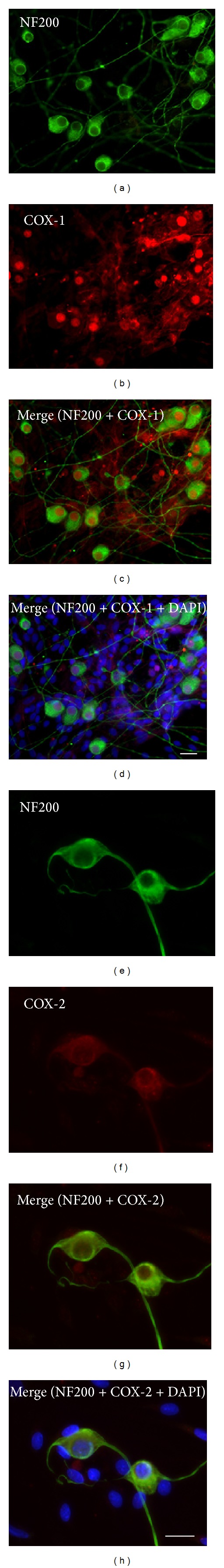
Immunofluorescence of COX-1 and COX-2 in VGNs. All of the neurons identified as NF200-positive (a) expressed COX-1 in either the cytoplasm or nuclei (b) and had an increased signal in the nuclei as confirmed by the merged images ((c), (d)). In comparison, weak COX-2-specific staining was identified mainly in the cytoplasm of VGNs as shown in ((e), (f)) and was confirmed by the merged images ((g), (h)). Scale bar, 25 microns.