Abstract
Ediacara fossils are central to our understanding of animal evolution on the eve of the Cambrian explosion, because some of them likely represent stem-group marine animals. However, some of the iconic Ediacara fossils have also been interpreted as terrestrial lichens or microbial colonies. Our ability to test these hypotheses is limited by a taphonomic bias that most Ediacara fossils are preserved in sandstones and siltstones. Here we report several iconic Ediacara fossils and an annulated tubular fossil (reconstructed as an erect epibenthic organism with uniserial arranged modular units), from marine limestone of the 551–541 Ma Dengying Formation in South China. These fossils significantly expand the ecological ranges of several key Ediacara taxa and support that they are marine organisms rather than terrestrial lichens or microbial colonies. Their close association with abundant bilaterian burrows also indicates that they could tolerate and may have survived moderate levels of bioturbation.
The Ediacara biota, exemplified by fossils preserved in the Ediacara Member of South Australia, provides key information about the origin, diversification, and disappearance of a distinct group of soft-bodied, macroscopic organisms on the eve of the Cambrian diversification of marine animals1,2,3. Ediacara fossils have been reported from all major continents except Antarctica, but they are mostly restricted to 580–541 Ma sandstones and siltstones, with rare occurrences in carbonate rocks4,5,6,7,8 and black shales9,10. The origin of this taphonomic bias toward cast and mold preservation in sandstones and siltstones is probably related to the pervasive development of thick microbial mats in these facies11. This taphonomic pattern is opposite to that of Phanerozoic soft-bodied Lagerstätte which are mostly found in shales and carbonate rocks, and it may represent a taphonomic bias limiting a full appreciation of the ecological and taphonomic diversity of Ediacara fossils, which in turn hampers our understanding of the phylogenetic affinities and evolutionary dynamics of Ediacara organisms. This is highlighted in the recent debate on the proposition that many Ediacara fossils may represent terrestrial lichens or microbial colonies that lived on paleosols9,12,13,14,15,16, emphasizing the inadequate knowledge about the paleoecology of Ediacara fossils17,18. This deficiency also limits our ability to test various hypotheses about the evolutionary dynamics of Ediacara organisms, specifically whether their disappearance at the Ediacaran-Cambrian boundary was driven by an environmentally rooted mass extinction, ecological displacement by Cambrian metazoans, or closure of a unique taphonomic window3.
In this study, we examine the under-explored limestone taphonomic window in order to fully document the ecological range and to test various interpretations about the paleoecology and evolution of Ediacara organisms. We report several iconic Ediacara genera (Hiemalora, Pteridinium, Rangea, and Charniodiscus) and a new taxon (Wutubus annularis new genus and species) from bituminous limestone of the Shibantan Member of the Dengying Formation (551–541 Ma) in the Yangtze Gorges area of South China. The Shibantan limestone was deposited in a subtidal marine setting, between the fair weather and storm wave bases, and is also known to contain abundant trace fossils19,20 as well as other Ediacara-type fossils such as Paracharnia dengyingensis21 and Yangtziramulus zhangi5,6. The new data extend the stratigraphic, paleogeographic, environmental, and taphonomic ranges of Charniodiscus, Hiemalora, Pteridinium, and Rangea, and they suggest that these Ediacara organisms were marine organisms rather than terrestrial lichens or microbial colonies. Some of these fossils, particularly Wutubus annularis, are also closely associated with trace fossils, indicating that they could tolerate at least moderate levels of bioturbation.
Results
Pteridinium
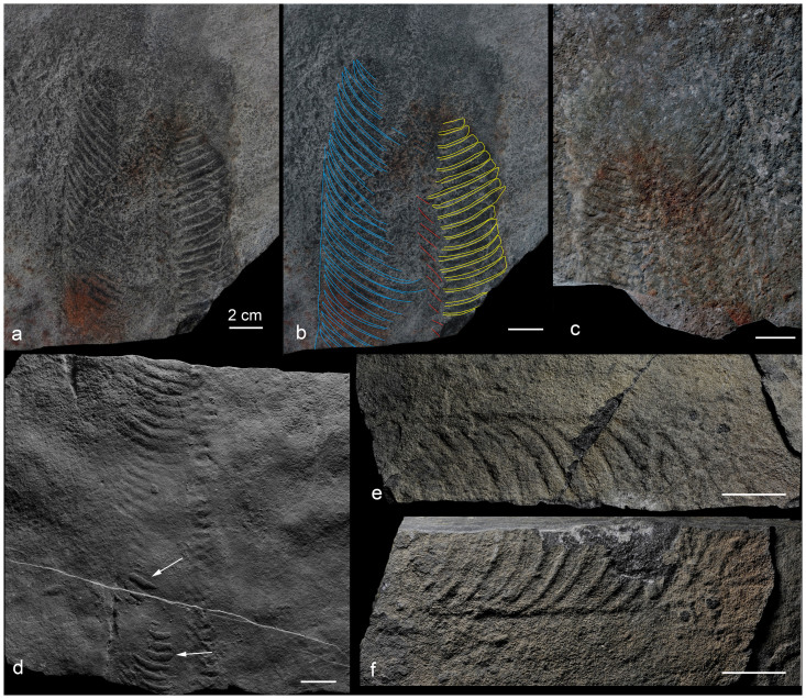
Six fragments of frondose fossils are assigned to this genus. The best preserved specimen, consisting a part and a counterpart (Fig. 2a–c), is a leaf-shaped petalodium, 110 mm in maximum width, 171 mm in preserved length, and consisting two lateral petaloids (or vanes) and a central petaloid. The two lateral petaloids are clearly visible, one of which with 26 well-preserved and gently curved segments (outlined in blue in Fig. 2b). The central petaloid is poorly preserved, with indication of segments only in the axial region of the fossil (outlined in red in Fig. 2b); these segments are oriented differently from those of lateral petaloids and are thus inferred to represent the central petaloid. The axial region of the specimen is poorly preserved; hence it cannot be determined whether the segments of different petaloids meet opposingly or alternately in the axial zone. Individual segments are elongate laterally, about 5 mm in axial width and 66 mm in maximum lateral length. Because the segments are curved distally, they also gently taper towards the margin of the specimen. The branching angle between segments and central axis varies at 70°–88°, and it systematically decreases toward one end of the petalodium (and presumably also decreases toward the other end, which is not preserved); as a result, the petalodium may taper toward both ends. The segments do not seem to be free from each other, and the boundary between neighboring segments is preserved as a ridge (about 1.5 mm wide) or a groove on the corresponding counterpart. The five other specimens are morphologically similar (Fig. 2d–f), although several segments on one of the specimens are preserved as internal molds (arrows in Fig. 2d), suggesting that the segments are tubular in nature.
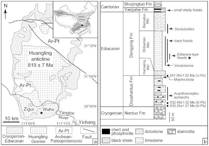
Figure 1. Geological map (a) and stratigraphic column (b).
Inset map in (a) shows simplified geological map of China and location of the Yangtze Craton. Star in (a) points to fossil locality near Wuhe, and star in (b) points to fossil horizon. Modified from ref. 19. Radiometric dates from ref. 22.
Figure 2. Pteridinium from the Shibantan Member.
(a), (c) Part and counterpart (NIGP159071). (b) Same as (a) with segments outlined (lateral petaloids in blue and yellow, and central petaloid in red). Arrows in (d; NIGP159072) point to internal molds of segments. (e–f) Part and counterpart (NIGP159073). Scale bars 20 mm. Photographs taken by authors.
The recurved segments of the Shibantan Pteridinium specimens are similar to those of P. carolinaense from the McManus Formation of North Carolina28 and Spitzkopf Formation in southern Namibia29, P. cf. simplex from the Ediacara Member30, and P. nenoxa from the White Sea area31, which may all be synonymous28,29. The Shibantan specimens are here referred to as Pteridinium sp., although detailed systematic work may prove them to be P. carolinaense.
Rangea
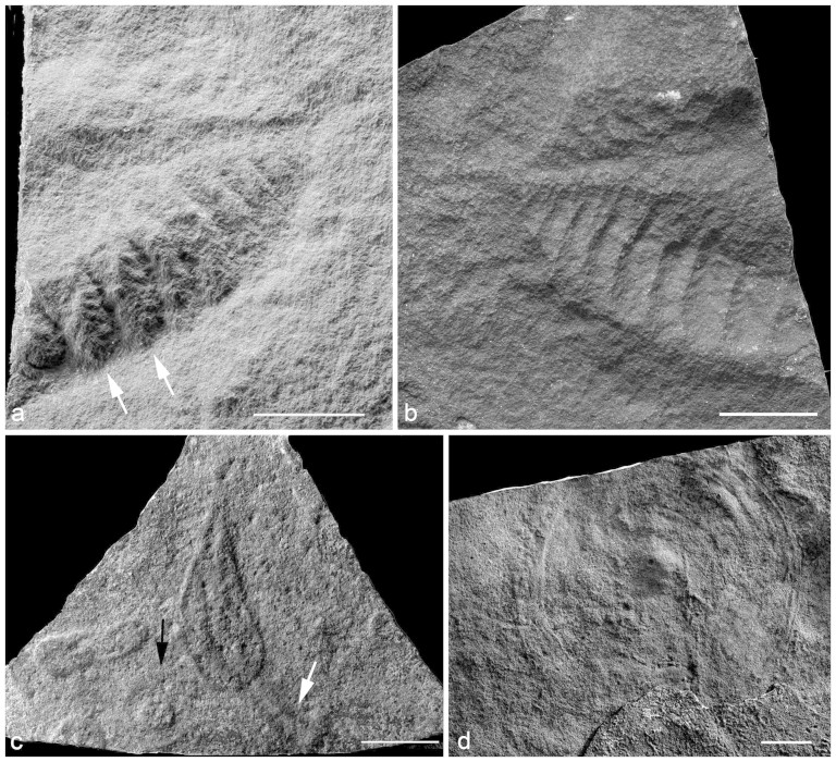
A single frondose specimen, with part and counterpart (Fig. 3a–b), is assigned to the genus Rangea. This is an incompletely preserved specimen with the apical part of a petaloid (or vane) bearing at least nine primary branches or frondlets, which are sutured tightly and emanate from axial zone at 90°. The axial width of the primary branches decreases from 4.7 mm to 2.5 mm and the lateral length decreases from 16.1 mm to 6.1 mm toward the apical end of the petaloid. Two of the primary branches (arrows in Fig. 3a) bear fully visible, biserially arranged rangid-type secondary branches (0.7–1.1 mm in width), so that they can be described as double-sided32 or unfurled and fully displayed primary branches33. Because the axial part of the petaloid is poorly preserved, it is uncertain whether there are any subsidiary primary branches34.
Figure 3. Rangea (a–b; NIGP159074), Charniodiscus (c; NIGP159075), and Spriggia-type holdfast (d; NIGP159076).
(a–b) Part and counterpart, with arrows in (a) pointing to frondlets with secondary branches. White arrow in (c) points to a prominent stem and black arrow in (c) points to an isolate holdfast. Scale bars 10 mm in (a–b) and 20 mm in (c–d). Photographs taken by authors.
Among all described rangeomorphs32,33, Rangea appears to be the best generic home for the Shibantan specimen. This taxonomic assignment is based on the overall frond morphology and the tightly sutured, rangid-type primary branches. However, the fragmentary preservation does not allow a confident determination of the number of petaloids, the full number of frondlets per petaloids, and the morphology of the basal part of the petalodium. Thus, although the current taxonomic assignment at the genus level is likely, determination at the species level is impossible.
Charniodiscus
A single frondose specimen with a prominent stem (white arrow in Fig. 3c) and a central stalk is described under the genus Charniodiscus. The specimen is 73 mm in length and 20 mm in maximum width, consisting of a stem (27 mm long and 7.1 mm wide) and a lanceolate frond (50.1 mm long) with no subdivisions discernible. Although a discoidal holdfast is not directly attached to the stem, several discoidal fossils are preserved next to the frond (black arrow in Fig. 3c) and they are presumably the holdfast of Charniodiscus. Large discoidal fossils, some of which have a central boss surrounded by concentric rings and are similar to Spriggia that has been synonymized with Aspidella35, are common in the Shibantan Member (Fig. 3d), and they could well be holdfasts of frondose fossils.
The overall frond shape and the presence of a prominent stalk of this frondose specimen are characteristic of Charniodiscus. The Shibantan specimen is similar in overall morphology to C. procerus36,37, but its poor preservation gives no detail of frond subdivisions or primary branches. Thus, it is tentatively identified as a Charniodiscus specimen.
Hiemalora
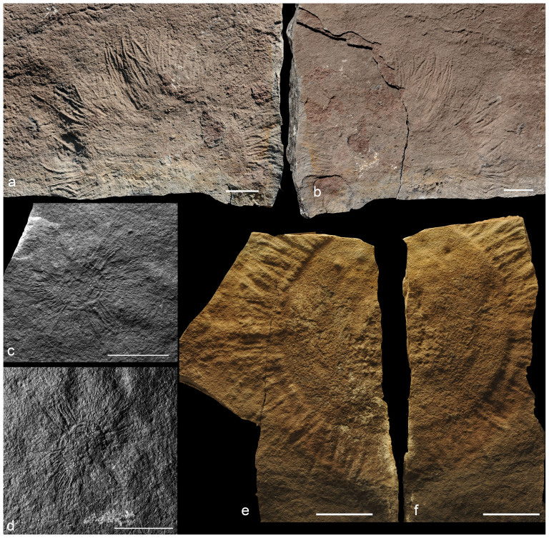
Nine discoidal fossils, six with part and counterpart, bear slender appendages characteristic of the genus Hiemalora (Fig. 4). They have a central disc with a highly variable diameter (5–100 mm) and defined by an outer rim or groove, although one specimen lacks a well-defined central disc. Numerous radiating appendages are attached to the outer rim of the central disc. The appendages are densely arranged, flexible, distally tapering, relatively long (60–100% of central disc diameter), and ~2 mm in basal width. Some appendages are apparently bifid, but this could be an artifact due to superimposed appendages.
Figure 4. Hiemalora pleiomorpha.
(a–b; NIGP159077), (c–d; NIGP159078), and (e–f; NIGP159079) Three pairs of part and counterpart, showing densely arranged appendages. Scale bars 20 mm. Photographs taken by authors.
The Shibantan specimens are similar in appendage density and relative length to Hiemalora pleiomorpha from the Khatyspyt Formation in northern Siberia38, although H. pleiomorpha has been considered as a junior synonym of the type species H. stellaris, with the differences between them regarded as intraspecific and taphonomic39. Hiemalora has been interpreted as a hydrozoan polyp40 or a body fossil with unknown affinities41. However, recent investigations have shown that it is likely a discoidal holdfast with radiating root-like rays39,42; this interpretation is supported by the discovery of Hiemalora-like structures attached to the fronds of Primocandelarbum hiemaloranum39. Because Hiemalora is reconstructed as a hemispherical holdfast buried within sediments, taphonomic factors (e.g., impression of the holdfast at different levels) can account for the variable diameter and the complete absence of a central disc in some specimens43.
Annulated tubes
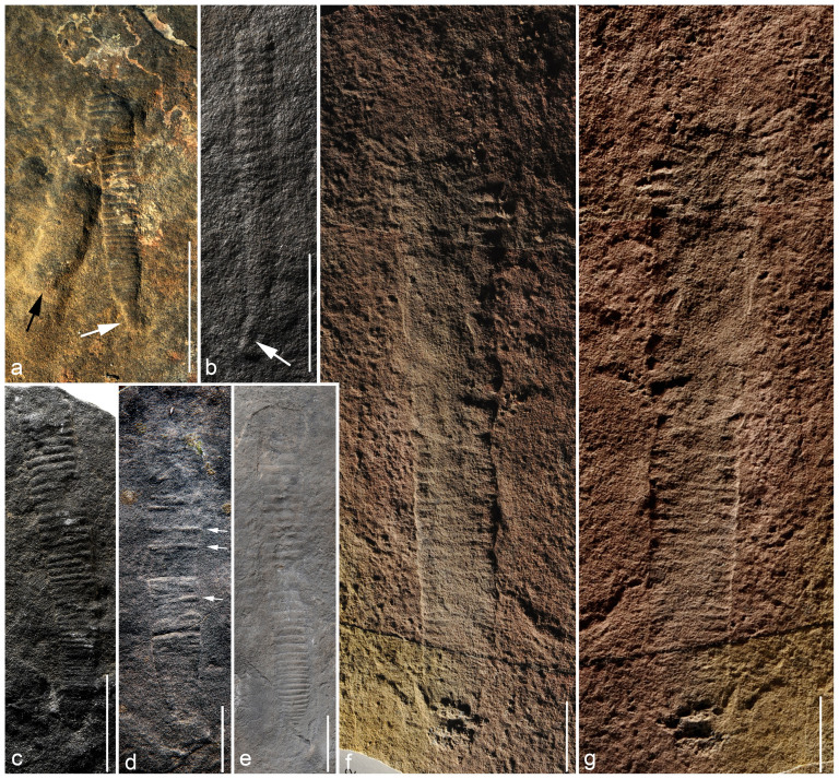
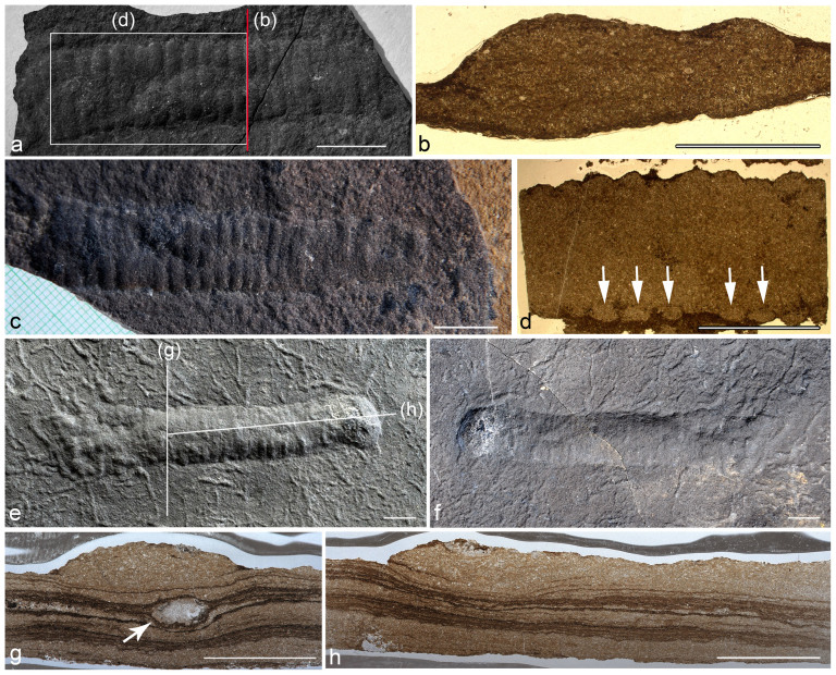
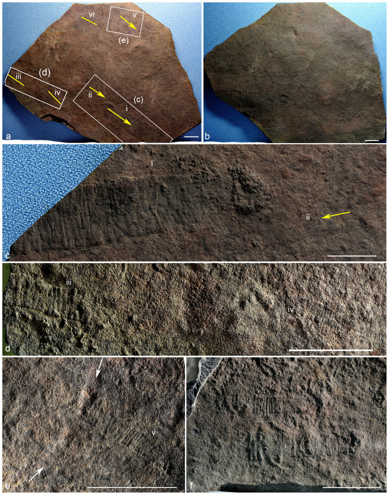
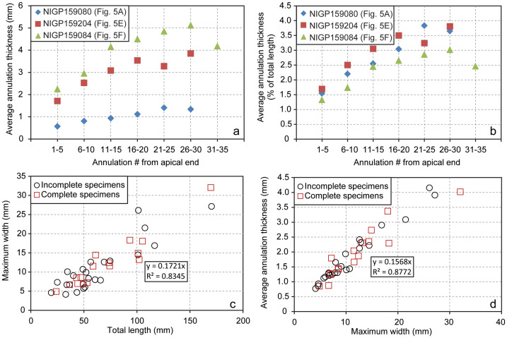
More than one hundred specimens of annulated tubular fossils (Figs. 5,6,78), many with parts and counterparts, are here described as Wutubus annularis new genus and species (see “Systematic Paleontology”). Most specimens are compressed to various degrees (Fig. 5), although a few show the three-dimensional nature of the tubes (Fig. 6). When multiple specimens occur on the same bedding surface, they tend to be preferentially aligned (Fig. 7). The most completely preserved specimens have a narrower conical end (referred to as the apex; white arrows in Fig. 5a–b) and a slightly widening but more or less cylindrical tube with a truncated abapical end; often there is a sharp change in width between the apex and the tube (Fig. 5a–b). The apex is smoother than the tube, which is ornamented by transverse annulations. The fossils are up to 180 mm in total length, 3–32 mm in maximum width, whereas the apex is ~5 mm in length and 2 mm in width. Annulae are 0.5–4.5 mm in thickness. In most specimens, the annulae thicken abapically (Fig. 5a–e; 8a–b); in a few others, thick annulae appear to be interspersed between thin ones (Fig. 5d), probably a taphonomic artifact because the apparently thicker annulae are preserved three-dimensionally between deflated annulae. There are positive correlations between total length and maximum width (Fig. 8c) and between maximum width and mean annular thickness (Fig. 8d). Pustular structures about 2–4 mm in size are present at the apical end of several specimens (Fig. 5f–g), but their biological vs. taphonomic origin, biological association with the tubular fossils, and ecological functions (e.g., holdfast) are uncertain.
Figure 5. Wutubus annularis.
White arrows in (a–b) point to apex, and black arrow in (a) points to a burrow underlying the tubular fossil. Arrows in (d) point to inflated annulae, with deflated ones in between. Note pustular structures at apical end in (f–g; part and counterpart). (a) NIGP159080; (b) NIGP159081; (c) NIGP159082; (d) NIGP159083; (e) NIGP159204; (f–g) NIGP159084. Scale bars 20 mm. Photographs taken by authors.
Figure 6. Thin section observations of Wutubus annularis.
(a, c) and (e–f) Two pairs of part and counterpart. (b) Transverse section cut perpendicular to bedding surface along red line labeled “b” in (a), showing tube infillings. (d) Longitudinal section cut parallel to bedding surface along white rectangle labeled “d” in (a), showing annulae in cross section (arrows). (g) Transverse section cut perpendicular to bedding surface along line labeled “g” in (e), showing a burrow (arrow) underlying Wutubus annularis tube. (h, apex to the left) Longitude section cut perpendicular to bedding surface along line labeled “h” in (e, apex to the right). Note abundant burrows in (e–f). Scale bars 5 mm for (b) and 10 mm for all others. Photographs taken by authors.
Figure 7. Alignment of Wutubus annularis tubes.
(a–b) Part and counterpart (NIGP159085), with yellow lines marking orientation of tubes (arrow heads pointing away from apex when preserved, or pointing to inferred down-current direction). Labeled rectangles in (a) outline areas enlarged in (c–e), and labeled specimens (i–v) are enlarged in (c–e). Arrow in (c) show orientation of a small specimen just above the arrow, and arrows in (e) point to a burrow beneath W. annularis tube. (f) Two specimens on the same bedding surface as (a). NIGP159086. Scale bar 20 mm. Photographs taken by authors.
Figure 8. Measurements of Wutubus annularis.
(a–b) Plots showing abapical thickening of annulae in three completely preserved specimens of different size. Annulation thickness is averaged for every five annulae. When annulation thickness is normalized against total length of specimens (b), the trend of abapical thickening is more similar among the three specimens, indicating inflational growth. (c–d) Correlation among total length, maximum width, and average annulation thickness (averaged for each specimen). Regression and correlation statistics are based on pooled measurements of both complete and incomplete specimens.
Thin sections cut transversely and longitudinally (both parallel and perpendicular to bedding plane) show that the interior of W. annularis is filled with peloids and intraclasts cemented by calcite (Fig. 6b, d, g, h), similar to the surrounding matrix. One of the longitudinal thin sections cut parallel to bedding plane shows that the annulae appear to correspond to faintly preserved circles at the margin of the tube (arrows in Fig. 6d). Thus, the annulae may represent hollow rings around the W. annularis tube. This interpretation is consistent with the observation that some annulae can be deflated and preserved as lower relief features than others (Fig. 5d).
Numerous W. annularis specimens of different sizes can be preserved on the same bedding plane, and they are preferentially oriented with their apices pointing to the same direction (Fig. 7a). Sometimes they are found on bedding surface with abundant trace fossils (Figs. 5a, 6e–f, 7e), which are interpreted as undermat burrows19,20. It is clear from both bedding surface and thin section observations that co-existing burrows do not penetrate W. annularis tubes; instead, W. annularis tubes overlap burrows, and both are compressed slightly and depress underlying microlaminae (Fig. 6g) that are interpreted as microbial mats19,20.
Wutubus annularis is reconstructed as an erect benthic tubular organism living on the sediment surface (Fig. 9). The non-penetration relationship with co-existing burrows is consistent with, although not a proof of, W. annularis being an erect benthic organism. Moreover, the non-penetration relationship indicates that, after death, the erect tubes of W. annularis fell upon and superimposed on the burrows. The preferential orientation of W. annularis tubes was probably due to current alignment, suggesting that the apex was tethered to the substrate and that W. annularis was preserved in-situ rather than transported.
Figure 9. Reconstruction of Wutubus annularis in life position.
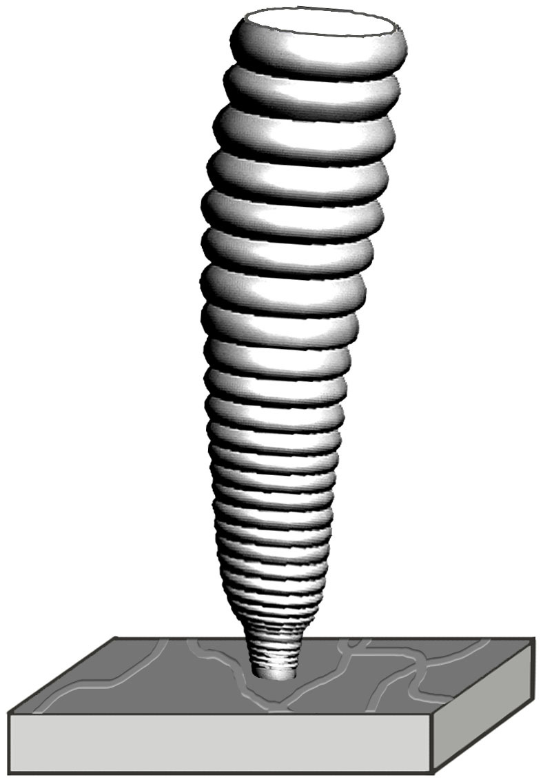
The more or less smooth apex could be the embryonic test separated from the tube by an abrupt change in growth dynamics. The tube seems to have grown through initial accretionary growth to a set number of annulae, followed by isometric inflation, as evidenced by the observation that small and large tubes, when completely preserved, have similar number of annulae (e.g., specimen in Fig. 5a is 37 mm long with 37 annulae; specimen in Fig. 5e is 107 mm long with 37 annulae; specimen in Fig. 5f is 170 mm long with ~35 annulae). The inflational growth can also explain the isometric relationship between total length, maximum width, and annular thickness (Fig. 8c–d). It is possible that W. annularis had an apertural opening at the abapical end, but it is uncertain whether or what organisms occupied the tube.
Annulated tubular fossils are very common in Ediacaran successions, often preserved as casts/molds in sandstones/siltstones44,45,46 or as carbonaceous compressions in shales or carbonate rocks4,47,48. In particular, W. annularis is somewhat similar to an annulated tubular fossil reported from the Khatyspyt Formation in Siberia (Fig. 3G of ref. 4), although the latter is much larger. Considering that the annulae of W. annularis could be hollow rings that are arranged uniserially to make an annulated tube, however, it is more appropriate to make a comparison with Ediacara-type fossils with a uniserial modular construction. The annulae of W. annularis can be compared with and may be homologous to the serial elements in Funisia dorothea49, Palaeopascichnus50, Cloudina51,52, and Conotubus53, although the morphologies of the serial elements vary between these taxa. These similarities highlight the prevalence of modular organisms in the Ediacara biota54,55. Compared with other Ediacara-type fossils that are uniserially modular, however, W. annularis can be distinguished by a differentiated apex that may represent an embryonic test.
The phylogenetic affinity of Wutubus annularis is unknown, largely because anatomical details within the tube are not preserved. Other uniserially modular organisms, such as Funisia dorothea49, Palaeopascichnus50,54, and Cloudina51, have been interpreted as protists (e.g., xenophyophores), sponges, cnidarians, or bilaterians. Although these divergent interpretations may provide a guide to the phylogenetic placement of W. annularis, currently available evidence does not constrain its phylogenetic affinity.
Discussion
Charniodiscus, Hiemalora, Pteridinium, and Rangea have wide geographic distribution and long stratigraphic ranges2,3. For example, Charniodiscus ranges from ~580 Ma to ~555 Ma, and has been known as a common member of the Avalon assemblage in Newfoundland and England, as well as in the White Sea assemblage in Russia, Ukraine, and South Australia3. Pteridinium ( = Onegia) first appears in the 558 Ma Verkhovka Formation in Russia17 and survived as one of the youngest Ediacara fossils until 543 Ma29; it is a member of the White Sea assemblage (in Russia, Ukraine, and South Australia) and the Nama assemblage of southern Namibia, and possibly also present in the deep-water Blueflower Formation in the Mackenzie Mountains of northwestern Canada3,7. Rangea has been found in White Sea, South Australia, and Namibia, ranging from 558 Ma to 549 Ma. Hiemalora probably ranges from 565 Ma to 550 Ma, and has been reported from Newfoundland39,41, White Sea40,42, Ukraine56, northern Siberia4,38,43, Mackenzie Mountains of northwestern Canada57, northern Norway58, and possibly the Jodhpur Sandstone in Rajasthan of western India59.
The new occurrence of Hiemalora, Pteridinium, Rangea, and Charniodiscus in the 551–541 Ma Shibantan Member does not significantly change the known stratigraphic range of these genera, except for Charniodiscus which may be extended to <551 Ma. However, the new data do broaden the already wide geographic distribution of these genera. More importantly, among the four genera listed above, only Hiemalora has been previously known from both carbonate and siliciclastic facies, and the others were known exclusively from siliciclastic facies. Thus, the new data from the Shibantan Member do extend the environmental and taphonomic ranges of Charniodiscus, Pteridinium, and Rangea. Indeed, the wide geographic and environmental distribution of some Ediacara fossils now forces us to recognize their unusual capability of environmental tolerance and ecological dispersal across large expanses of Ediacaran oceans3. However, even these widely distributed Ediacara organisms must also be restricted by environmental factors, and it is highly unlikely that Charniodiscus, Pteridinium, and Rangea could have crossed the immense environmental boundary between subaqueous and subaerial regimes. Because Charniodiscus, Pteridinium, and Rangea are also present in the Ediacara Member of South Australia18, their occurrence in marine limestone of the Shibantan Member implies that they were marine organisms9 rather than terrestrial lichens or microbial colonies13, and that the fossiliferous rocks of the Ediacara Member are marine sediments14 rather than terrestrial paleosols12.
Although it has been recognized that environmental factors played a role in controlling the distribution of Ediacara fossils17,18, it is undeniable that the Ediacara biota did evolve and eventually went extinct3. Thus, the three Ediacara assemblages recognized by Waggoner60—the Avalon, White Sea, and Nama assemblages—still provide a useful framework to view the evolutionary pattern of the Ediacara biota, even though new discoveries will constantly modify our views about the environmental and temporal ranges of individual taxa. Individually, Charniodiscus, Hiemalora, Pteridinium, and Rangea do not make any unique assignment of the Shibantan assemblage to one of the three assemblage recognized by Waggoner60. The overall taxonomic makeup of the Shibantan assemblage, however, is similar to the Nama assemblage; affinity with the Nama assemblage is also supported by the presence of Cloudina and Sinotubulites, the absence of bilateralomorphs that are so common in the White Sea assemblage, the abundance of trace fossils19, and the age of the Dengying Formation (551–541 Ma). Thus, the Shibantan fossils described here indicate that elements of the Nama assemblages survived to the late Ediacaran Period in both high-energy distributary channel systems of the Nama Group and low-energy subtidal environments in the Shibantan Member. The presence of Charniodiscus in the Shibantan Member suggests that this genus rivals Charnia as one of the longest-ranging Ediacara fossils, present in all three assemblages as recognized by Waggoner60.
The Shibantan Member is characterized by abundant trace fossils, particularly undermat burrows19,20. Although the depth of burrows is relatively shallow (within a few millimeters), there is a moderate level of bedding surface bioturbation intensity in the Shibantan Member. In burrowed beds of the Shibantan Member, the percentage of bedding plane area covered by trace fossils can be as much as 20–40%, comparable to similar measurements in pre-trilobite Cambrian carbonate rocks20. Hiemalora, Pteridinium, and Rangea are sometimes found in bioturbated beds. Wutubus annularis, in particular, can co-exist with abundant burrows without being toppled by bioturbation (Figs. 5a, 6e–f, 7e). Thus, Ediacara taxa such as Wutubus annularis could tolerate moderate levels of bioturbation. Broader investigation of ecological interaction between Ediacara fossils and trace fossils is an important step toward a critical test whether animal bioturbation might have driven Ediacara organisms to extinction at the Ediacaran-Cambrian boundary3 and if so, whether there is any ecological selectivity in this extinction event.
Conclusions
Previous reports of Ediacara fossils in the Shibantan Member are tantalizing but inconclusive5,21. The new discovery of unambiguous Ediacara fossils, including Hiemalora, Pteridinium, Rangea, and Charniodiscus, adds to our understanding of the geographic, stratigraphic, environmental, and taphonomic distribution of these iconic genera. The stratigraphic range of Charniodiscus is extended to be <551 Ma, making it one of the longest living Ediacara genera. Charniodiscus, Pteridinium, and Rangea were previously known only from siliciclastic successions, and they are now known from marine carbonate successions as well. These three genera are also present in the Ediacara Member of South Australia, and their occurrence in marine limestones of the Shibantan Member indicates that they are marine organisms rather than terrestrial lichens or microbial colonies. A new annulated tubular fossil, Wutubus annularis new genus and species, is described from the Shibantan Member. Although the phylogenetic affinity of this new fossil remains unresolved, it is possible that it is a uniserially modular organism that is similar in body construction to many other Ediacara-type organisms such as Funisia, Palaeopascichnus, Cloudina, and Conotubus, highlighting the importance of modular body construction in the Ediacaran biota. Even though it has only a simple apex that could represent an embryonic test and may have functioned as a holdfast, Wutubus annularis was able to tolerate and may have survived moderate levels of bioturbation.
Systematic paleontology
Wutubus new genus. Etymology: Genus name derived from the fossil locality near the village of Wuhe (Wu River) and from Latin tubus (tube).
Type species: Wutubus annularis new genus and species.
Diagnosis: As for type species.
Wutubus annularis new genus and species.
Etymology: Species epithet derived from Latin, annularis, with reference to the transverse annulae on the tube.
Holotype: NIGP159080 (Fig. 5a), reposited at the Nanjing Institute of Geology and Paleontology.
Diagnosis: Annulated conotubular fossil, straight or slight curved, millimeters to centimeters in maximum width, and consisting of an conical apex and a slightly expanding but more or less cylindrical tube. Apex is more or less smooth, but tube is ornamented with tightly arranged transverse annulae that are millimeters in thickness and sometimes thicken abapically.
Description: Fossils 20–180 mm in total length (apex and tube) and 3–32 mm in maximum width. Apex ~5 mm in length and 2 mm in width. Annulae 0.5–4.5 mm in thickness, and thicken abapically. Total length, maximum width, and mean annular thickness are positively correlated.
Occurrence: The Shibantan Member of the Dengying Formation at Wuhe, Yangtze Gorges area, South China.
Nomenclature Note: The electronic edition of this article is published in a journal with an ISSN (2045–2322), and has been archived in the following publically available repositories: PubMed Central, Nanjing Institute of Geology and Paleontology library, and Virginia Tech library. Taxonomic nomenclature published in this article conforms to the requirements of the amended International Code of Zoological Nomenclature (ICZN), and hence is available under ICZN. This publication and the nomenclatural acts it contains have been registered in ZooBank (www.zoobank.org). The ZooBank LSID (Life Science Identifier) for this publication is urn:lsid:zoobank.org:pub:1C531183-DABB-4295-8564-86D3CFA74679.
Geological setting and methods
The Dengying Formation in the Yangtze Gorges area was deposited in a shallow water carbonate platform, and it is underlain by the Doushantuo Formation and overlain by the Yanjiahe Formation (Fig. 1). Insofar as the uppermost Doushantuo Formation was dated at 551.09 ± 1.02 Ma22 and the Yanjiahe Formation contains early Cambrian small shelly fossils and acritarchs23,24,25, the Dengying Formation is constrained between 551 Ma and 541 Ma. It can be divided into, in ascending order, the Hamajing, Shibantan, and Baimatuo members. The Hamajing Member consists of peritidal dolostone with dissolution vugs and tepees, the Shibantan Member of dark grey thin-bedded fetid bituminous limestone deposited in a subtidal environment, and the Baimatuo Member of light grey massive peritidal dolostone with dissolution vugs20. The fossiliferous Shibantan Member is characterized with thin-bedded limestones consisting of thin layers of peloidal grainstones interbedded with silty-muddy microlaminae. Unlike the Hamajing and Baimatuo members, the Shibantan Member shows no evidence of intertidal sedimentation (e.g., dissolution vugs and tepees). Parallel bedding is the dominant sedimentary structures, although low-angle cross-bedding and rip-up clasts are occasionally present, indicating that the Shibantan Member was deposited in subtidal environments between fair-weather and storm wave bases.
The Shibantan Member in the Yangtze Gorges area contains abundant trace fossils19,20,26, the macroalgal fossil Vendotaenia26, as well as Ediacara-type fossils Paracharnia21 and Yangtziramulus5,6. The tubular fossil Sinotubulites has been reported from the uppermost Shibantan and lower Baimatuo members26. In southern Shaanxi of South China, the Dengying Formation contains tubular fossils such as Shaanxilithes, Conotubus, Gaojiashania, Sinotubulites, and Cloudina27, the latter of which is considered as an index fossil of the late Ediacaran Period.
Ediacara fossils described in this paper were collected from the middle Shibantan Member, with known stratigraphic orientation, at two freshly excavated quarries near the Wuhe village in the Yangtze Gorges area19. Additional specimens were collected as floats in the Wuhe area, and their stratigraphic orientation is either undetermined or inferred indirectly from sedimentary structures. Selected specimens were cut in slabs or thin sections in order to determine whether there are internal structures.
Author Contributions
Z.C., S.X., X.Y. and C.Z. designed research. Z.C., C.G., H.H., W.W., S.X., X.Y. and C.Z. carried out field excavation. Z.C. and S.X. prepared all figures. Z.C. and S.X. prepared the manuscript, with input from C.G., H.H., W.W., X.Y. and C.Z.
Acknowledgments
This research was supported by Chinese Ministry of Science and Technology, National Natural Science Foundation of China, U.S. National Science Foundation, and Chinese Academy of Sciences.
References
- Narbonne G. M. The Ediacara Biota: Neoproterozoic origin of animals and their ecosystems. Ann. Rev. Earth Planet. Sci. 33, 421–442 (2005). [Google Scholar]
- Xiao S. & Laflamme M. On the eve of animal radiation: Phylogeny, ecology and evolution of the Ediacara biota. Trends Ecol. Evol. 24, 31–40 (2009). [DOI] [PubMed] [Google Scholar]
- Laflamme M., Darroch S. A. F., Tweedt S. M., Peterson K. J. & Erwin D. H. The end of the Ediacara biota: Extinction, biotic replacement, or Cheshire Cat? Gondwana Res. 23, 558–573 (2013). [Google Scholar]
- Grazhdankin D. V., Balthasar U., Nagovitsin K. E. & Kochnev B. B. Carbonate-hosted Avalon-type fossils in Arctic Siberia. Geology 36, 803–806 (2008). [Google Scholar]
- Xiao S., Shen B., Zhou C., Xie G. & Yuan X. A uniquely preserved Ediacaran fossil with direct evidence for a quilted bodyplan. Proc. Natl. Acad. Sci. USA 102, 10227–10232 (2005). [DOI] [PMC free article] [PubMed] [Google Scholar]
- Shen B., Xiao S., Zhou C. & Yuan X. Yangtziramulus zhangi new genus and species, a carbonate-hosted macrofossil from the Ediacaran Dengying Formation in the Yangtze Gorges area, South China. J. Paleontol. 83, 575–587 (2009). [Google Scholar]
- Narbonne G. M. & Aitken J. D. Ediacaran fossils from the Sekwi Brook area, Mackenzie Mountains, northwestern Canada. Palaeontology 33, 945–980 (1990). [Google Scholar]
- MacNaughton R. B., Narbonne G. M. & Dalrymple R. W. Neoproterozoic slope deposits, Mackenzie Mountains, northwestern Canada: Implications for passive-margin development and Ediacaran faunal ecology. Can. J. Earth Sci. 37, 997–1020 (2000). [Google Scholar]
- Xiao S. et al. Affirming life aquatic for the Ediacara biota in China and Australia. Geology 41, 1095–1098 (2013). [Google Scholar]
- Zhu M., Gehling J. G., Xiao S., Zhao Y.-L. & Droser M. Eight-armed Ediacara fossil preserved in contrasting taphonomic windows from China and Australia. Geology 36, 867–870 (2008). [Google Scholar]
- Gehling J. G. Microbial mats in terminal Proterozoic siliciclastics: Ediacaran death masks. Palaios 14, 40–57 (1999). [Google Scholar]
- Retallack G. J. Were Ediacaran siliciclastics of South Australia coastal or deep marine? Sedimentology 59, 1208–1236 (2012). [Google Scholar]
- Retallack G. J. Ediacaran life on land. Nature 493, 89–92 (2013). [DOI] [PubMed] [Google Scholar]
- Callow R. H. T., Brasier M. D. & McIlroy D. Discussion: “Were the Ediacaran siliciclastics of South Australia coastal or deep marine?”. Sedimentology 60, 624–627 (2013). [Google Scholar]
- Xiao S. & Knauth L. P. Fossils come in to land. Nature 493, 28–29 (2013). [DOI] [PubMed] [Google Scholar]
- Antcliffe J. B. & Hancy A. Critical questions about early character acquisition: Comment on Retallack 2012: “Some Ediacaran fossils lived on land”. Evol. Dev. 15, 225–227 (2013). [DOI] [PubMed] [Google Scholar]
- Grazhdankin D. Patterns of distribution in the Ediacaran biotas: facies versus biogeography and evolution. Paleobiology 30, 203–221 (2004). [Google Scholar]
- Gehling J. G. & Droser M. L. How well do fossil assemblages of the Ediacara Biota tell time? Geology 41, 447–450 (2013). [Google Scholar]
- Chen Z. et al. Trace fossil evidence for Ediacaran bilaterian animals with complex behaviors. Precambrian Res. 224, 690–701 (2013). [Google Scholar]
- Meyer M. et al. Interactions between Ediacaran animals and microbial mats: insights from Lamonte trevallis, a new trace fossil from the Dengying Formation of South China. Palaeogeogr. Palaeoclimatol. Palaeoecol. 396, 62–74 (2014). [Google Scholar]
- Sun W. Late Precambrian pennatulids (sea pens) from the eastern Yangtze Gorge, China: Paracharnia gen. nov. Precambrian Res. 31, 361–375 (1986). [Google Scholar]
- Schmitz M. D. Appendix 2—Radiometric ages used in GTS2012. In: The Geologic Time Scale 2012. (Gradstein, F., Ogg, J., Schmitz, M. D. & Ogg, G. eds.) 1045–1082 (Elsevier, Boston, 2012). [Google Scholar]
- Dong L. et al. Basal Cambrian microfossils from the Yangtze Gorges area (South China) and the Aksu area (Tarim Block, northwestern China). J. Paleontol. 83, 30–44 (2009). [Google Scholar]
- Jiang G., Wang X., Shi X. & Xiao S. The origin of decoupled carbonate and organic carbon isotope signatures in the early Cambrian (ca. 542-520 Ma) Yangtze platform. Earth Planet. Sci. Lett. 317–318, 96–110 (2012). [Google Scholar]
- Guo J., Li Y. & Li G. Small shelly fossils from the early Cambrian Yanjiahe Formation, Yichang, Hubei, China. Gondwana Res. 25, 999–1007 (2014). [Google Scholar]
- Zhao Z. et al. The Sinian System of Hubei (China University of Geosciences Press, Wuhan, 1988). [Google Scholar]
- Cai Y., Hua H., Xiao S., Schiffbauer J. D. & Li P. Biostratinomy of the late Ediacaran pyritized Gaojiashan Lagerstätte from southern Shaanxi, South China: Importance of event deposits. Palaios 25, 487–506 (2010). [Google Scholar]
- Gibson G. G., Teeter S. A. & Fedonkin M. A. Ediacarian fossils from the Carolina slate belt, Stanly County, North Carolina. Geology 12, 387–390 (1984). [Google Scholar]
- Narbonne G. M., Saylor B. Z. & Grotzinger J. P. The youngest Ediacaran fossils from southern Africa. J. Paleontol. 71, 953–967 (1997). [DOI] [PubMed] [Google Scholar]
- Glaessner M. F. W. & Mary The late Precambrian fossils from Ediacara, South Australia. Palaeontology 9, 599–628 (1966). [Google Scholar]
- Keller B. M., Menner V. V., Stepanov V. A. & Chumakov N. M. New findings of Metazoan in the Vendian of the Russian Platform. Izvestiya Akademii Nauk SSSR, Seriya Geologicheskaya 12, 130–134 (1974). [Google Scholar]
- Narbonne G. M., Laflamme M., Greentree C. & Trusler P. Reconstructing a lost world: Ediacaran rangeomorphs from Spaniard's Bay, Newfoundland. J. Paleontol. 83, 503–523 (2009). [Google Scholar]
- Brasier M. D., Antcliffe J. B. & Liu A. G. The architecture of Ediacaran fronds. Palaeontology 55, 1105–1124 (2012). [Google Scholar]
- Vickers-Rich P. et al. Reconstructing Rangea: new discoveries from the Ediacaran of southern Namibia. J. Paleontol. 87, 1–15 (2013). [Google Scholar]
- Gehling J. G., Narbonne G. M. & Anderson M. M. The first named Ediacaran body fossil, Aspidella terranovica. Palaeontology 43, 427–456 (2000). [Google Scholar]
- Laflamme M. & Narbonne G. M. Ediacaran fronds. Palaeogeogr. Palaeoclimatol. Palaeoecol. 258, 162–179 (2008). [Google Scholar]
- Laflamme M., Narbonne G. M. & Anderson M. M. Morphometric analysis of the Ediacaran frond Charniodiscus from the Mistaken Point Formation, Newfoundland. J. Paleontol. 78, 827–837 (2004). [Google Scholar]
- Vodanjuk S. A. Ostatki besskeletnykh Metazoa iz khatyspytskoi svity Olenekskogo podniatia. In: Pozdnii dokembrii i rannii paleozoi Sibiri, Aktualnye voprosy stratigrafii. (Khomentovsky, V. V. & Sovetov, Y. K. eds.) 61–74 (Institut geologii i geofiziki Sibirskogo otdelenia Akademii Nauk SSSR, Novosibirsk, 1989). [Google Scholar]
- Hofmann H. J., O'Brien S. J. & King A. F. Ediacaran biota on Bonavista Peninsula, Newfoundland, Canada. J. Paleontol. 82, 1–36 (2008). [Google Scholar]
- Fedonkin M. A. New Precambrian Coelenterata in the north of the Russian Platform. Paleontol. J. 14(2), 1–10 (1980). [Google Scholar]
- Anderson M. M. & Morris S. C. A review, with descriptions of four unusual forms, of the soft-bodied fauna of the Conception and St. John's Groups (Late Precambrian), Avalon Peninsula, Newfoundland. Proc. Third North Am. Paleontol. Convention 1, 1–18 (1982). [Google Scholar]
- Serezhnikova E. A. Attachments of Vendian fossils: preservation, morphology, morphotypes, and possible morphogenesis. Paleontol. J. 47, 231–143 (2013). [Google Scholar]
- Serezhnikova E. A. Vendian Hiemalora from Arctic Siberia interpreted as holdfasts of benthic organisms. In: The Rise and Fall of the Ediacaran Biota. (Vickers-Rich, P. & Komarower, P. eds.) 331–337 (Geological Society of London Special Publications 286, 2007). [Google Scholar]
- Fedonkin M. A., Gehling J. G., Grey K., Narbonne G. M. & Vickers-Rich P. The Rise of Animals: Evolution and Diversification of the Kingdom Animalia 326 (Johns Hopkins University Press, Baltimore, 2007). [Google Scholar]
- Sappenfield A., Droser M. L. & Gehling J. G. Problematica, trace fossils, and tubes within the Ediacara Member (South Australia): Redefining the Ediacaran trace fossil record one tube at a time. J. Paleontol. 85, 256–265 (2011). [Google Scholar]
- Meyer M., Schiffbauer J. D., Xiao S., Cai Y. & Hua H. Taphonomy of the upper Ediacaran enigmatic ribbon-like fossil Shaanxilithes. Palaios 27, 354–372 (2012). [Google Scholar]
- Sokolov B. S. Drevneyshiye pognofory [The oldest Pogonophora]. Doklady Akademii Nauk SSSR 177, 201–204 (English translation, page 252–255) (1967). [Google Scholar]
- Xiao S., Yuan X., Steiner M. & Knoll A. H. Macroscopic carbonaceous compressions in a terminal Proterozoic shale: A systematic reassessment of the Miaohe biota, South China. J. Paleontol. 76, 347–376 (2002). [Google Scholar]
- Droser M. L. & Gehling J. G. Synchronous aggregate growth in an abundant new Ediacaran tubular organism. Science 319, 1660–1662 (2008). [DOI] [PubMed] [Google Scholar]
- Antcliffe J. B., Gooday A. J. & Brasier M. Testing the protozoan hypothesis for Ediacaran fossils: A developmental analysis of Palaeopascichnus. Palaeontology 54, 1157–1175 (2011). [Google Scholar]
- Hua H., Chen Z., Yuan X., Zhang L. & Xiao S. Skeletogenesis and asexual reproduction in the earliest biomineralizing animal Cloudina. Geology 33, 277–280 (2005). [Google Scholar]
- Cortijo I., Martí Mus M., Jensen S. & Palacios T. A new species of Cloudina from the terminal Ediacaran of Spain. Precambrian Res. 176, 1–10 (2009). [Google Scholar]
- Cai Y., Schiffbauer J. D., Hua H. & Xiao S. Morphology and paleoecology of the late Ediacaran tubular fossil Conotubus hemiannulatus from the Gaojiashan Lagerstätte of southern Shaanxi Province, South China. Precambrian Res. 191, 46–57 (2011). [Google Scholar]
- Seilacher A., Grazhdankin D. & Legouta A. Ediacaran biota: The dawn of animal life in the shadow of giant protists. Paleontol. Res. 7, 43–54 (2003). [Google Scholar]
- Narbonne G. M. Modular construction of early Ediacaran complex life forms. Science 305, 1141–1144 (2004). [DOI] [PubMed] [Google Scholar]
- Gureev Y. A. Besskeletnaya fauna venda. In: Biostratigrafiya i paleogeograficheskie reconstruktzii dokembriya Ukrainy. (Ryabenko, V. A., Aseeva, E. A. & Furtes, V. V. eds.) 65–81 (Naukova dumka, Kiev, 1988). [Google Scholar]
- Narbonne G. M. New Ediacaran fossils from the Mackenzie Mountains, northwestern Canada. J. Paleontol. 68, 411–416 (1994). [Google Scholar]
- Farmer J. et al. Ediacaran fossils from the Innerelv Member (late Proterozoic) of the Tanafjorden area, northeastern Finnmark. Geol. Mag. 129, 181–195 (1992). [Google Scholar]
- Kumar S. & Pandey S. K. Note on the occurrence of Arumberia banksi and associated fossils from the Jodhpur sandstone, Marwar Supergroup, western Rajasthan. J. Palaeontol. Soc. India 54, 171–178 (2009). [Google Scholar]
- Waggoner B. The Ediacaran biotas in space and time. Integr. Comp. Biol. 43, 104–113 (2003). [DOI] [PubMed] [Google Scholar]


