Abstract
Previous studies demonstrated that L-Fabp KO mice are more susceptible to lithogenic diet (LD)-induced gallstones because of altered hepatic cholesterol metabolism and increased canalicular cholesterol secretion. Other studies demonstrated that liver-specific deletion of microsomal triglyceride transfer protein (Mttp-LKO) reduced LD-induced gallstone formation by increasing biliary phospholipid secretion. Here we show that mice with combined deletion (i.e., DKO mice) are protected from LD-induced gallstone formation. Following 2 weeks of LD feeding, 73% of WT and 100% of L-Fabp KO mice developed gallstones versus 18% of Mttp-LKO and 23% of DKO mice. This phenotype was recapitulated in both WT and L-Fabp KO mice treated with an Mttp antisense oligonucleotide (M-ASO). Biliary cholesterol secretion was increased in LD-fed L-Fabp KO mice and decreased in DKO mice. However, phospholipid secretion was unchanged in LD-fed Mttp-LKO and DKO mice as well as in M-ASO-treated mice. Expression of the canalicular export pump ABCG5/G8 was reduced in LD-fed DKO mice and in M-ASO-treated L-Fabp KO mice. We conclude that liver-specific Mttp deletion not only eliminates apical lipoprotein secretion from hepatocytes but also attenuates canalicular cholesterol secretion, which in turn decreases LD-induced gallstone susceptibility.
Keywords: canalicular cholesterol, bile acid, phospholipid, ATP binding cassette transporter, hepatic cholesterol metabolism
Gallstone disease and its associated complications are an ongoing healthcare challenge in the US, both in terms of hospital admissions as well as the resulting economic impact (1). Susceptibility to cholesterol gallstone disease has been linked to both genetic and environmental risk factors, with considerable attention focused on the pathways that coordinate canalicular secretion of bile acids, phospholipids, and cholesterol (2, 3). Studies have demonstrated a predictable relationship between obesity, type 2 diabetes, and cholesterol gallstone disease, which are linked to alterations in hepatic cholesterol metabolism via pathways that reflect insulin resistance and the associated metabolic syndrome (4, 5). Other studies, using inbred mouse strains, have advanced our understanding of the genetic factors in cholesterol gallstone disease with the identification of quantitative trait loci and corresponding positional candidate genes (6, 7).
Studies in mice using a high-fat high-cholesterol cholic acid-supplemented lithogenic diet (LD) have taken an independent candidate gene approach to identify modifiers of gallstone susceptibility. Such studies demonstrated that mice with conditional liver-specific deletion of microsomal triglyceride transfer protein (Mttp) were protected against LD-induced cholesterol gallstone disease through mechanisms that led to increased phospholipid secretion, which in turn reduced biliary cholesterol saturation (8). Mttp is a resident endolumenal endoplasmic reticulum (ER) protein that functions as a heterodimer (with protein disulfide isomerase) in regulating the vectorial assembly and secretion of VLDLs across the apical pole of hepatocytes (9, 10). However, a definitive role for Mttp in canalicular lipid secretion was not previously appreciated. Those findings assumed additional importance with the observation that hepatic Mttp activity was increased in humans with cholesterol gallstone disease (11). Among the mechanisms proposed for the increased canalicular phospholipid secretion in mice with liver-specific deletion of Mttp (Mttp-LKO mice) was increased availability of phosphatidylcholine as a result of the block in VLDL secretion (8).
Recent studies have demonstrated that deletion of L-Fabp, the dominant lipid binding protein expressed in mammalian liver (12), renders mice highly susceptible to LD-induced gallstone formation, at least in part through augmented canalicular cholesterol secretion (13). Those observations, considered together with earlier findings suggesting that hepatic L-Fabp and Mttp transcription are coordinately regulated (14), led us to ask whether increasing biliary phospholipid secretion as a result of hepatic Mttp deletion might offset increased canalicular cholesterol secretion in mice with combined deletion (Mttp-LKO, L-Fabp−/−, i.e., DKO mice) and thereby attenuate gallstone susceptibility.
Here we show that DKO mice are indeed protected against LD-induced cholesterol gallstone formation, not, however, because of augmented biliary phospholipid secretion, but rather through alterations in canalicular cholesterol secretion.
MATERIALS AND METHODS
Animals and diets
All animals were maintained in a C57BL/6 background and housed on a 12 h light-dark cycle in a full-barrier facility. Male mice (10–12 weeks old) were fed either standard rodent chow (PicoLab Rodent Diet 20, fat 4.5%, cholesterol 0.015%) or a LD (Research Diet #960393, fat 18.8%, cholesterol 1.2%, and cholic acid 0.5%) for 2–4 weeks, as indicated. Liver-specific Mttp KO mice [Mttpflox/flox Mx1Cre mice (originally generated by Stephen Young (15)] were obtained from Richard Blumberg (Brigham and Women's Hospital, Boston, MA). Mttpflox/flox Mx1Cre mice were crossed with L-Fabp−/− mice (12) to generate the compound line of L-Fabp−/− Mttpflox/flox Mx1Cre mice. Male Mttpflox/flox Mx1Cre and L-Fabp−/− Mttpflox/flox Mx1Cre mice at 6–8 weeks of age were injected intraperitoneally with 500 μg polyinosinic-polycytidylic RNA (pI-pC) (Sigma, St. Louis, MO) every other day for 11 days to generate liver-specific Mttp KO mice (Mttp-LKO and L-Fabp−/− Mttp-LKO, hereafter referred to as DKO mice). Age-matched male Mttpflox/flox (Mttpf/f) mice and L-Fabp−/− mice were injected with pI-pC as above and used as controls. Inhibition of hepatic Mttp expression was also accomplished by antisense oligonucleotide (ASO) injection: 8-week-old male C57BL/6 WT (Jackson Lab) and L-Fabp−/− mice were injected intraperitoneally twice a week with an ASO targeted against Mttp ASO (M-ASO) or a nonspecific control (C-ASO) for 4–6 weeks as detailed in the figure and table legends (ISIS). LD feeding was started after 2 weeks of ASO injection and continued for a further 2–4 weeks as indicated in the figure and table legends. All animal protocols followed National Institutes of Health guidelines and were approved by the Washington University Animal Studies Committee (IACUC # A3381-01).
Serum, hepatic lipid, and alanine aminotransferase determinations
Animals were euthanized after an overnight fast. Liver and serum were collected and frozen at −80°C until analyzed. Serum triglyceride and cholesterol were measured using reagents from Wako (Neuss, Germany), L-type triglyceride M kits (catalog numbers 465-09791 and 461-09891), and a cholesterol E kit (catalog number 439-17501). Hepatic lipid was extracted using chloroform:methanol (2:1) and triglyceride, cholesterol, free cholesterol, FFAs, or phospholipids were measured enzymatically with the indicated Wako reagent kits: L-type triglyceride M kit, cholesterol E kit, free cholesterol E kit (catalog number 435-35801), HR series NRFA-HR2 kits (catalog numbers 991-34891 and 995-34791), and phospholipid C kit (catalog number 433-36201). Serum alanine aminotransferase (ALT) was analyzed by Teco ALT set (catalog number A526-120; Teco Diagnostics, Anaheim, CA). Serum from four to five mice per group was pooled and lipoproteins fractionated by fast-performance liquid chromatography using tandem Superose 6 columns. Cholesterol content in each lipoprotein fraction was analyzed using Wako enzymatic kits.
Gallstone analysis and gallbladder bile cholesterol saturation index
Animals were fed a LD for 2 or 4 weeks (as indicated in the figure legends) and euthanized after an overnight fast. Gallbladder bile was collected either from intact gallbladders or from the first 15 min of bile collection following cannulation for biliary secretion studies. Fresh gallbladder bile was immediately analyzed by polarizing microscopy using a visual scale with the following criteria: 0 = absence of cholesterol monohydrate crystals (ChMCs), 1 = small number of ChMCs (<3/high power field); 2 = many ChMCs (≥3/high power field); 3 = aggregated ChMCs; 4 = presence of “sandy” light-translucent stones or “solid” light-opaque stones (6, 7). Gallbladder bile from mice fed LD for 4 weeks was used for cholesterol saturation index (CSI) calculation. Gallbladder bile cholesterol content was measured enzymatically after chloroform:methanol (2:1) extraction. Gallbladder bile phospholipids and bile acid content were analyzed enzymatically using a phospholipid C kit and total bile acids assay kit (BQ 092A-EALD; BQ Kits, San Diego, CA). Cholesterol saturation indices in gallbladder bile were calculated using published parameters (16).
Biliary lipid secretion
Mice were fed a LD for 2 weeks and anesthetized after an overnight fast. An external bile fistula was established surgically and bile samples collected during the first 15 min were used for gallstone analysis. Bile samples were collected for another 60 min and used for biliary lipid secretion analysis. Hepatic bile volume was determined gravimetrically assuming a density of 1 g·ml−1. Biliary phospholipids, cholesterol, and total bile acid content were determined enzymatically. Biliary lipid secretion values are presented as nmol/min/g liver weight.
Gene expression analysis
Hepatic total RNA was extracted and cDNA prepared using an ABI high capacity cDNA reverse transcription kit with 1 μg of total RNA. Real-time quantitative PCR (qPCR) used cDNAs from four animals per group and was performed in triplicate on an ABI Step-One-Plus sequence detection system using SYBR Green PCR Master Mix (Applied Biosystems) and primer pairs (provided on request) designed by Primer Express software (Applied Biosystems). Relative mRNA abundance is expressed as fold change compared with Mttpflox/flox mice, normalized to GAPDH. Hepatic membrane protein was prepared as described previously (17). In brief, a total of 100–200 mg liver was homogenized by polytron in 1.2 ml buffer A [250 mM sucrose, 2 mM MgCl2, and 20 mM Tris-HCL (pH 7.5)] containing protease inhibitors (complete protease inhibitor mixture; Roche Diagnostics). The crude preparation was centrifuged at 2,000 g for 10 min at 4°C. The supernatant was collected and recentrifuged at 120,000 g for 45 min at 4°C. The membrane pellet was resuspended in buffer B [80 mM NaCl, 2 mM CaCl2, 1% Triton X-100, 50 mM Tris-HCL (pH 8), and protease inhibitors]. Membrane proteins (50 μg) were separated on 8% SDS-PAGE gel, transferred to Immobilon®-P membrane (Milllipore, Billerica, MA), immunoblotted with 1:1,000 monoclonal mouse anti-mouse ABCG8 antibody (NBP1-71706; Novus Biological, Littleton, CO), and quantitated by Kodak Image software.
Statistical analysis
Statistical significance was determined with one-way ANOVA for multiple group comparisons and t-testing as post hoc test for comparing different pairs using GraphPad Prism 4 software (GraphPad, San Diego, CA). Data are expressed as the mean ± SE unless otherwise noted.
RESULTS
Alterations in serum and hepatic lipid profile following deletion of hepatic Mttp in chow-fed and LD-fed L-Fabp−/− mice
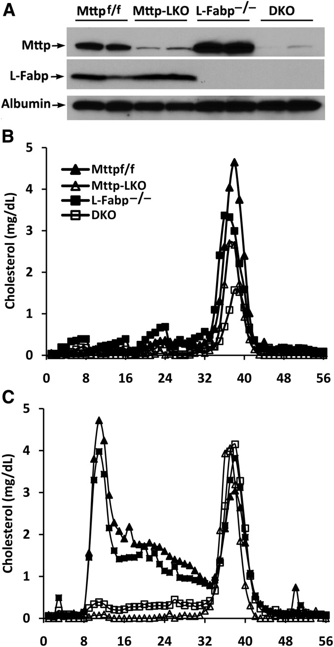
Chow-fed Mttp-LKO mice (Fig. 1A) revealed the expected reductions in serum lipid (cholesterol and triglyceride) levels and lipoprotein distribution in both L-Fabp-sufficient and L-Fabp−/− backgrounds (Table 1, Fig. 1B). In addition, liver-specific Mttp deletion completely prevented the increase in VLDL and LDL cholesterol following 2 weeks of LD feeding (Fig. 1C, Table 2). There were no differences in ALT levels among the genotypes in chow-fed animals and while ALT levels increased in all groups fed the LD for 2 weeks, hepatic Mttp deletion was associated with lower ALT levels in both L-Fabp-sufficient and L-Fabp−/− mice (Tables 1, 2). We will return to the implications of the ALT findings in a later section.
Fig. 1.
Serum lipoprotein cholesterol distribution in mice with liver-specific inactivation of Mttp. A: Confirmation of L-Fabp and Mttp deletion in the indicated genotypes by Western blot. B, C: Sera pooled from 4–5 mice per group were separated by fast-performance liquid chromatography (see Materials and Methods). Cholesterol content in lipoprotein fraction (500 μl per fraction, 56 fractions) was determined in animals fed either a chow diet (B) or a LD (C) for 2 weeks. Fractions 6–16 correspond to VLDLs, fractions 20–30 correspond to LDLs, and fractions 32–42 correspond to HDLs. Solid triangles, Mttpf/f; open triangles, Mttp-LKO; solid squares, L-Fabp−/−; open squares, DKO.
TABLE 1.
Serum lipids and ALT levels in mice of the indicated genotype on chow
| TG (mg/dl) | TC (mg/dl) | ALT (IU/l) | |
| Mttpf/f (n = 9) | 35.7 ± 2.2a | 64.5 ± 2.5a | 19.2 ± 6.0a |
| Mttp-LKO (n = 17) | 11.8 ± 1.1b | 39.2 ± 3.2b | 51.4 ± 4.4a |
| L-Fabp−/− (n = 4) | 42.6 ± 4.7a | 64.3 ± 7.9a | 47.1 ± 19.6a |
| DKO (n = 17) | 10.4 ± 2.5b | 26.3 ± 2.8c | 51.4 ± 10.7a |
Mice from four experimental groups were fed a chow diet. Sera were collected after fasting overnight and cholesterol, triglyceride, and ALT were analyzed (see Materials and Methods). The difference between values associated with different superscript letters for the parameters indicated in each vertical column is statistically significant (P < 0.05). TG, triglyceride; TC, cholesterol.
TABLE 2.
Serum lipids and ALT levels in mice of the indicated genotype on LD
| TG (mg/dl) | TC (mg/dl) | ALT (IU/l) | |
| Mttpf/f (n = 11) | 15.8 ± 1.9a | 169.7 ± 8.9a | 285.3 ± 79.4a |
| Mttp-LKO (n = 8) | 8.4 ± 1.6a | 83.1 ± 14.4b | 99.3 ± 10.7b |
| L-Fabp−/− (n = 5) | 31.1 ± 8.5b | 183.5 ± 16.7a | 281.4 ± 59.4a |
| DKO (n = 17) | 14.8 ± 2.0a | 81.1 ± 5.9b | 111.6 ± 13.4b |
Mice from four experimental groups were fed a LD for 2 weeks. Sera were collected after fasting overnight and cholesterol, triglyceride, and ALT were analyzed (see Materials and Methods). The difference between values associated with different superscript letters for the parameters indicated in each vertical column is statistically significant (P < 0.05). TG, triglyceride; TC, cholesterol.
Hepatic lipid determinations revealed the expected increase (15) in triglyceride content following deletion of hepatic Mttp in chow-fed mice (Table 3). Hepatic cholesterol content (both free and esterified) increased with 2 weeks of LD feeding in all genotypes, but the magnitude of this increase was significantly attenuated with hepatic Mttp deletion in both L-Fabp-sufficient and L-Fabp−/− mice (Table 4). In particular, there was a 2.6-fold increase in hepatic free cholesterol content in LD-fed L-Fabp−/− mice, compared with 1.5- to 1.8-fold increases in the other three genotypes (Tables 3, 4). These findings suggest that hepatic Mttp deletion modifies the changes in hepatic cholesterol metabolism observed in LD-fed L-Fabp−/− mice.
TABLE 3.
Hepatic lipids content in mice of the indicated genotype on chow
| TG | TC | FC | CE | PL | FFA | |
| Mttpf/f (n = 9) | 62.5 ± 5.8a | 18.1 ± 0.7a | 13.3 ± 0.5ab | 4.9 ± 0.4a | 69.1 ± 4.0a | 38.8 ± 2.7ab |
| Mttp-LKO (n = 5) | 161.4 ± 13.2b | 24.0 ± 2.7bc | 11.1 ± 1.5a | 12.9 ± 3.0b | 64.6 ± 1.9a | 42.3 ± 2.6bc |
| L-Fabp−/− (n = 4) | 48.7 ± 4.0a | 19.8 ± 1.2ac | 14.6 ± 0.4b | 5.7 ± 1.2a | 77.3 ± 6.9a | 33.9 ± 3.7ab |
| DKO (n = 17) | 111.2 ± 13.8b | 19.6 ± 0.7ac | 13.4 ± 0.5ab | 6.2 ± 0.4a | 72.3 ± 2.4a | 64.2 ± 4.6c |
Mice from four experimental groups were fed a chow diet. Hepatic lipids were extracted and analyzed (see Materials and Methods). Hepatic lipid content is expressed as mean ± SE (μg/mg protein), except FFA (nmol/mg protein). The difference between values associated with different superscript letters for the parameters indicated in each vertical column is statistically significant (P < 0.05). TG, triglyceride; TC, cholesterol; FC, free cholesterol; CE, cholesterol ester; PL, phospholipids.
TABLE 4.
Hepatic lipids in mice of the indicated genotype on LD
| TG | TC | FC | CE | PL | FFA | |
| Mttpf/f (n = 11) | 84.3 ± 5.8a | 122.7 ± 15.4a | 23.9 ± 1.9a | 98.7 ± 13.4a | 74.3 ± 3.3a | 42.6 ± 4.8a |
| Mttp-LKO (n = 8) | 101.9 ± 12.1a | 71.1 ± 5.5b | 19.6 ± 1.8a | 51.6 ± 5.3b | 56.1 ± 2.4b | 37.9 ± 3.1a |
| L-Fabp−/− (n = 5) | 119.9 ± 14.4a | 98.4 ± 6.7ab | 38.7 ± 2.5b | 59.8 ± 6.4ab | 76.5 ± 8.3a | 41.4 ± 2.8a |
| DKO (n = 17) | 98.8 ± 4.8a | 74.7 ± 3.7b | 20.9 ± 0.7a | 53.7 ± 3.6b | 62.6 ± 2.2b | 39.3 ± 4.6a |
Mice from four experimental groups were fed a LD (C, D) for 2 weeks. Hepatic lipids were extracted and analyzed (see Materials and Methods). Hepatic lipid content is expressed as mean ± SE (μg/mg protein), except FFA (nmol/mg protein). The difference between values associated with different superscript letters for the parameters indicated in each vertical column is statistically significant (P < 0.05). TG, triglyceride; TC, cholesterol; FC, free cholesterol; CE, cholesterol ester; PL, phospholipids.
Hepatic Mttp deletion reverses gallstone susceptibility in L-Fabp−/− mice
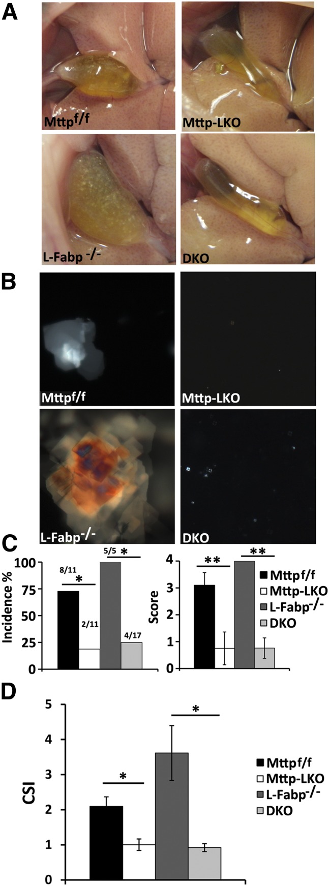
Based on the findings above and earlier observations suggesting that Mttp-LKO mice are protected against LD-induced gallstones (8), we asked whether hepatic Mttp deletion in L-Fabp−/− mice would attenuate gallstone formation after 2 or 4 weeks of LD feeding. The gross visual appearance of the gallbladder in the different genotypes (Fig. 2A) confirmed this expectation even after 2 weeks of LD feeding, which was verified by the absence of polarizing cholesterol crystals (Fig. 2B) and the reduced incidence of gallstones and lower gallstone score (Fig. 2C). We extended those findings to 4 weeks of LD feeding [to replicate the timing used by Amigo et al. (8)], which revealed virtually identical findings (zero gallstone incidence with hepatic Mttp deletion in either L-Fabp-sufficient or L-Fabp−/− background, data not shown) and also reduced the biliary CSI (Fig. 2D).
Fig. 2.
Biliary cholesterol crystals and gallstone formation in the indicated genotypes. A–C: Mice (n = 5–17 per group) were fed a LD for 2 weeks. A: Representative images of gross appearance of gallbladder. B: Representative images of freshly isolated gallbladder bile viewed under polarizing microscope. Aggregated cholesterol crystals were observed in bile from Mttpf/f and L-Fabp−/− mice, while only rare Maltese crosses were present in bile from Mttp-LKO and DKO mice. C: Gallstone incidence presented as percentage of animals with solid gallstones (left panel). Gallstone score was obtained using polar microscopy and quantitated as detailed in the Materials and Methods (right panel). The bars represent the mean ± SE of 5–17 mice per group. D: Biliary CSI in gallbladder bile from mice fed a LD for 4 weeks was calculated as detailed in the Materials and Methods. The bars represent the mean ± SE of 3–7 mice per group *P < 0.05, **P < 0.01.
Hepatic Mttp deletion decreases biliary cholesterol secretion
In order to examine the mechanisms underlying the attenuated gallstone susceptibility in L-Fabp−/− mice following hepatic Mttp deletion, we measured biliary lipid secretion in the different genotypes. The findings reveal comparable secretion rates of cholesterol, phospholipids, and bile acids in chow-fed mice and no difference in bile flow (Table 5), suggesting that canalicular lipid output is unchanged at baseline among the genotypes. Biliary lipid secretion increased in all the genotypes following LD feeding, as expected, with no changes in bile flow by genotype (Table 6). In addition, the findings revealed increased biliary cholesterol secretion in LD-fed L-Fabp−/− mice and further demonstrated that hepatic Mttp deletion led to decreased biliary cholesterol secretion in mice with combined L-Fabp deletion (Table 6). There was a trend to reduced biliary cholesterol secretion following hepatic Mttp deletion alone, but this did not reach statistical significance (Table 6). However, we could not confirm the earlier observation that hepatic Mttp deletion leads to increased biliary phospholipid secretion (8). Our findings, by contrast, reveal that biliary phospholipid secretion, if anything, tended to be reduced following hepatic Mttp deletion (Table 6). These results suggest that the protection against cholesterol gallstone formation mediated by hepatic Mttp deletion likely involves alterations in canalicular cholesterol secretion rather than phospholipid output.
TABLE 5.
Biliary lipid secretion in mice of the indicated genotype on chow
| Cholesterol | Phospholipids | Bile Acids | Bile Flow | |
| Mttpf/f (n = 7) | 0.7 ± 0.2a | 7.0 ± 0.8a | 33.2 ± 6.3a | 1.4 ± 0.2a |
| Mttp-LKO (n = 5) | 0.5 ± 0.1a | 6.2 ± 1.1a | 27.2 ± 6.7a | 1.0 ± 0.2a |
| L-Fabp−/− (n = 6) | 0.7 ± 0.1a | 6.8 ± 0.4a | 21.3 ± 2.8a | 1.4 ± 0.1a |
| DKO (n = 14) | 0.5 ± 0.1a | 5.6 ± 0.4a | 24.0 ± 2.6a | 1.3 ± 0.1a |
Mice from four experimental groups were fed a chow diet. Bile samples were collected for 1 h. Biliary cholesterol, phospholipids, and bile acids were analyzed (see Materials and Methods). Flow rate is expressed as μl/min/g liver weight. Biliary lipid secretion is expressed as nmol/min/g liver weight. The difference between values associated with different superscript letters for the parameters indicated in each vertical column is statistically significant (P < 0.05).
TABLE 6.
Biliary lipid secretion in mice of the indicated genotype on a LD
| Cholesterol | Phospholipid | Bile Acid | Bile Flow | |
| Mttpf/f (n = 11) | 4.3 ± 0.9a | 21.7 ± 3.3a | 81.0 ± 8.7a | 1.5 ± 0.3a |
| Mttp-LKO (n = 8) | 3.8 ± 0.4a | 17.4 ± 1.6a | 74.7 ± 11.7a | 1.8 ± 0.4a |
| L-Fabp−/− (n = 7) | 9.9 ± 2.5c | 28.2 ± 6.1a | 92.1 ± 12.9a | 1.4 ± 0.1a |
| DKO (n = 17) | 2.7 ± 0.4b | 19.4 ± 2.0a | 64.7 ± 5.4a | 1.8 ± 0.4a |
Mice from four experimental groups were fed a LD for 2 weeks. Bile samples were collected for 1 h. Biliary cholesterol, phospholipids, and bile acids were analyzed (see Materials and Methods). Flow rate is expressed as μl/min/g liver weight. Biliary lipid secretion is expressed as nmol/min/g liver weight. The difference between values associated with different superscript letters for the parameters indicated in each vertical column is statistically significant (P < 0.05).
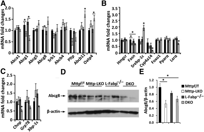
We explored the possible mechanisms for altered canalicular cholesterol secretion following LD feeding using RNA profiling of candidate transporter genes as a first step in identifying the pathways involved. The findings show decreased mRNA abundance of the intracellular cholesterol transporter Abcg1 (18) in Mttp-LKO and DKO mice and increased abundance of bile acid transporters Abcb11 (19) and Oatp4 (20) in DKO mice, but no changes in mRNA expression of the canalicular cholesterol efflux transporter Abcg5/g8 or of Srb1 (Fig. 3A). We also observed no changes in mRNA abundance of transcriptional regulators of hepatic cholesterol flux, including FoxO1 and PPARα, and we found that LXRα and Cyp4a14 mRNA abundance [the latter a downstream target of PPARα (21, 22)] were reduced in DKO mice (Fig. 3B). We found no increase in mRNA abundance for markers of ER stress in the setting of Mttp deletion (Fig. 3C), findings in keeping with previous observations in mice (23) and generally consistent with the findings noted above which revealed ALT levels were lower in Mttp-LKO mice. In view of other work showing that the expression of Abcg5/g8 is regulated at the posttranscriptional level (24, 25), we examined the expression of Abcg8 protein in the various genotypes as a surrogate marker of the heterodimeric canalicular transporter. Those findings demonstrated reduced Abcg8 protein abundance in membranes prepared from LD-fed Mttp-LKO and DKO mice compared with Mttpflox/flox mice (Fig. 3D, E).
Fig. 3.
A, B: mRNA expression of genes related to cholesterol, phospholipid, and bile acid metabolism and transport. Hepatic mRNA was extracted from mice fed a LD for 2 weeks. mRNA expression of indicated gene was quantitated by real-time qPCR and the value expressed as fold change relative to Mttpf/f mice, which was defined as 1. C: mRNA expression of genes related to ER stress. The bars represent the mean ± SE (n = 4–8 per group). D, E: Protein expression of cholesterol transporter Abcg8. Membrane protein (50 μg) from the indicated genotypes of mice was separated by 8% SDS-PAGE. Abcg8 expression was determined by Western blot and quantitated by Kodak Image software, with β-actin as loading control. D: The representative image. E: The bar graph of relative Abcg8 abundance (mean ± SE of 4 mice per group) after normalizing expression to Mttpf/f mice (n = 4 per group). *P < 0.05.
Short-term ASO-mediated hepatic Mttp knockdown attenuates LD-induced gallstone formation by reducing canalicular cholesterol secretion
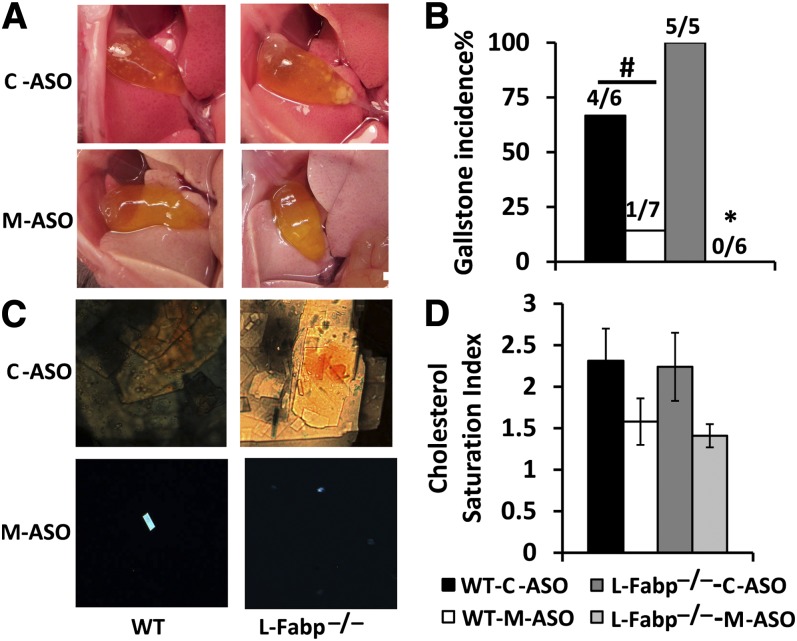
We next sought to examine the changes associated with liver-specific Mttp knockdown in LD-fed mice with a complementary approach using ASO-mediated knockdown, as recently described (26). This approach confirmed that Mttp knockdown (M-ASO) led to a visual reduction in gallstone incidence, even after 4 weeks of LD feeding (Fig. 4A, B), eliminated bile cholesterol crystals (Fig. 4C), and tended to reduce the biliary CSI (Fig. 4D).
Fig. 4.
Biliary cholesterol crystals and gallstone formation following ASO-mediated Mttp knockdown. Mice (4–7 mice per group) underwent either C-ASO or M-ASO administration for 6 weeks and were fed a LD for the final 4 weeks. A: Representative images of solid gallstones in the indicated genotype. B: Gallstone incidence presented as percentage of animals with solid gallstones C: Representative images of freshly isolated gallbladder bile viewed under polarizing microscopy. Aggregated cholesterol crystals were observed in gallbladder bile from C-ASO-treated mice in both WT and L-Fabp−/− backgrounds, while only a few Maltese crosses or single ChMCs were present in M-ASO-treated mice. D: CSI was calculated as described in the Materials and Methods. The bars represent the mean ± SE of 4–7 mice per group. *P < 0.05 versus C-ASO treated mice; # P = 0.06.
Treatment with the M-ASO produced the expected decrease in serum cholesterol in both LD-fed WT and L-Fabp−/− mice (Table 7). Hepatic lipid content determination revealed an increase in triglycerides and cholesterol esters in the M-ASO-treated animals, but no increase in free cholesterol (Table 8). Turning to biliary lipid secretion measurements, we found a trend toward decreased cholesterol secretion in M-ASO-treated WT mice which became significant in M-ASO-treated L-Fabp−/− mice (Table 9). Biliary bile acid secretion was higher in L-Fabp−/− mice, but there was no effect of ASO treatment in either the WT or L-Fabp−/− background (Table 9). In addition, there was no significant effect of M-ASO treatment on biliary phospholipid secretion rates (Table 9). These findings reinforce the earlier conclusions that the protective effect of Mttp knockdown is largely mediated through alterations in canalicular cholesterol secretion.
TABLE 7.
Serum lipids and ALT level in ASO-treated mice of the indicated genotype
| TG (mg/dl) | TC (mg/dl) | ALT (IU/l) | |
| WT-C-ASO | 15.5 ± 4.9 | 134.8 ± 7.2a | 142.9 ± 14.2a |
| WT-M-ASO | 12.8 ± 2.7 | 93.4 ± 10.5b | 182.0 ± 18.0a |
| L-Fabp−/−-C-ASO | 9.7 ± 1.9 | 183.7 ± 12.7c | 158.9 ± 20.5a |
| L-Fabp−/−-M-ASO | 14.5 ± 5.7 | 90.0 ± 7.7b | 163.09 ± 7.8a |
WT and L-Fabp−/− mice received either control-ASO or Mttp-ASO for four weeks and were fed a LD for the final 2 weeks, while continuing ASO injection (n = 4–5 per group). Sera were collected after overnight fasting and serum lipids and ALT assayed. The difference between values associated with different superscript letters for the parameters indicated in each vertical column is statistically significant (P < 0.05). TG, triglyceride; TC, cholesterol.
TABLE 8.
Hepatic lipid content in ASO-treated mice of the indicated genotype
| TG | TC | FC | CE | PL | FFA | |
| WT-C-ASO | 86.6 ± 8.3a | 93.3 ± 12.3a | 26.2 ± 3.3a | 67.0 ± 11.7ac | 65.9 ± 6.9a | 49.0 ± 11.1a |
| WT-M-ASO | 133.1 ± 10.3b | 124.2 ± 14.9a | 30.3 ± 3.2a | 93.9 ± 12.0ab | 64.4 ± 2.9a | 70.7 ± 3.1a |
| L-Fabp−/−-C-ASO | 63.9 ± 9.1a | 92.1 ± 8.3a | 34.3 ± 6.3a | 57.8 ± 5.4c | 87.0 ± 12.1a | 25.3 ± 9.6b |
| L-Fabp−/−-M-ASO | 167.6 ± 31.6b | 135.9 ± 21.6a | 32.6 ± 5.4a | 103.3 ± 16.5b | 82.5 ± 8.9a | 86.3 ± 8.2a |
WT and L-Fabp−/− mice received either control-ASO or Mttp-ASO for four weeks and were fed a LD for the final 2 weeks, while continuing ASO injection (n = 4–5 per group). Hepatic lipids were extracted and expressed as μg/mg protein except FFA (expressed as nmol/mg protein). The difference between values associated with different superscript letters for the parameters indicated in each vertical column is statistically significant (P < 0.05). TG, triglyceride; TC, cholesterol; FC, free cholesterol; CE, cholesterol ester; PL, phospholipids.
TABLE 9.
Biliary lipid secretion in ASO-treated mice of the indicated genotype
| TC | PL | BA | |
| WT-C-ASO | 2.7 ± 0.5a | 11.4 ± 1.0a | 79.2 ± 6.4a |
| WT-M-ASO | 2.4 ± 0.4a | 11.8 ± 1.2a | 64.8 ± 7.1a |
| L-Fabp−/−-C-ASO | 5.5 ± 0.9b | 21.9 ± 3.5a | 158.3 ± 32.0b |
| L-Fabp−/−-M-ASO | 2.9 ± 0.4a | 15.3 ± 0.4a | 117.9 ± 10.5ab |
WT and L-Fabp−/− mice received either control-ASO or Mttp-ASO for four weeks and were fed a LD for the final 2 weeks, while continuing ASO injection (n = 4–5 per group). Bile was collected and biliary lipid secretion determined and expressed as nmol/min/g liver weight. The difference between values associated with different superscript letters for the parameters indicated in each vertical column is statistically significant (P < 0.05). TC, cholesterol; PL, phospholipids; BA, bile acids.
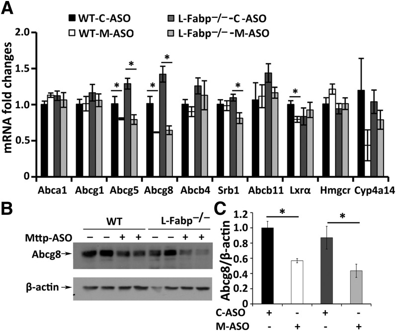
We next examined canalicular transporter expression, which revealed a significant decrease in mRNA abundance for both Abcg5 and Abcg8 in M-ASO-treated mice but no change in Abcb4 or Abcb11 (Fig. 5A). There was also a small but significant decrease in Srb1 mRNA expression in M-ASO-treated L-Fabp−/− mice and a trend to reduced mRNA abundance of Cyp4a14 (Fig. 5A). The decrease in Abcg5/g8 mRNA expression in M-ASO-treated mice was accompanied by decreased protein expression for Abcg8, particularly in L-Fabp−/− mice (Fig. 5B, C). These findings support the conclusion that Mttp knockdown alters canalicular cholesterol secretion in conjunction with decreased expression of the cholesterol transporter Abcg5/g8.
Fig. 5.
A: mRNA expression of genes related to cholesterol, phospholipid, and bile acid metabolism and transport. Hepatic mRNA was extracted from mice treated with the indicated ASO for 4 weeks and fed a LD for the final 2 weeks. The mRNA expression of indicated genes was quantitated by real-time qPCR and the value is expressed as fold change related to C-ASO-treated WT mice, which was defined as 1. The bars represent the mean ± SE (n = 4 per group). B, C: Protein expression of cholesterol transporter Abcg8. Membrane protein (50 μg) from the above mice was separated by 8% SDS-PAGE. Abcg8 protein expression was determined by Western blot and quantitated by Kodak Image software, with β-actin as loading control. B: A representative image. C: A bar graph of relative Abcg8 abundance (mean ± SE of 4 mice per group) after normalizing expression to C-ASO-treated WT mice. *P < 0.05.
DISCUSSION
The major observations from this study suggest that reducing the expression of hepatic Mttp, either by genetic deletion or by ASO-mediated knockdown, attenuates gallstone formation in both WT and L-Fabp−/− mice, through pathways that result in decreased biliary cholesterol secretion. The findings suggest that the critical functions of Mttp in the export of hepatocyte lipid extend beyond a role in VLDL-dependent pathways of apical secretion and include the regulation of canalicular cholesterol secretion pathways. Several aspects of these findings and conclusions merit additional discussion.
Previous studies demonstrated that liver-specific Mttp deletion protected mice from LD-induced gallstone formation (8). Those studies demonstrated compensatory alterations in hepatic cholesterol metabolism in Mttp-LKO mice including increased cholesterol ester content and decreased cholesterogenesis, which in turn was postulated to limit free cholesterol availability and attenuate biliary cholesterol secretion in LD-fed mice (8). In addition, and critical to the mechanisms postulated, those authors found no decrease in biliary cholesterol secretion rates in LD-fed Mttp-LKO mice but rather demonstrated increased phospholipid secretion (8). Our findings differ from those data (8) in several key respects. First, we observed that hepatic cholesterol ester content in LD-fed Mttp-LKO and DKO mice was significantly reduced rather than increased as reported by those workers. Second, we observed that biliary cholesterol secretion rates were decreased in Mttp-LKO and DKO mice. Third, we did not observe increased biliary phospholipid secretion in Mttp-LKO and DKO mice as reported by those workers, but rather found (if anything) slightly reduced secretion rates. The sources of the discrepancies between the current report and those earlier findings may reside in a combination of factors, including the genetic background of the mice [C57BL/6 as used in the current study versus a 50/50 mixed C57BL/6 and 129/SvJae background as used in that report (8)] and the different facilities, which conceivably could include differences in microbial communities. We evaluated different durations of LD feeding (2–4 weeks in the current study versus 4–8 weeks in those earlier studies) which yielded virtually identical outcomes, suggesting that the timing of LD exposure is not a factor.
The observation that biliary phospholipid secretion was unaltered in the setting of hepatic Mttp deletion should not be construed as implying that there is no effect of hepatic Mttp on hepatic phospholipid metabolism. Indeed, Hussain and colleagues defined both neutral and polar lipid transfer activities of Mttp based on the properties of human versus Drosophila proteins and have demonstrated distinct effects on whole body lipid metabolism and in the apical secretion of hepatic VLDLs (27). However, those studies did not address the question of whether canalicular phospholipid secretion is altered following adenoviral Mttp expression. In addition, we did not specifically examine the secretion rates of individual phospholipid species, which could conceivably represent yet another level of regulation of biliary cholesterol saturation.
Other studies in Mttp-LKO mice demonstrated increased hepatic free cholesterol with decreased cholesterol ester, which was consistent with a role for Mttp in relieving product inhibition of cholesterol esterification by transferring neutral lipid (28). Our findings are consistent with that report and show that hepatic cholesterol ester content decreases in Mttp-LKO and DKO mice following LD feeding (Tables 1–4). However, we did not find the same results in LD-fed mice treated with the Mttp-ASO. In those experiments, we found that hepatic cholesterol ester content increased in both WT and L-Fabp−/− mice when they were treated with the Mttp-ASO (Tables 7–9). The reasons for this discrepancy are not immediately apparent because we achieved comparable reduction of hepatic Mttp expression (>80%) following knockdown and genetic deletion experiments (data not shown), but it remains possible that differences in ASO versus genetic deletion approaches may be at least partially responsible. It should be noted that the genetic backgrounds of the mice used for the ASO and genetic deletion experiments are not truly identical (even though the studies were performed in C57BL/6 mice), because the control mice used in the genetic deletion experiments contained a “floxed” allele (Mttpflox/flox) and also received pI-pC injections over several days. As alluded to above, earlier studies in LD-fed Mttp-LKO mice also showed increased hepatic cholesterol ester content (8).
Our findings suggest a plausible mechanism by which hepatic Mttp deletion leads to attenuated canalicular cholesterol secretion, specifically via alterations in the expression of the heterodimeric cholesterol transporter Abcg5/g8, which represents one of the dominant pathways for canalicular cholesterol export (17, 29–31). The findings suggest decreased expression of Abcg8 in LD-fed Mttp-LKO and DKO mice and also following Mttp-ASO treatment of WT and L-Fabp−/− mice. The findings in the Mttp-ASO-treated mice revealed more robust evidence (compared with the genetic deletion experiments) for decreased expression of both Abcg5 and Abcg8 mRNA and also demonstrated decreased Abcg8 protein abundance. The explanation for this subtle difference may again lie in the liver-specific Mttp knockdown approaches, which we speculate may result in less adaptation or compensation. Previous studies have demonstrated that the Abcg5/g8 heterodimer is regulated by alterations in leptin signaling as well as by chemical chaperones, suggesting that posttranscriptional events may represent an important mode of functional regulation (25). In this scenario, we speculate that interactions of Abcg5/g8 with endolumenal ER proteins may promote the functional dimerization and directed trafficking of the heterodimeric cholesterol transporter. The earlier findings of Graf et al. (30), coupled with more recent findings alluded to above (25) raise the testable possibility that Mttp functions as a chaperone for Abcg5/g8 assembly within the hepatocyte, conceivably in a manner analogous to that described for its chaperone function in relation to the initial steps in the cotranslational processing of apoB (10). That particular suggestion will require further examination to resolve the role of Mttp in the posttranslational processing, assembly, and vectorial delivery of the Abcg5/g8 heterodimer.
Notwithstanding the observations above, it remains possible that other pathways are involved in Mttp-dependent modulation of canalicular cholesterol secretion. These include Srb1-dependent pathways of canalicular cholesterol secretion where a role for Abcg5/g8 independent cholesterol transport has been invoked (32, 33). While we found no evidence to support such a role for Srb1 in the current study, the possibility merits further examination. An additional consideration is that liver-specific Mttp deletion might conceivably promote hepatic cholesterol mobilization through nonbiliary pathways (34–36), a possibility that will require evaluation. In summary, the current findings demonstrate an expanded role for hepatic Mttp in the regulation of canalicular cholesterol secretion and illustrate the complexity in orchestrating its metabolic compartmentalization.
Acknowledgments
The authors are grateful to all members of the Davidson laboratory for their helpful comments and support.
Footnotes
Abbreviations:
- ALT
- alanine aminotransferase
- ASO
- antisense oligonucleotide
- C-ASO
- non-specific control antisense oligonucleotide
- ChMC
- cholesterol monohydrate crystal
- CSI
- cholesterol saturation index
- DKO
- mice with combined deletion
- ER
- endoplasmic reticulum
- LD
- lithogenic diet
- M-ASO
- microsomal triglyceride transfer protein antisense oligonucleotide
- Mttp
- microsomal triglyceride transfer protein
- Mttp-LKO
- liver-specific deletion of microsomal triglyceride transfer protein
- pI-pC
- polyinosinic-polycytidylic RNA
- qPCR
- quantitative PCR
N.O.D. was supported by National Institutes of Health Grants HL38180, DK56260, and DK52574 (Murine and Advanced Imaging Cores).
REFERENCES
- 1.Peery A. F., Dellon E. S., Lund J., Crockett S. D., McGowan C. E., Bulsiewicz W. J., Gangarosa L. M., Thiny M. T., Stizenberg K., Morgan D. R., et al. 2012. Burden of gastrointestinal disease in the United States: 2012 update. Gastroenterology. 143: 1179–1187 [DOI] [PMC free article] [PubMed] [Google Scholar]
- 2.Lammert F., Sauerbruch T. 2005. Mechanisms of disease: the genetic epidemiology of gallbladder stones. Nat. Clin. Pract. Gastroenterol. Hepatol. 2: 423–433 [DOI] [PubMed] [Google Scholar]
- 3.Di Ciaula A., Wang D. Q., Garruti G., Wang H. H., Grattagliano I., de Bari O., Portincasa P. Therapeutic reflections in cholesterol homeostasis and gallstone disease. A review. Curr. Med. Chem. Epub ahead of print. September 16, 2013. [DOI] [PubMed] [Google Scholar]
- 4.Biddinger S. B., Haas J. T., Yu B. B., Bezy O., Jing E., Zhang W., Unterman T. G., Carey M. C., Kahn C. R. 2008. Hepatic insulin resistance directly promotes formation of cholesterol gallstones. Nat. Med. 14: 778–782 [DOI] [PMC free article] [PubMed] [Google Scholar]
- 5.Biddinger S. B., Hernandez-Ono A., Rask-Madsen C., Haas J. T., Aleman J. O., Suzuki R., Scapa E. F., Agarwal C., Carey M. C., Stephanopoulos G., et al. 2008. Hepatic insulin resistance is sufficient to produce dyslipidemia and susceptibility to atherosclerosis. Cell Metab. 7: 125–134 [DOI] [PMC free article] [PubMed] [Google Scholar]
- 6.Lyons M. A., Wittenburg H. 2006. Cholesterol gallstone susceptibility loci: a mouse map, candidate gene evaluation, and guide to human LITH genes. Gastroenterology. 131: 1943–1970 [DOI] [PubMed] [Google Scholar]
- 7.Lyons M. A., Wittenburg H., Li R., Walsh K. A., Churchill G. A., Carey M. C., Paigen B. 2003. Quantitative trait loci that determine lipoprotein cholesterol levels in DBA/2J and CAST/Ei inbred mice. J. Lipid Res. 44: 953–967 [DOI] [PubMed] [Google Scholar]
- 8.Amigo L., Castro J., Miquel J. F., Zanlungo S., Young S., Nervi F. 2006. Inactivation of hepatic microsomal triglyceride transfer protein protects mice from diet-induced gallstones. Gastroenterology. 131: 1870–1878 [DOI] [PubMed] [Google Scholar]
- 9.Hussain M. M., Rava P., Pan X., Dai K., Dougan S. K., Iqbal J., Lazare F., Khatun I. 2008. Microsomal triglyceride transfer protein in plasma and cellular lipid metabolism. Curr. Opin. Lipidol. 19: 277–284 [DOI] [PubMed] [Google Scholar]
- 10.Hussain M. M., Rava P., Walsh M., Rana M., Iqbal J. 2012. Multiple functions of microsomal triglyceride transfer protein. Nutr. Metab. (Lond.). 9: 14. [DOI] [PMC free article] [PubMed] [Google Scholar]
- 11.Castro J., Amigo L., Miquel J. F., Galman C., Crovari F., Raddatz A., Zanlungo S., Jalil R., Rudling M., Nervi F. 2007. Increased activity of hepatic microsomal triglyceride transfer protein and bile acid synthesis in gallstone disease. Hepatology. 45: 1261–1266 [DOI] [PubMed] [Google Scholar]
- 12.Newberry E. P., Xie Y., Kennedy S., Han X., Buhman K. K., Luo J., Gross R. W., Davidson N. O. 2003. Decreased hepatic triglyceride accumulation and altered fatty acid uptake in mice with deletion of the liver fatty acid-binding protein gene. J. Biol. Chem. 278: 51664–51672 [DOI] [PubMed] [Google Scholar]
- 13.Xie Y., Newberry E. P., Kennedy S. M., Luo J., Davidson N. O. 2009. Increased susceptibility to diet-induced gallstones in liver fatty acid binding protein knockout mice. J. Lipid Res. 50: 977–987 [DOI] [PMC free article] [PubMed] [Google Scholar]
- 14.Spann N. J., Kang S., Li A. C., Chen A. Z., Newberry E. P., Davidson N. O., Hui S. T., Davis R. A. 2006. Coordinate transcriptional repression of liver fatty acid-binding protein and microsomal triglyceride transfer protein blocks hepatic very low density lipoprotein secretion without hepatosteatosis. J. Biol. Chem. 281: 33066–33077 [DOI] [PubMed] [Google Scholar]
- 15.Raabe M., Veniant M. M., Sullivan M. A., Zlot C. H., Bjorkegren J., Nielsen L. B., Wong J. S., Hamilton R. L., Young S. G. 1999. Analysis of the role of microsomal triglyceride transfer protein in the liver of tissue-specific knockout mice. J. Clin. Invest. 103: 1287–1298 [DOI] [PMC free article] [PubMed] [Google Scholar]
- 16.Carey M. C. 1978. Critical tables for calculating the cholesterol saturation of native bile. J. Lipid Res. 19: 945–955 [PubMed] [Google Scholar]
- 17.Yu L., Gupta S., Xu F., Liverman A. D., Moschetta A., Mangelsdorf D. J., Repa J. J., Hobbs H. H., Cohen J. C. 2005. Expression of ABCG5 and ABCG8 is required for regulation of biliary cholesterol secretion. J. Biol. Chem. 280: 8742–8747 [DOI] [PubMed] [Google Scholar]
- 18.Tarling E. J. 2013. Expanding roles of ABCG1 and sterol transport. Curr. Opin. Lipidol. 24: 138–146 [DOI] [PubMed] [Google Scholar]
- 19.Henkel A. S., Kavesh M. H., Kriss M. S., Dewey A. M., Rinella M. E., Green R. M. 2011. Hepatic overexpression of abcb11 promotes hypercholesterolemia and obesity in mice. Gastroenterology. 141: 1404–1411 [DOI] [PMC free article] [PubMed] [Google Scholar]
- 20.Csanaky I. L., Lu H., Zhang Y., Ogura K., Choudhuri S., Klaassen C. D. 2011. Organic anion-transporting polypeptide 1b2 (Oatp1b2) is important for the hepatic uptake of unconjugated bile acids: studies in Oatp1b2-null mice. Hepatology. 53: 272–281 [DOI] [PMC free article] [PubMed] [Google Scholar]
- 21.Kroetz D. L., Yook P., Costet P., Bianchi P., Pineau T. 1998. Peroxisome proliferator-activated receptor alpha controls the hepatic CYP4A induction adaptive response to starvation and diabetes. J. Biol. Chem. 273: 31581–31589 [DOI] [PubMed] [Google Scholar]
- 22.Patsouris D., Reddy J. K., Muller M., Kersten S. 2006. Peroxisome proliferator-activated receptor alpha mediates the effects of high-fat diet on hepatic gene expression. Endocrinology. 147: 1508–1516 [DOI] [PubMed] [Google Scholar]
- 23.Liao W., Hui T. Y., Young S. G., Davis R. A. 2003. Blocking microsomal triglyceride transfer protein interferes with apoB secretion without causing retention or stress in the ER. J. Lipid Res. 44: 978–985 [DOI] [PubMed] [Google Scholar]
- 24.Sabeva N. S., Liu J., Graf G. A. 2009. The ABCG5 ABCG8 sterol transporter and phytosterols: implications for cardiometabolic disease. Curr. Opin. Endocrinol. Diabetes Obes. 16: 172–177 [DOI] [PMC free article] [PubMed] [Google Scholar]
- 25.Sabeva N. S., Rouse E. J., Graf G. A. 2007. Defects in the leptin axis reduce abundance of the ABCG5-ABCG8 sterol transporter in liver. J. Biol. Chem. 282: 22397–22405 [DOI] [PubMed] [Google Scholar]
- 26.Lee R. G., Fu W., Graham M. J., Mullick A. E., Sipe D., Gattis D., Bell T. A., Booten S., Crooke R. M. 2013. Comparison of the pharmacological profiles of murine antisense oligonucleotides targeting apolipoprotein B and microsomal triglyceride transfer protein. J. Lipid Res. 54: 602–614 [DOI] [PMC free article] [PubMed] [Google Scholar]
- 27.Khatun I., Zeissig S., Iqbal J., Wang M., Curiel D., Shelness G. S., Blumberg R. S., Hussain M. M. 2012. Phospholipid transfer activity of microsomal triglyceride transfer protein produces apolipoprotein B and reduces hepatosteatosis while maintaining low plasma lipids in mice. Hepatology. 55: 1356–1368 [DOI] [PMC free article] [PubMed] [Google Scholar]
- 28.Iqbal J., Rudel L. L., Hussain M. M. 2008. Microsomal triglyceride transfer protein enhances cellular cholesteryl esterification by relieving product inhibition. J. Biol. Chem. 283: 19967–19980 [DOI] [PMC free article] [PubMed] [Google Scholar]
- 29.Graf G. A., Li W. P., Gerard R. D., Gelissen I., White A., Cohen J. C., Hobbs H. H. 2002. Coexpression of ATP-binding cassette proteins ABCG5 and ABCG8 permits their transport to the apical surface. J. Clin. Invest. 110: 659–669 [DOI] [PMC free article] [PubMed] [Google Scholar]
- 30.Graf G. A., Yu L., Li W. P., Gerard R., Tuma P. L., Cohen J. C., Hobbs H. H. 2003. ABCG5 and ABCG8 are obligate heterodimers for protein trafficking and biliary cholesterol excretion. J. Biol. Chem. 278: 48275–48282 [DOI] [PubMed] [Google Scholar]
- 31.Yu L., Hammer R. E., Li-Hawkins J., Von Bergmann K., Lutjohann D., Cohen J. C., Hobbs H. H. 2002. Disruption of Abcg5 and Abcg8 in mice reveals their crucial role in biliary cholesterol secretion. Proc. Natl. Acad. Sci. USA. 99: 16237–16242 [DOI] [PMC free article] [PubMed] [Google Scholar]
- 32.Dikkers A., Freak de Boer J., Annema W., Groen A. K., Tietge U. J. 2013. Scavenger receptor BI and ABCG5/G8 differentially impact biliary sterol secretion and reverse cholesterol transport in mice. Hepatology. 58: 293–303 [DOI] [PubMed] [Google Scholar]
- 33.Wiersma H., Gatti A., Nijstad N., Oude Elferink R. P., Kuipers F., Tietge U. J. 2009. Scavenger receptor class B type I mediates biliary cholesterol secretion independent of ATP-binding cassette transporter g5/g8 in mice. Hepatology. 50: 1263–1272 [DOI] [PubMed] [Google Scholar]
- 34.Temel R. E., Sawyer J. K., Yu L., Lord C., Degirolamo C., McDaniel A., Marshall S., Wang N., Shah R., Rudel L. L., et al. 2010. Biliary sterol secretion is not required for macrophage reverse cholesterol transport. Cell Metab. 12: 96–102 [DOI] [PMC free article] [PubMed] [Google Scholar]
- 35.van der Velde A. E., Vrins C. L., van den Oever K., Kunne C., Oude Elferink R. P., Kuipers F., Groen A. K. 2007. Direct intestinal cholesterol secretion contributes significantly to total fecal neutral sterol excretion in mice. Gastroenterology. 133: 967–975 [DOI] [PubMed] [Google Scholar]
- 36.van der Velde A. E., Vrins C. L., van den Oever K., Seemann I., Oude Elferink R. P., van Eck M., Kuipers F., Groen A. K. 2008. Regulation of direct transintestinal cholesterol excretion in mice. Am. J. Physiol. Gastrointest. Liver Physiol. 295: G203–G208 [DOI] [PubMed] [Google Scholar]


