
Figure 1. Loss of Kcnq1 promotes tumor progression.
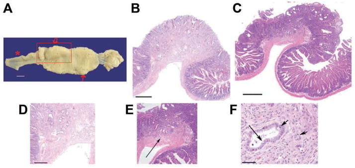
Panel 1A. Duodenum from ApcMin Kcnq1+/− mouse with stomach at right. Bar = 5 mm. Broad area of hyperplastic tissue with embedded tumors extends from pylorus-dudoenum juncture past the ampulla to ~ the duodenal jejunal flexure. Asterisk indicates area of normal duodenum-proximal jejunum. Box encloses a region of adenocarcinoma tissue of > 2 cm. Sections from this lesion were taken from various regions and all showed evidence of invasion. Two of these adenocarcinoma sections are depicted in panels B through E. Smaller arrow depicts normal large adenoma of ~ 5 mm that is embedded in hyperplastic tissue. Panels B&C, bars = 500 μm; invasion into muscularis mucosa, panels D&E, bar = 250 μm; prominent scirrhous response to invading tubules that exhibit differentiated goblet cells (long arrow) and signet ring cells (short arrows) panel E, bar = 50 μm.