Abstract
Purpose
Humans differ from other great ape species in their propensity to develop tears of the rotator cuff. The aim of this study was to compare the anatomical risk factors for subacromial impingement and rotator cuff tears amongst the great apes and to determine which features may be accentuated in humans and therefore play a more significant role in disease aetiology.
Methods
Orthogonal digital photographs of 22 human, 17 gorilla, 13 chimpanzee and 12 orangutan dry bone scapula specimens oriented in the glenoid plane were taken. Anatomical measurements were preformed using a calibrated digital image technique and the results scaled according to scapula vertebral border length.
Results
Of the ten anatomical features associated with subacromial impingement and rotator cuff tears in humans, none were shown to be accentuated and significantly different to the other species studied. However the human supraspinatus fossa was shown to be significantly smaller.
Conclusions
These results indicate that an alternative primary aetiological factor for rotator cuff tears must exist. A reduction in the size of the supraspinatus fossa in human scapulae suggests that structural insufficiency of the supraspinatus or a change in rotator cuff force vectors could play a role.
Keywords: Evolution, Impingement, Rotator cuff, Shoulder, Subacromial
Introduction
In 1972, Neer first popularised the idea that subacromial impingement and rotator cuff tears develop secondary to attrition of the cuff beneath the coracoacromial arch [1]. This mechanism, known as the external impingement theory, has since been supported by several studies associating features of scapula morphology with impingement symptoms and rotator cuff tears. These associated features refer to the size and shape of the coracoacromial arch and the orientation of the glenoid, and theoretically lead to a reduction in the functional supraspinatus outlet area [2–14]. Arthroscopic acromioplasty, which forms the mainstay of surgical management for subacromial impingement syndrome, has been developed in accordance with the external impingement theory. However there is evidence that rotator cuff disease often originates from within or on the articular side of the supraspinatus tendon, which is inconsistent with an external impingement mechanism [15]. In view of the potential clinical risks and costs of surgical treatment therefore, determining the primary aetiological factor of subacromial impingement is of significant clinical and economical importance.
The Hominidae family (great apes), consisting of humans, chimpanzees, gorillas and orangutans, share a common ancestor that lived approximately 14 million years ago. Since this time each species has diversified in accordance with Darwin’s theory of evolution and demonstrate different locomotive behaviours. Orangutans are predominantly arboreal in contrast to the more terrestrial activities of both chimpanzees and gorillas. However the adoption of an orthograde posture and the requirement for vertical climbing remain a common feature. Scapula morphology appears to reflect this shared function with an elongated and laterally projecting acromion together with a stabilising coracoacromial ligament that helps to maximise deltoid power. Interestingly, evidence of rotator cuff tears is completely devoid in the non-human primate literature [16, 17]. In addition, the osteological changes associated with impingement and cuff tears have been shown to be much rarer indicating that human evolution has been associated with factors that increase disease risk [17].
The aim of this study was therefore to compare the anatomical features reported to be associated with subacromial impingement and rotator cuff tears in the medical literature between the great ape species. We hypothesise that features accentuated or unique to humans may play a more significant role in disease aetiology.
Materials and methods
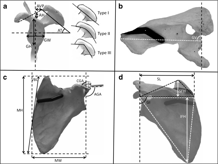
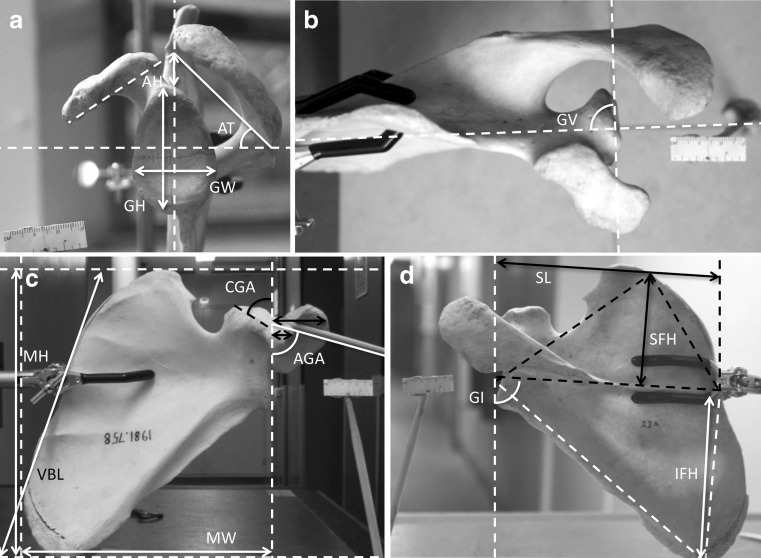
Dry bone specimens of 22 human (Homo sapien), 17 gorilla (Gorilla gorilla), 13 chimpanzee (Pan troglodytes), and 12 orangutan (Pongo pigmaeus/abelii) scapula were analysed. Each specimen was secured in a retort stand (Edu-Lab, Norfolk, UK) and orientated with the superior and inferior rim of the glenoid parallel to the vertical. Orthogonal photographs were taken (Canon Digital IXUS 9515–10.0MPx) in the lateral, anterior, posterior and superior planes with a 3-cm ruler for scale (Figs. 1 and 2).
Fig. 1.
Photographs of a human scapula and measurements. a Lateral with Bigliani classification. b Superior. c Anterior (white arrows: acromion and coracoid lateral projections). d Posterior
Fig. 2.
Setup of a gorilla scapula and measurements. a Lateral. b Superior. c Anterior (black arrows: acromion and coracoid lateral projections). d Posterior
A literature review identified ten anatomical features of the scapula associated with subacromial impingement and rotator cuff tears in humans (Table 1). Anatomical measurements were preformed using Fiji ImageJ software (National Institutes of Health, USA) (Figs. 1 and 2). Precision of the technique expressed as the mean relative standard deviation of angular and linear measurements was evaluated with five independent repetitive tests. Due to differences in body size between species, all measurements were scaled to the mean equivalent human size according to vertebral border length, which has been shown to be proportional to body size amongst higher primates [18]. Mean measurements were compared between species using the students T-test. A p-value of less than 0.05 was considered significant.
Table 1.
Measurements performed for each scapula orientation. References are shown for those measurements associated with impingement or rotator cuff tears
| Refers to figure parts in Figs. 1 and 2 | Measurement | Description | References |
|---|---|---|---|
| A | AH | Coracoacromial arch height | Edelson et al. [5] |
| AT | Acromion tilt angle | Edelson et al. [5], Gohlke et al. [7], Prato et al. [10], Zuckerman et al. [14] | |
| AVP | Acromion ventral projection | Gohlke et al. [7], Zuckerman et al. [14] | |
| Bigliani classification | I (flat), II (curved), III (hooked) | Bigliani et al. [4] | |
| GH | Glenoid height | ||
| GW | Glenoid width | ||
| B | GV | Glenoid version | Tetreault et al. [11], Tokgoz et al. [12] |
| C | White / Black Arrows | Coracoid lateral projection | Anetzberger et al. [2] |
| White / Black Arrows | Acromion lateral projection | Anetzberger et al. [2], Nyffeler et al. [9] | |
| CGA | Coracoglenoid angle | Anetzberger et al. [2], Edelson et al. [5] | |
| AGA | Acromioglenoid angle | Banas et al. [3], Tetreault et al. [11] | |
| MH | Maximum height | ||
| MW | Maximum width | ||
| VBL | Vertebral border length | ||
| D | GI | Glenoid inclination | Anetzberger et al. [2], Flieg et al. [6], Hughes et al. [8], Tetreault et al. [11], Wong et al. [13] |
| SL | Spine length | ||
| SFH | Supraspinatus fossa height | ||
| IFH | Infraspinatus fossa height |
Results
The setup intra-observer mean relative standard deviation was 1.1 % for all angular measurements and 1.7 % for all linear measurements. The mean results for each measurement and species are shown in Table 2.
Table 2.
Mean results for each angular (degrees) and linear (cm) measurement per species
| Measurement | Human | Orangutan | Chimpanzee | Gorilla |
|---|---|---|---|---|
| Mean specimen measurements | ||||
| Vertebral border length (VBL) | 13.55 | 13.69 | 14.08 | 20.61 |
| Supraspinatus fossa area | 17.34 | 21.34 | 23.18 | 69.88 |
| Infraspinatus fossa area | 50.75 | 53.77 | 37.66 | 89.09 |
| Fossa ratio | 0.34 | 0.40 | 0.62 | 0.79 |
| Mean angular measurements | ||||
| Acromion morphology I/II/III | 3/17/2 | 0/8/4 | 1/11/1 | 4/13/0 |
| Acromion tilt (AT) | 47.7 | 44.2 | 36.8 | 40.2 |
| Acromioglenoid angle (AGA) | 82.1 | 78.6 | 78.4 | 69.2 |
| Coracoglenoid angle (CGA) | 76.6 | 41.8 | 49.4 | 52.1 |
| Glenoid version (GV) | −2.7 | −2.8 | −6.0 | 0.3 |
| Glenoid inclination (GI) | 92.4 | 100.6 | 87.8 | 92.3 |
| Mean scaled linear measurements | ||||
| Acromion ventral projection (AVP) | −0.73 | −0.59 | 0.20 | −0.02 |
| Acromion lateral projection | 2.78 | 2.41 | 2.65 | 3.01 |
| Coracoid lateral projection | 1.17 | 0.85 | 1.06 | 1.29 |
| Coracoacromial arch height (AH) | 1.03 | 0.72 | 1.25 | 0.79 |
Values in bold represent results for humans that are significantly different from each of the other species
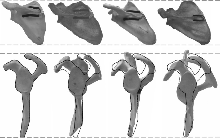
The coracoglenoid angle was shown to be significantly different in humans from each of the other species but against the direction reported to increase impingement risk (Fig. 3) [2]. As a result none of the ten anatomical risk factors were shown to be accentuated and significantly different in humans compared to other great apes. Interestingly, the fossa ratio was significantly smaller in humans than in the other species studied and appears to be secondary to a relative reduction in the supraspinatus fossa area (Figs. 3 and 4).
Fig. 3.
Graphs demonstrating human results significantly different from other great ape species
Fig. 4.
Comparison of scapula scaled to vertebral border length demonstrating a relative reduction in the size of the human supraspinatus fossa. From left to right: human, orangutan, chimpanzee, gorilla. Outline of the lateral human scapula is superimposed
Discussion
Since Neer popularised the concept that impingement and rotator cuff tears were secondary to an external abrasive mechanism, several studies have reported an association between this condition and the structure and orientation of the coracoacromial arch and glenoid. Acromion morphology was first classified by Bigliani et al. as either flat, curved or hooked with the latter associated with rotator cuff tears [4]. However, this classification has been criticised for poor inter-observer correlation due to the confounding effects of acromion orientation and the presence of age-related acromion spurs [19]. In addition acromion spurs may develop secondary to rotator cuff pathology and superior migration of the humeral head, explaining this association in some studies [20].
Features that reduce the height of the coracoacromial arch have also been implicated in subacromial impingement. Several cadaveric studies have associated a low coracoacromial arch, acute anterior tilt of the acromion and increasing acromion ventral projection with subacromial degenerative changes and rotator cuff tears [5, 7, 14]. An acute acromion tilt angle has also been correlated with cuff pathology in vivo [10]. Other factors potentially reducing the supraspinatus outlet area include a reduction in acromioglenoid angle and coracoid angle in the coronal plane. Both Edelson and Taitz, and Anetzberger et al. associated these factors with rotator cuff tears in cadaveric specimens [2, 5]. However, results from in vivo magnetic resonance imaging (MRI) studies have not always supported this association [3, 11, 12].
Active shoulder abduction may also affect the supraspinatus outlet area by reducing the distance between the greater tuberosity of the humerus and the coracoacromial arch. In accordance with this theory, Anetzberger et al. demonstrated an association between lateral projection of both the acromion and coracoid with rotator cuff tears [2]. Similarly, the acromion index, which is expressed as a ratio between the distance from the plane of the glenoid and the lateral edge of the acromion and proximal humerus, has been linked with full thickness rotator cuff tears [9]. Superior migration of the humeral head during abduction may further reduce the supraspinatus outlet area, and has been linked with increasing glenoid inclination according to both biomechanical and computer modelling studies [6, 13]. In support of this mechanism some studies have associated glenoid inclination with rotator cuff tears [2, 8, 11]. However, osteoarthritis of the glenohumeral joint secondary to rotator cuff pathology, which most commonly affects the antero-superior glenoid, could be an important confounding factor [8].
Finally, glenoid retroversion has been associated with rotator cuff tears by contributing to a reduction in the subacromial space [11, 12]. Once again however this association is not always a consistent finding [21].
Although there are several reported associations between scapular morphology and rotator cuff disease in humans, we were unable to identify any features that were accentuated and significantly different to other great ape species. This suggests that an alternative primary aetiological factor must have developed during our evolution. Our results are in general agreement with those of Potau et al. who compared the subacromial space area between humans, chimpanzees and gorillas [16]. Despite measuring subacromial space dimensions that have not been directly associated with rotator cuff tears in humans, they too could not identify any significant differences between the species [16]. This evidence therefore does not support the external impingement theory. However, we observed a reduction in the supraspinatus fossa area in humans, which may support the intrinsic theory of rotator cuff tear aetiology. It is possible that a reduction in the supraspinatus muscle mass during our evolution has increased the risk of tendon failure. In support of this hypothesis, Nakajima et al. have demonstrated that collagen fibres on the bursal side of the supraspinatus tendon have a greater cross-sectional area than those on the articular side [22]. Since the strain within the tendon increases with glenohumeral elevation [23], those fibres on the articular side may be more vulnerable to exceeding their ultimate tensile strength correlating with the location of articular sided cuff tears in vivo [15]. An alternative hypothesis is that a reduction in the supraspinatus muscle mass has compromised the net glenoid centralising force vector on the humeral head resulting in secondary superior migration and subacromial attrition. This hypothesis correlates with studies that demonstrate increased humeral head translation in the presence of rotator cuff fatigue [24]. Further anatomical and in-vivo dynamic studies comparing subjects with different rotator cuff muscle mass would be of interest.
This study has several limitations. First, it is recognised that measurements of the proximal humerus have not been included due to the limited availability of paired human specimens and difficulties in accurately estimating articular cartilage thickness from dry bone specimens. As a result, calculating the acromion index as described by Nyffeler et al. or the supraspinatus outlet area described by Zuckerman et al. was not possible [9, 14]. Nevertheless, the characteristics of great ape proximal humerii are similar with regard to humeral head size, head version and the orientation of the greater tuberosity [17]. In addition it is important to note that although the anatomical factors analysed are unlikely to be primary aetiological factors in subacromial impingement and rotator cuff tears, a secondary role cannot be excluded. Indeed Soslowski et al. demonstrated in a rat model that rotator cuff tears were primarily associated with an overuse phenomenon and although external compression was not a direct aetiological factor it did seem to exacerbate tears when they occurred [25].
Conclusions
Ten anatomical risk factors have been reported to be associated with subacromial impingement and rotator cuff tears in the medical literature. Amongst the great ape species, these features are not unique to humans indicating that an alternative primary aetiological factor must have developed during our evolution. A reduction in the size of the supraspinatus suggests that muscular insufficiency or a change in rotator cuff kinetics could play a role.
Acknowledgments
Mr Roberto Portela Miguez — Curator of the mammal collections of the Natural History Museum of London. All primate scapula images © Natural History Museum, London.
Dr Matthew Szarko — Academic Director and Lecturer in Anatomy, St. George’s University of London.
Conflict of interest
The authors declare that they have no conflict of interest.
References
- 1.Neer CS. Anterior acromioplasty for the chronic impingement syndrome in the shoulder: a preliminary report. J Bone Joint Surg Am. 1972;54:41–50. [PubMed] [Google Scholar]
- 2.Anetzberger H, Maier M, Zysk S, et al. The architecture of the subacromial space after full thickness supraspinatus tears. Z Orthop Ihre Grenzgeb. 2004;142:221–227. doi: 10.1055/s-2004-818780. [DOI] [PubMed] [Google Scholar]
- 3.Banas MP, Miller RJ, Totterman S. Relationship between the lateral acromion angle and rotator cuff disease. J Shoulder Elb Surg. 1995;4:454–461. doi: 10.1016/S1058-2746(05)80038-2. [DOI] [PubMed] [Google Scholar]
- 4.Bigliani LU. The morphology of the acromion and its relationship to rotator cuff tears. Orthop Trans. 1986;10:228. [Google Scholar]
- 5.Edelson JG, Taitz C. Anatomy of the coraco-acromial arch. Relation to degeneration of the acromion. J Bone Joint Surg Br. 1992;74:589–594. doi: 10.1302/0301-620X.74B4.1624522. [DOI] [PubMed] [Google Scholar]
- 6.Flieg NG, Gatti CJ, Doro LC, et al. A stochastic analysis of glenoid inclination angle and superior migration of the humeral head. Clin Biomech (Bristol, Avon) 2008;23:554–561. doi: 10.1016/j.clinbiomech.2008.01.001. [DOI] [PMC free article] [PubMed] [Google Scholar]
- 7.Gohlke F, Barthel T, Gandorfer A. The influence of variations of the coracoacromial arch on the development of rotator cuff tears. Arch Orthop Trauma Surg. 1993;113:28–32. doi: 10.1007/BF00440591. [DOI] [PubMed] [Google Scholar]
- 8.Hughes RE, Bryant CR, Hall JM et al. (2003) Glenoid inclination is associated with full-thickness rotator cuff tears. Clin Orthop Relat Res 407:86–91 [DOI] [PubMed]
- 9.Nyffeler RW, Werner CML, Sukthankar A, et al. Association of a large lateral extension of the acromion with rotator cuff tears. J Bone Joint Surg Am. 2006;88:800–805. doi: 10.2106/JBJS.D.03042. [DOI] [PubMed] [Google Scholar]
- 10.Prato N, Peloso D, Franconeri A, et al. The anterior tilt of the acromion: radiographic evaluation and correlation with shoulder diseases. Eur Radiol. 1998;8:1639–1646. doi: 10.1007/s003300050602. [DOI] [PubMed] [Google Scholar]
- 11.Tétreault P, Krueger A, Zurakowski D, Gerber C. Glenoid version and rotator cuff tears. J Orthop Res. 2004;22:202–207. doi: 10.1016/S0736-0266(03)00116-5. [DOI] [PubMed] [Google Scholar]
- 12.Tokgoz N, Kanatli U, Voyvoda NK, et al. The relationship of glenoid and humeral version with supraspinatus tendon tears. Skelet Radiol. 2007;36:509–514. doi: 10.1007/s00256-007-0290-x. [DOI] [PubMed] [Google Scholar]
- 13.Wong AS, Gallo L, Kuhn JE, et al. The effect of glenoid inclination on superior humeral head migration. J Shoulder Elb Surg. 2003;12:360–364. doi: 10.1016/S1058-2746(03)00026-0. [DOI] [PubMed] [Google Scholar]
- 14.Zuckerman JD, Kummer FJ, Cuomo F, et al. The influence of coracoacromial arch anatomy on rotator cuff tears. J Shoulder Elb Surg. 2009;1:4–14. doi: 10.1016/S1058-2746(09)80010-4. [DOI] [PubMed] [Google Scholar]
- 15.Ozaki J, Fujimoto S, Nakagawa Y, et al. Tears of the rotator cuff of the shoulder associated with pathological changes in the acromion. A study in cadavera. J Bone Joint Surg Am. 1988;70:1224–1230. [PubMed] [Google Scholar]
- 16.Potau JM, Bardina X, Ciurana N. Subacromial space in African great apes and subacromial impingement syndrome in humans. Int J Primatol. 2007;28:865–880. doi: 10.1007/s10764-007-9167-z. [DOI] [Google Scholar]
- 17.Roberts AM (2008) Rotator cuff disease in humans and apes: a palaeopathological and evolutionary perspective on shoulder pathology. Dissertation, University of Bristol
- 18.Selby M (2012) Evolution of the hominoid forelimb skeleton from miocene to present. Dissertation, Kent State University
- 19.Edelson JG. The“hooked” acromion revisited. J Bone Joint Surg Br. 1995;77:284–287. [PubMed] [Google Scholar]
- 20.Chambler A, Bull A, Reilly P, et al. Coracoacromial ligament tension in vivo. J Shoulder Elb Surg. 2003;12:365–367. doi: 10.1016/S1058-2746(03)00031-4. [DOI] [PubMed] [Google Scholar]
- 21.Dogan M, Cay N, Tosun O, et al. Glenoid axis is not related with rotator cuff tears–a magnetic resonance imaging comparative study. Int Orthop (SICOT) 2012;36:595–598. doi: 10.1007/s00264-011-1356-x. [DOI] [PMC free article] [PubMed] [Google Scholar]
- 22.Nakajima T, Rokuuma N, Hamada K, et al. Histologic and biomechanical characteristics of the supraspinatus tendon: reference to rotator cuff tearing. J Shoulder Elb Surg. 1994;3:79–87. doi: 10.1016/S1058-2746(09)80114-6. [DOI] [PubMed] [Google Scholar]
- 23.Bey MJM, Song HKH, Wehrli FWF, Soslowsky LJL. Intratendinous strain fields of the intact supraspinatus tendon: the effect of glenohumeral joint position and tendon region. J Orthop Res. 2002;20:869–874. doi: 10.1016/S0736-0266(01)00177-2. [DOI] [PubMed] [Google Scholar]
- 24.Chen SK, Simonian PT, Wickiewicz TL, et al. Radiographic evaluation of glenohumeral kinematics: a muscle fatigue model. J Shoulder Elb Surg. 1999;8:49–52. doi: 10.1016/S1058-2746(99)90055-1. [DOI] [PubMed] [Google Scholar]
- 25.Soslowsky LJ, Thomopoulos S, Tun S, et al. Neer award 1999. Overuse activity injures the supraspinatus tendon in an animal model: a histologic and biomechanical study. J Shoulder Elb Surg. 2000;9:79–84. doi: 10.1067/mse.2000.101962. [DOI] [PubMed] [Google Scholar]


