Abstract
Left ventricular (LV) thrombus is a life-threatening complication of severe LV dysfunction. Ventriculotomy has been a commonly performed procedure for LV thrombus; however, it often further decrease LV function after surgery. We present an alternative approach to thrombectomy in order to minimize the postoperative LV dysfunction. A 37-year-old female with a postpartum cardiomyopathy found to have poor LV function and a large left ventricular apical thrombus (3 cm × 3 cm) attached to the apex by a narrow stalk. Given her severe LV dysfunction, the LV thrombus was approached via left atriotomy under cardiopulmonary bypass. The LV thrombus was easily extracted with gentle traction via the mitral valve. Postoperatively, the patient was discharged home without any embolization event or inotropic support. LV thrombectomy via left atriotomy through the mitral valve could be an alternative option for the patients with poor LV function with a mobile LV thrombus.
Keywords: Left ventricular thrombus, Atriotomy, Cardiomyopathy, Surgical thrombectomy, Pedunculated thrombus
Core tip: We successfully treated the patient of a large pedunculated left ventricular (LV) thrombus with poor LV function via left atriotomy. Compared to conventional ventriculotomy, left atrial approach would be more suitable for emergency LV thrombectomy for highly mobile thrombi because the left atriotomy may not further decrease the LV function and would preserve the LV apex for future ventricular assist device placement.
INTRODUCTION
Left ventricular (LV) thrombus is a life-threatening complication of severe left ventricular dysfunction. Possible treatment options include anticoagulation, thrombolysis and surgical thrombectomy[1,2]. Small immobile thrombi can be safely managed with anticoagulation; however, treatment for large mobile thrombi is often problematic. LV thrombus is usually associated with poor LV function[3]. Therefore, surgical approaches such as left ventriculotomy, which potentially cause further deterioration of LV function, should be avoided if possible. We present an alternative approach of LV thrombectomy in order to preserve the remaining LV function.
CASE REPORT
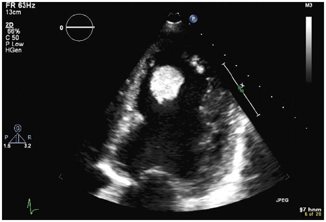
A 37-year-old female with a history of postpartum cardiomyopathy and multiple pulmonary embolisms in the past presented to an outside hospital with worsening dyspnea on exertion and fatigue 3 mo after her second delivery. She stated that she ran out of her medications for several weeks including rivaroxaban which was prescribed for her possible hypercoagulable state. At the outside hospital she complained of chest pain and underwent right and left heart catheterization, which disclosed a cardiac index of 1.5 L/min per square meter and non-obstructive coronary disease. She was initiated on inotropic support for low output cardiac failure. Her transthoracic echocardiogram followed by transesophageal echocardiogram showed worsening LV function with an ejection fraction of 10%, which was previously 20%, and a large apical pedunculated LV thrombus measuring 3 cm × 3 cm (Figure 1). She was therefore transferred to our hospital for further management.
Figure 1.

Transthoracic echocardiogram demonstrated a large left ventricular thrombus. It was highly mobile and the risk of embolization was considered to be high.
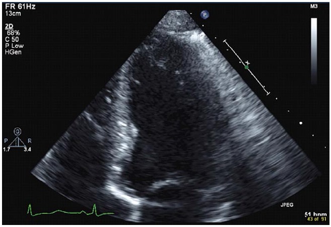
Considering a narrow stalk and large size of the LV thrombus, the risk of embolization was considered to be high. Emergent surgery was thus undertaken. Under cardioplegic cardiac arrest and cardiopulmonary support, the left atrium was opened at Waterson’s groove. A Cosgrove retractor was placed to optimize the exposure of the mitral valve. The LV thrombus was visualized through the mitral valve and was located at the apex connected to the ventricular wall only with a small stalk, which was divided at the base of the papillary muscle. The thrombus was extracted with gentle traction through the mitral valve without difficulty. The LV cavity was extensively irrigated, and then the left atrium was closed. The cross clamp time was 20 min and there was no issue weaning from cardiopulmonary bypass. Postoperatively, the patient was on minimal inotropic support which was successfully weaned off by postoperative day 4. She developed transient atrial fibrillation on postoperative day 3, which was converted to sinus rhythm by medical therapy. The postoperative echocardiogram revealed a small residual mural thrombus measuring 3 mm × 4 mm with left ventricular function of 40% (Figure 2). Hematology was consulted for workup of a possible hypercoagulable state, however all studies were negative. She was placed on coumadin therapy for the residual LV thrombus and was discharged home on postoperative day 10 without an embolic event. Pathologic workup of the mass revealed a large thrombus without any malignant component. Throughout her postoperative course, she has remained symptom free 6 mo after surgery.
Figure 2.

Postoperative transthoracic echocardiogram demonstrated only minimally remaining mural thrombus.
DISCUSSION
First line of treatment for a LV thrombus is anticoagulation; however, a large mobile thrombus as is in this case often requires urgent surgical thrombectomy. The concern with surgical removal of a large LV thrombus is ventricular function, since it is often seen in patients with poor LV function. The conventional approach to LV thrombus is left ventriculotomy[4,5]. Ventriculotomy provides direct visualization of the thrombus; thus it has been considered the standard approach for complete removal of the thrombus. This may be best utilized for mural thrombus which is adhered to the ventricular wall. However, LV ventriculotomy often causes further deterioration of the LV function[6], and should be avoided in cases of poor LV function if possible. Furthermore, if once the left ventriculotomy was performed, future placement of the ventricular assist device in case of further deterioration of the LV function would be more complicated. Another possible approach is thrombus extraction via aortotomy. This trans-aortic approach has been reported in conjunction with the video-assisted thoracoscopy to facilitate visualization[7]. However, the size of the thrombus is often the limiting factor in this approach and was too large to pass through the aortic valve in this case.
A left atrial approach does not require incision to the LV, thus theoretically preserves the remaining LV function. This approach also provides adequate visualization of the thrombus and trans-mitral valve extraction allows extraction of a larger thrombus than the trans-aortic approach[8-10]. The potential disadvantage of the left atrial approach would be limited room for maneuvering of the thrombus and should be reserved only for one that is loosely connected to the ventricular wall with a narrow stalk, which is exactly the case that emergent thrombectomy is usually indicated. When surgical thrombectomy is indicated for mural thrombi, which can usually be managed with anticoagulation, the left atrial approach should not be selected because extensive debridement is expected. These cases should be operated semi-electively with a standby left ventricular assist device since further deterioration of the LV function is expected after ventriculotomy. Therefore, we advocate left atriotomy as an alternative approach for emergent LV thrombectomy.
COMMENTS
Case characteristics
A 37-year-old female with a history of postpartum cardiomyopathy presented with chest pain and dyspnea 3 mo after her second delivery.
Clinical diagnosis
Echocardiogram showed worsening left ventricular (LV) dysfunction (ejection fraction of 10%) with a large left ventricular apical thrombus (3 cm × 3 cm) attached to the apex with a narrow stalk.
Differential diagnosis
Differential diagnosis included intracardiac thrombus, primary or metastatic tumor and cardiac myxoma.
Laboratory diagnosis
The patient’s cardiac index was 1.5 L/min per square meter and ejection fraction of 10%.
Imaging diagnosis
Echocardiogram showed a large left ventricular apical thrombus (3 cm × 3 cm) attached to the apex with a narrow stalk.
Pathological diagnosis
The removed mass was found to be a thrombus.
Treatment
The left ventricular thrombus was removed via the right-sided left atrium though the mitral valve.
Related reports
Conventional LV thrombectomy by left ventriculotomy; may this decrease the left ventricular function after surgery.
Experiences and lessons
Compared to conventional ventriculotomy, left atrial approach would be more suitable for emergency LV thrombectomy for highly mobile thrombi because the left atriotomy may not have any effect on the left ventricular function and would preserve the left ventricular apex for future ventricular assist device placement.
Peer review
Left ventricular thrombectomy via the left atrium though the mitral valve would be most feasible for the thrombus connected to the left ventricle with a narrow stalk.
Footnotes
P- Reviewers: Paraskevas KI, Satoh S, Said SAM, Vermeersch P S- Editor: Gou SX L- Editor: A E- Editor: Wu HL
References
- 1.Lee JM, Park JJ, Jung HW, Cho YS, Oh IY, Yoon CH, Suh JW, Chun EJ, Choi SI, Youn TJ, et al. Left ventricular thrombus and subsequent thromboembolism, comparison of anticoagulation, surgical removal, and antiplatelet agents. J Atheroscler Thromb. 2013;20:73–93. doi: 10.5551/jat.13540. [DOI] [PubMed] [Google Scholar]
- 2.Leick J, Szardien S, Liebetrau C, Willmer M, Fischer-Rasokat U, Kempfert J, Nef H, Rolf A, Walther T, Hamm C, et al. Mobile left ventricular thrombus in left ventricular dysfunction: case report and review of literature. Clin Res Cardiol. 2013;102:479–484. doi: 10.1007/s00392-013-0565-2. [DOI] [PubMed] [Google Scholar]
- 3.Stratton JR, Lighty GW, Pearlman AS, Ritchie JL. Detection of left ventricular thrombus by two-dimensional echocardiography: sensitivity, specificity, and causes of uncertainty. Circulation. 1982;66:156–166. doi: 10.1161/01.cir.66.1.156. [DOI] [PubMed] [Google Scholar]
- 4.Yadava OP, Yadav S, Juneja S, Chopra VK, Passey R, Ghadiok R. Left ventricular thrombus sans overt cardiac pathology. Ann Thorac Surg. 2003;76:623–625. doi: 10.1016/s0003-4975(03)00198-x. [DOI] [PubMed] [Google Scholar]
- 5.Suzuki R, Kudo T, Kurazumi H, Takahashi M, Shirasawa B, Mikamo A, Hamano K. Transapical extirpation of a left ventricular thrombus in Takotsubo cardiomyopathy. J Cardiothorac Surg. 2013;8:135. doi: 10.1186/1749-8090-8-135. [DOI] [PMC free article] [PubMed] [Google Scholar]
- 6.DiBernardo LR, Kirshbom PM, Skaryak LA, Quarterman RL, Johnson RL, Davies MJ, Gaynor JW, Ungerleider RM. Acute functional consequences of left ventriculotomy. Ann Thorac Surg. 1998;66:159–165. doi: 10.1016/s0003-4975(98)00376-2. [DOI] [PubMed] [Google Scholar]
- 7.Tsukube T, Okada M, Ootaki Y, Tsuji Y, Yamashita C. Transaortic video-assisted removal of a left ventricular thrombus. Ann Thorac Surg. 1999;68:1063–1065. doi: 10.1016/s0003-4975(99)00662-1. [DOI] [PubMed] [Google Scholar]
- 8.Kuh JH, Seo Y. Transatrial resection of a left ventricular thrombus after acute myocarditis. Heart Vessels. 2005;20:230–232. doi: 10.1007/s00380-004-0811-7. [DOI] [PubMed] [Google Scholar]
- 9.Lutz CJ, Bhamidipati CM, Ford B, Swartz M, Hauser M, Kyobe M, Dilip K. Robotic-assisted excision of a left ventricular thrombus. Innovations (Phila) 2007;2:251–253. doi: 10.1097/IMI.0b013e31815cea73. [DOI] [PubMed] [Google Scholar]
- 10.Early GL, Ballenger M, Hannah H, Roberts SR. Simplified method of left ventricular thrombectomy. Ann Thorac Surg. 2001;72:953–954. doi: 10.1016/s0003-4975(00)02607-2. [DOI] [PubMed] [Google Scholar]


