Abstract
Purpose
To evaluate the efficacy and stability of the wrap-clipping methods as a reconstructive strategy in the treatment of unclippable cerebral aneurysms.
Materials and Methods
Twenty four patients who had undergone wrap-clipping microsurgery were retrospectively reviewed. Type and morphology of the treated aneurysm, utilized technique for wrap-clip procedure, and clinical outcome with angiographic results at their last follow-up were evaluated.
Results
Of 24 patients, eleven patients had internal carotid artery (ICA) blister-like aneurysms, three had dissecting type aneurysms, and ten had fusiform aneurysms. The follow-up period for the late clinical and angiographic results ranged from 10 to 75 months (mean 35 months). Wrap-clipping was performed in eleven, wrap-holding clipping was in ten, and combination of wrap-clip and wrap-holding clip was in three cases. At the last angiographic follow-up study, twelve aneurysms (50%) were found to have completely healed, and nine aneurysms (38%) were at least stable. However, wrap-holding clip for the elongated blister type of ICA aneurysm was found failed, leading to fatal rebleeding in one case, and two cases of combination of wrap-clip-wrap-holding clip revealed delayed branch occlusion and marked regrowing, respectively.
Conclusion
Wrap-clipping strategy could be an easy and safe alternative for unclippable aneurysms. The wrapped aneurysm mostly disappeared, or at least remained stationary, after a long-term period. However, surgeons should be aware of that the wrapped aneurysm might become worse. Therefore, follow-up surveillance for an extended period should be mandatory.
Keywords: Cerebral aneurysms, unclippable, wrap-clipping, follow-up
INTRODUCTION
Although recent advances in microsurgical and endovascular techniques have allowed many challenging cerebral aneurysms to be successfully treated, a small subset are still not amenable to any definitive treatment. Friable blister-like aneurysms and fusiform aneurysms lacking an identifiable neck are representative of such challenging cases, for which many surgeons occasionally employ substitutional surgical techniques rather than direct neck clippings.1-6
Various surgical methods for unclippable aneurysms include parent artery sacrification, trapping with or without arterial bypass, aneurysmectomy following direct suture, and wrapping with reinforcements. Among them, wrapping may be the simplest way for surgeons to prevent further bleeding and simultaneously save normal arterial flow. Nonetheless, because of case rarity, disregard for incomplete extirpation, and extemporaneous manner of technique, previous reports are not readily available in regard with currently used techniques and the delicate nuances thereof. Furthermore, the fate of a wrapped aneurysm requires further long term evaluation of its efficacy and stability. Therefore, the present study attempted to clarify the detailed surgical techniques thereof and investigate the clinical and angiographic follow-up results for 24 consecutive patients who underwent wrap-clipping microsurgery using cellulose cotton fabric.
MATERIALS AND METHODS
Yonsei Aneurysm Database, which is prospectively maintained by two affiliated hospitals (Shinchon and Gangnam Severance Hospitals), were searched by using the query of 'wrap'. Cases of simple wrapping alone or wrapping with topical agents without additional clip appliance were excluded. Patients who did not undergo any post-operative angiographic study during the late follow-up were also excluded. As a result, a total of twenty four cases treated by the wrap-clip method were collected. All angiographic outcomes were evaluated by catheter based digital subtraction angiography (DSA). Data from the clinical and operative records were evaluated for the type and morphology of the aneurysm treated, technique utilized for wrap-clip method, and clinical outcome with angiographic results at the last follow-up.
Techniques
In all cases, cellulose cotton fabric was used as the wrapping agent. A thin layer peeled away from a microsurgical cotton pad (Bemsheet®, Kawamoto, Osaka, Japan) was prepared to the proper length and width. On occasion, tailoring strips into a Y-shape was useful to accommodate side branches and perforators. To encase the aneurysmal segment of the parent artery circumferentially with the cotton strip, ample room to allow free manipulation with micro forceps and angled micro dissectors was essential. In cases of subarachnoid hemorrhage, care must be taken not to violate the clot scab around the rupture point during circumferential dissection. If the lesion is buried in the cortical surface, subpial dissection must be facilitated.
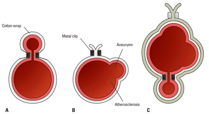
After enveloping the entire aneurysm segment, the portion of the aneurysm body that appears acceptable to approximate is clip-ligated as much as possible over the wrapped material (Wrap-Clip). A wrapped surface may help the clip to remain stationary and prevent tearing at the weakest spot. Wrap-Clipping is the authors' preferred technique. However, applying a clip parallel to the target artery and saving branches and perforators is not always possible. Then, the free ends of cotton wrapping are secured with an angled clip to tighten the wrapped construct (Wrap-Holding Clip). Sometimes, the two techniques can be used in combination: the most vulnerable portion of the lesion is wrap-clipped first, and the remainder is double reinforced by a second wrap-holding clipping (Wrap-Clip-Wrap-Holding Clip) (Fig. 1).
Fig. 1.
Schematic diagram of wrap-clip, wrap-holding clip, and combination of two. (A) After enveloping the entire aneurysm segment of the parent artery with a cotton strip, the portion of the aneurysm body that appears acceptable to approximate is clip-ligated as much as possible over the wrapped material (Wrap-Clip). (B) If the clip ligation to the aneurysm body is deemed difficult and risky in such a case with laterally projecting aneurysm or thick atherosclerosis of the parent artery, the free ends of cotton wrapping are secured with a clip to tighten the wrapped construct (Wrap-Holding Clip). (C) Occasionally, the two techniques can be used in combination. The most vulnerable portion of the lesion is wrap-clipped first, and the remainder is double reinforced by a second wrap-holding clipping (Wrap-Clip-Wrap-Holding Clip).
RESULTS
A total of twenty four cases included were composed of eleven internal carotid artery (ICA) blister-like aneurysms, three dissecting aneurysms, and ten fusiform aneurysms. They were eighteen women and six men with a mean age of 48 years (range from 29 to 72 years). The follow-up period for late clinical and angiographic results ranged from 10 to 75 months (mean 35 months).
Wrap-Clipping was performed in four of eleven ICA blister-like aneurysms and seven of ten fusiform aneurysms. Wrap-Holing Clipping was performed in seven of eleven ICA blister-like aneurysms, two of three dissecting aneurysms, and one of ten fusiform aneurysm. The combination of Wrap-Clip-Wrap-Holding Clipping was performed in one dissecting aneurysm and two fusiform aneurysms.
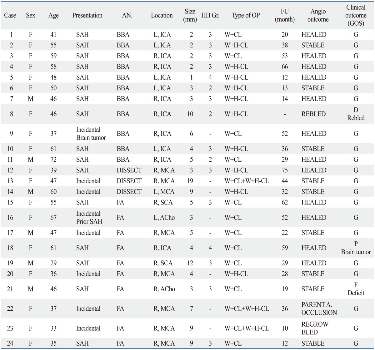
At the patients' last angiographic follow-up, twelve (50%) aneurysms (seven ICA blister-like aneurysms, one dissecting aneurysm, and four fusiform aneurysms) were no longer present, and DSA showed no evidence of recurrence or undesirable changes around the wrapped segment of the parent artery (defined as HEALED). In nine (38%) aneurysms (three ICA blister-like aneurysms, two dissecting aneurysms, and four fusiform aneurysms), DSA showed no identifiable changes in size and shape of the wrapped aneurysm (defined as STABLE), although minor contrast filling in the aneurysms was still noted. One patient harboring a broad based ICA blister-like aneurysm had undergone Wrap-Holding Clipping. However, the patient developed rebleeding and subsequently died during hospitalization (case 8). Two patients harboring globe-shaped fusiform dilatation of the middle cerebral artery (MCA) trunk were treated by combination of Wrap-Clip-Wrap-Holding Clip method. One patient showed delayed asymptomatic occlusion of the MCA inferior branch at the 36-month follow-up DSA (case 22). Another patient unexpectedly presented with mural hemorrhage in rapidly regrown fusiform dilatation of the whole MCA segment ten months after the initial combination wrap-clipping treatment, and underwent MCA trapping with radial artery interposition graft bypass from the cervical external carotid artery to MCA distal branch (case 23). Overall, 21 patients (87.5%) were scored as good according to the Glasgow Outcome Scale at their last visit, one patient was fair due to effects of the initial insult, one patient was poor due to a combined malignant brain tumor, and one patient died of rebleeding as described above. Details are summarized in Table 1.
Table 1.
Summary of 24 Patients Who Underwent Wrap-Clipping Microsurgery

AN, aneurysm; HH Gr, initial Hunt and Hess grade; OP, operation; FU, follow-up; GOS, Glasgow Outcome Scale (G, good; F, fair; P, poor; D, dead); SAH, subarachnoid hemorrhage; BBA, Blood Blister-like Aneurysm; DISSECT, dissecting aneurysm; FA, fusiform aneurysm; L, left; R, right; ICA, internal carotid artery; MCA, middle cerebral artery; SCA, superior cerebellar artery; ACho, anterior choroidal artery; W+CL, wrap-clipping; W+H-CL, wrap-holding clipping (refer to the text); A., artery; CASE 9, meningioma at right cerebellopontine angle; CASE 18, recurred oligodendroglioma at right frontal lobe; Deficit, remaining initial insult due to subarachnoid hemorrhage.
Illustrative case 6
A 50-year-old woman presented with subarachnoid hemorrhage (SAH). Catheter angiography revealed a 3-mm-sized tit-like aneurysm at the left ICA (Fig. 2). Microsurgery was performed via left pterional craniotomy combined with lumbar drainage and cervical ICA exposure. The aneurysm was thinly covered with a localized clot. The posterior communicating artery and anterior choroidal artery were discerned to have originated from the opposite wall of the ICA. Due to the dorso-medial direction of the aneurysm and the curvature of the aneurysm segment of the ICA, to attempt parallel clipping would have been difficult and risky, therefore, a cellulose cotton sheet was inserted and wrapped around the ICA, taking care not to compromise the posterior communicating artery or anterior choroidal artery. An L-shaped clip was then applied to tightly hold the wrapped aneurysmal segment of the ICA. The patient recovered fully and was discharged without any neurological deficit. Catheter angiography 13 months later showed that the aneurysm remained unchanged and stable.
Fig. 2.

Illustrative case 6. (A) Catheter angiography revealing a 3-mm-sized tit-like aneurysm at the left ICA (arrow). (B) Intraoperative photo of the aneurysm covered with a localized clot. (C) Wrap-Holding Clipping of the aneurysmal segment of the ICA. (D) Catheter angiography taken after 13 months showed the remaining aneurysm without significant changes (arrow). ICA, internal carotid artery.
Illustrative case 7
A 46-year-old man was admitted via the emergency room due to sudden headache and drowsy mental status. Computed tomography scan confirmed SAH, and catheter angiography showed a 3-mm-sized sessile aneurysm arising at the non-branching site of the right ICA (Fig. 3). On the day of admission, microsurgery for the aneurysm was performed through pterional craniotomy. The cervical ICA was exposed and prepared for proximal control. Lumbar drainage was initiated before the dural incision. After a wide sylvian dissection, the ICA segment containing a clot-capped aneurysm was visualized. The aneurysm arose at the dorso-medial aspect of the ICA, mostly hiding under the ipsilateral optic nerve. The aneurysmal segment of ICA was circumferentially dissected and encircled by an approximately 5×10-mm-sized cotton sheet. Next, an L-shaped clip was advanced to secure the encircling cotton sheet. The patient recovered slowly without any event and was discharged in good condition. Catheter angiography taken 14 months later showed no preexisting aneurysm and normal appearance of the ICA.
Fig. 3.

Illustrative case 7. (A) Catheter angiography showed a 3-mm-sized sessile aneurysm arising at the non-branching site of the right ICA (arrow). (B) The ICA segment containing a clot-capped aneurysm. (C) The aneurysm was wrapped with a holding clip. (D) Catheter angiography taken 14 months later showed no preexisting aneurysm (arrow) and normal appearance of the ICA (arrow). ICA, internal carotid artery.
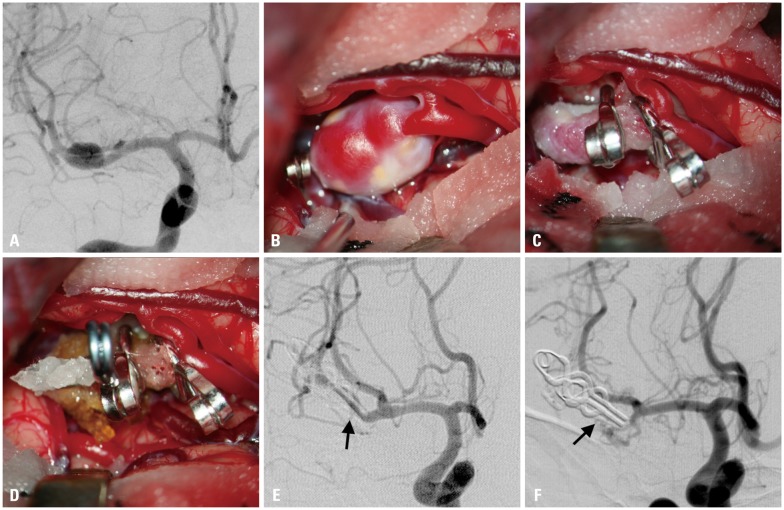
Illustrative case 22
A 37-year-old woman visited the outpatient clinic for an incidentally found cerebral aneurysm on magnetic resonance angiography during evaluation for dizziness. The patient had been suffering from panic disorder for years. After a thorough discussion with her and her family, a microsurgical treatment was chosen. A catheter angiography showed diffuse aneurysmal bulging without an identifiable neck measuring 7 mm at the inferior MCA branch (Fig. 4). During the operation via a right pterional craniotomy, the aneurysm was observed to have a cylindrical shape, encompassing 360 degrees around the MCA parent artery. First, wrapping with a cotton sheet was performed, and then most of the aneurysmal dilatation was clip-ligated using two angled fenestrated clips, reconstructing adequate lumen for normal blood flow. A second wrapping was added over the wrap-clipped construct, and a final clip was advanced to secure the second wrapping. Immediate postoperative angiography showed satisfactory obliteration of the original fusiform dilatation, and effective reconstruction of the parent artery. However, during the patient's routine follow-up after 36 months, catheter angiography revealed total occlusion of the previously wrap-reconstructed parent artery. Fortunately, she had no relevant symptoms and signs.
Fig. 4.
Illustrative case 22. (A) Catheter angiography showed diffuse aneurysmal bulging without an identifiable neck measuring 7 mm at the inferior MCA branch. (B) Intraoperative photo of the fusiform aneurysm. (C) Wrap-Clipping for most of the aneurysmal dilatation with reconstruction of adequate lumen for normal blood flow. (D) An additional Wrap-Holding Clipping over theWrap-Clipped construct. (E) Immediate postoperative angiography showing satisfactory obliteration of the original fusiform dilatation and satisfactory reconstruction of the parent artery (arrow). (F) After 36 months, an angiography revealed total occlusion of the previously wrap-reconstructed parent artery (arrow). MCA, middle cerebral artery.
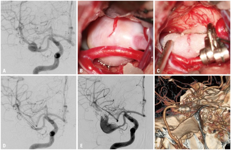
Illustrative case 23
A 33-year-old woman was transferred for a known aneurysm found during a health checkup. A microsurgery was planned because the patient was eager for treatment. Three-dimensional angiography revealed a globe-shaped fusiform aneurysm measuring 9 mm at the right MCA main trunk (Fig. 5). Through a pterional approach, a globe-shaped aneurysm without a clippable neck was identified. Wrap-clip plus subsequent wrap-holding clip was performed using two fenestrated angled clips. Postoperative angiography confirmed good reconstruction of the aneurysmal segment of the MCA. Ten months later, the patient presented with a sudden severe headache and vomiting. An emergent computerized tomography scan revealed localized scanty hemorrhage around the previously wrap-clipped right MCA. Catheter angiography showed a giant-sized fusiform dilatation of the right MCA, which appeared to have regrown along the MCA trunk proximal to the wrap-reconstructed segment. For treatment, the fusiform aneurysmal segment was trapped by clipping, followed by a radial artery graft bypass between the cervical external carotid artery and distal MCA. The patient recovered satisfactorily, and has remained in good condition.
Fig. 5.
Illustrative case 23. (A) Catheter angiography revealing a globe-shaped fusiform aneurysm at the right MCA main trunk. (B) Intraoperative photo showing aneurysmal dilatation without a clippable neck. (C) Wrap-Clipping plus subsequent Wrap-Holding Clipping using two fenestrated angled clips. (D) Postoperative angiography showing good reconstruction of the MCA. (E) Ten months later, catheter angiography revealed a giant-sized fusiform dilatation of the right MCA proximal to the wrap-reconstructed segment. (F) Computed tomographic angiography showing radial artery graft bypass between the cervical external carotid artery and distal MCA. MCA, middle cerebral artery.
DISCUSSION
Alternative methods for treatment of unclippable aneurysms may include parent artery occlusion with or without bypass, wrapping protection, and newer endovascular option such as flow-diverting stents. Among the alternatives, sacrificing the normal parent artery is not always safe, especially in the post hemorrhagic period of vasospasm, and is not always feasible when critical perforators or branches emanate from the body of the aneurysm.7,8 Even though many reports encourage the endovascular use of a recently developed flow diverter in various situations without further clinical experience, it remains too soon to accept the option as a safe and effective tool.9-12 Therefore, wrapping protection should remain as the only procedure to help prevent destructive parent artery occlusion or tentative insertion of endovascular devices.
The wrapping technique has been used for the treatment of cerebral aneurysms since the pre-microsurgical era,13,14 and a wide range of studies support its safety and durability.2 Nonetheless, an intuitive assumption of the inferiority of its protective effect makes surgeons hesitant to use wrapping, particularly in ruptured cases. The reason to adopt the wrap-clipping method in the present series was based mainly on the institutional policy for preferring reconstructive strategy whenever possible, in the management of cerebral aneurysms, rather than deconstructive treatments such as trap-bypass. Consistent with the fact that the wrapping strategy has been most commonly performed in the management of ICA blister-like aneurysms,3,4,15,16 the late angiographic findings revealing excellent lesion healing in the present case series suggest that focal, small blister-like aneurysms are the best candidates for the Wrap-Clip or Wrap-Holding Clip. Wrapped cotton enveloping the parent artery at the lesion site may play a crucial role in providing redundant leaflets to clip blades with a firm grip, and adding an artificial lining to promote the natural healing process (Fig. 3). Ideally, wrapping first and subsequent clipping to some part of the aneurysm or arterial tissue (Wrap-Clip) is the best substitute for bare direct clipping. However, the locations of blister-like aneurysms tend to be on the dorso-medial side of the ICA, which is toward the optic nerve, therefore, the most frequently used angled clip can be easily distorted (Fig. 2).17 Consequently, seven of eleven (64%) ICA blister-like aneurysms in the present series were wrapped circumferentially with a subsequent clip holding simply to tighten the wrapped construct (Wrap-Holding Clip). Mechanical reinforcement using a Wrap-Holding Clip was thought to be sufficiently protective to withstand short-term re-rupture of the lesion, and in the long-term, the wrapped cotton could induce inflammatory reactions, be replaced with connective tissue, and eventually remodel the weak fibrous layer lacking collagenous tissue of the blister-like lesion into a histologically competent structure.14
The Band-Aid effect of wrapping for aneurysm healing, however, did not always occur (Fig. 2). Nine of 24 wrapped aneurysms (38%) still remained, although they showed no significant changes in size and shape at the late follow-up angiography (STABLE). We assumed that differences in the histological composition of each aneurysm body might have affected the fate of the wrapped aneurysms. For example, if the aneurysm comprised a pseudo-wall like structure, wrapping may tend to promote scar healing processes. On the contrary, if the aneurysm body was lined with a relatively integrated wall structure, wrapping might not be able to occlude aneurysmal dilatation, though it could still play a role in preventing re-rupture or further growth.
For a blister-like or a fusiform aneurysm, if the lesion extends beyond the limited focal area of the parent artery, we suggest that wrapping with a tissue catching clip (Wrap-Clip) is more promising than Wrap-Holding Clip. As described above, the patient in case 8 who had segmental involvement of ICA aneurysms developed aggressive rebleeding after Wrap-Holding Clip, unlike aforementioned patients who had small and focal aneurysms treated by the same technique. On the contrary, Wrap-Clipping to bite as much of the thin-walled tissue as possible between the clip blades revealed successful results in the long-term regardless of aneurysm type.2 Although the number of cases is too limited to extrapolate comprehensive insight, it seems that the radial force of buttressing in the wrapped construct becomes shallower particularly at both ends as the wrapped construct lengthens. Furthermore, considering that the nature of elongated aneurysmal arteriopathy resembles arterial wall dissection which often progresses rapidly in the longitudinal direction along the parent artery, buttressing against radial expansion may be useless to stop propagating arterial pathology along its longitudinal axis.6
Such a potential pitfall of wrap-clipping method for a diseased artery was realized in cases 22 and 23. These cases had near identical morphology of circumferential fusiform dilatation in the MCA. Wrap-Clipping was performed first for the weakest area of bulging, and the remaining part was double reinforced by Wrap-Holding Clipping, as originally described by Kim, et al.18 Immediate postoperative angiography showed a nicely reconstructed parent artery and satisfactory obliteration of fusiform bulging. However, the major branching artery had been found occluded at the 36-month follow-up in case 22, and striking regrowth accompanied with mural hemorrhage occurred at ten month in case 23. Notably, the wrapped construct did not prevent expansile aneurysmal growth or progressive cylindrical deterioration along the parent artery proximal to the wrapped construct (Fig. 5). Because of complete vessel wall pathologies including dissection, disorders of collagen and elastin metabolism, and infective inflammations possibly involved in the formation of circumferential fusiform aneurysm,19,20 exo-vascular wrapping cannot be guaranteed to positively modify the endovascular pathologic progress, but rather seemed to be able to interfere with balance between initiation and stabilization of the natural healing process, and to boost aneurysm enlargement by the formation of vasa vasorum. Although we choose wrap-clip method because the patients were young and wanted to avoid parent artery sacrifice. However, looking back retrospectively, a conservative monitoring might be more reasonable in cases of non-symptomatic fusiform lesions, as recommended by several investigators.19-21 If the lesion becomes symptomatic, trapping the entire diseased segment of the parent artery with a distal bypass may then be better than a wrap-clip reconstruction policy.
Cellulose cotton sheets are excellent as a wrapping material and are most widely used.2,4,17 A thin layer peeled away from a cotton pad (Bemsheet®) adheres well to the vessel wall, is easy to tailor into any size or shape, and is believed not to be overly causative of foreign body reactions.2 Intense inflammatory reactions and evidence of vessel damage seen in older experimental reports were mainly due to coating materials such as cyanoacrylate glue, the use of which was recently withdrawn.22,23 Granuloma or toxic neuritis, well-known adverse effects of intracranially implanted cotton, was also reported in cases using coarsely-woven gauze.24 Other artificial materials such as Gore-tex, silastic sheet, Teflon, and Dacron have been proposed anecdotally; however, experiences are limited and long-term results are not known.18,25 One research using an elastase-induced rabbit aneurysm model advocated a combination of polyglycolic acid felt and fibrin glue to be superior to induce tight connective tissue formation without intimal thickening or neural tissue damage, compared to a combination of cellulose cotton sheet and fibrin glue;26 however, no clinical application has so far been reported.
Conclusions
Wrap-clipping strategy could be an easy and safe alternative for unclippable aneurysms. The wrapped aneurysm mostly disappeared, or at least remained stationary, after a long-term period. However, surgeons should be aware of the fact that the wrapped aneurysm might become worse. Therefore, follow-up surveillance for an extended period should be mandatory.
ACKNOWLEDGEMENTS
The authors thank Dong-Su Jang, BA (Research Assistant, Department of Anatomy, Yonsei University College of Medicine, Seoul, Korea), for his help with the figures.
Footnotes
The authors have no financial conflicts of interest.
References
- 1.Cossu M, Pau A, Turtas S, Viola C, Viale GL. Subsequent bleeding from ruptured intracranial aneurysms treated by wrapping or coating: a review of the long-term results in 47 cases. Neurosurgery. 1993;32:344–346. doi: 10.1227/00006123-199303000-00002. [DOI] [PubMed] [Google Scholar]
- 2.Deshmukh VR, Kakarla UK, Figueiredo EG, Zabramski JM, Spetzler RF. Long-term clinical and angiographic follow-up of unclippable wrapped intracranial aneurysms. Neurosurgery. 2006;58:434–442. doi: 10.1227/01.NEU.0000199158.02619.99. [DOI] [PubMed] [Google Scholar]
- 3.McLaughlin N, Laroche M, Bojanowski MW. Surgical management of blood blister-like aneurysms of the internal carotid artery. World Neurosurg. 2010;74:483–493. doi: 10.1016/j.wneu.2010.06.039. [DOI] [PubMed] [Google Scholar]
- 4.Ogawa A, Suzuki M, Ogasawara K. Aneurysms at nonbranching sites in the surpaclinoid portion of the internal carotid artery: internal carotid artery trunk aneurysms. Neurosurgery. 2000;47:578–583. doi: 10.1097/00006123-200009000-00008. [DOI] [PubMed] [Google Scholar]
- 5.Ohkuma H, Nakano T, Manabe H, Suzuki S. Subarachnoid hemorrhage caused by a dissecting aneurysm of the internal carotid artery. J Neurosurg. 2002;97:576–583. doi: 10.3171/jns.2002.97.3.0576. [DOI] [PubMed] [Google Scholar]
- 6.Todd NV, Tocher JL, Jones PA, Miller JD. Outcome following aneurysm wrapping: a 10-year follow-up review of clipped and wrapped aneurysms. J Neurosurg. 1989;70:841–846. doi: 10.3171/jns.1989.70.6.0841. [DOI] [PubMed] [Google Scholar]
- 7.Kamijo K, Matsui T. Acute extracranial-intracranial bypass using a radial artery graft along with trapping of a ruptured blood blister-like aneurysm of the internal carotid artery. Clinical article. J Neurosurg. 2010;113:781–785. doi: 10.3171/2009.10.JNS09970. [DOI] [PubMed] [Google Scholar]
- 8.Meling TR, Sorteberg A, Bakke SJ, Slettebø H, Hernesniemi J, Sorteberg W. Blood blister-like aneurysms of the internal carotid artery trunk causing subarachnoid hemorrhage: treatment and outcome. J Neurosurg. 2008;108:662–671. doi: 10.3171/JNS/2008/108/4/0662. [DOI] [PubMed] [Google Scholar]
- 9.Turowski B, Macht S, Kulcsár Z, Hänggi D, Stummer W. Early fatal hemorrhage after endovascular cerebral aneurysm treatment with a flow diverter (SILK-Stent): do we need to rethink our concepts? Neuroradiology. 2011;53:37–41. doi: 10.1007/s00234-010-0676-7. [DOI] [PubMed] [Google Scholar]
- 10.Cebral JR, Mut F, Raschi M, Scrivano E, Ceratto R, Lylyk P, et al. Aneurysm rupture following treatment with flow-diverting stents: computational hemodynamics analysis of treatment. AJNR Am J Neuroradiol. 2011;32:27–33. doi: 10.3174/ajnr.A2398. [DOI] [PMC free article] [PubMed] [Google Scholar]
- 11.Lubicz B, Collignon L, Raphaeli G, Pruvo JP, Bruneau M, De Witte O, et al. Flow-diverter stent for the endovascular treatment of intracranial aneurysms: a prospective study in 29 patients with 34 aneurysms. Stroke. 2010;41:2247–2253. doi: 10.1161/STROKEAHA.110.589911. [DOI] [PubMed] [Google Scholar]
- 12.Kim DJ, Suh SH, Kim BM, Kim DI, Huh SK, Lee JW. Hemorrhagic complications related to the stent-remodeled coil embolization of intracranial aneurysms. Neurosurgery. 2010;67:73–78. doi: 10.1227/01.NEU.0000370937.70207.95. [DOI] [PubMed] [Google Scholar]
- 13.Drake CG, Vanderlinden RG. The late consequences of incomplete surgical treatment of cerebral aneurysms. J Neurosurg. 1967;27:226–238. doi: 10.3171/jns.1967.27.3.0226. [DOI] [PubMed] [Google Scholar]
- 14.Mount LA, Antunes JL. Results of treatment of intracranial aneurysms by wrapping and coating. J Neurosurg. 1975;42:189–193. doi: 10.3171/jns.1975.42.2.0189. [DOI] [PubMed] [Google Scholar]
- 15.Abe M, Tabuchi K, Yokoyama H, Uchino A. Blood blisterlike aneurysms of the internal carotid artery. J Neurosurg. 1998;89:419–424. doi: 10.3171/jns.1998.89.3.0419. [DOI] [PubMed] [Google Scholar]
- 16.Nakagawa F, Kobayashi S, Takemae T, Sugita K. Aneurysms protruding from the dorsal wall of the internal carotid artery. J Neurosurg. 1986;65:303–308. doi: 10.3171/jns.1986.65.3.0303. [DOI] [PubMed] [Google Scholar]
- 17.Lee JW, Choi HG, Jung JY, Huh SK, Lee KC. Surgical strategies for ruptured blister-like aneurysms arising from the internal carotid artery: a clinical analysis of 18 consecutive patients. Acta Neurochir (Wien) 2009;151:125–130. doi: 10.1007/s00701-008-0165-5. [DOI] [PubMed] [Google Scholar]
- 18.Kim LJ, Klopfenstein JD, Spetzler RF. Clip reconstruction and sling wrapping of a fusiform aneurysm: technical note. Neurosurgery. 2007;61(3 Suppl):79–80. doi: 10.1227/01.neu.0000289717.03050.65. [DOI] [PubMed] [Google Scholar]
- 19.Day AL, Gaposchkin CG, Yu CJ, Rivet DJ, Dacey RG., Jr Spontaneous fusiform middle cerebral artery aneurysms: characteristics and a proposed mechanism of formation. J Neurosurg. 2003;99:228–240. doi: 10.3171/jns.2003.99.2.0228. [DOI] [PubMed] [Google Scholar]
- 20.Park SH, Yim MB, Lee CY, Kim E, Son EI. Intracranial Fusiform Aneurysms: It's Pathogenesis, Clinical Characteristics and Managements. J Korean Neurosurg Soc. 2008;44:116–123. doi: 10.3340/jkns.2008.44.3.116. [DOI] [PMC free article] [PubMed] [Google Scholar]
- 21.Lanzino G, Kaptain G, Kallmes DF, Dix JE, Kassell NF. Intracranial dissecting aneurysm causing subarachnoid hemorrhage: the role of computerized tomographic angiography and magnetic resonance angiography. Surg Neurol. 1997;48:477–481. doi: 10.1016/s0090-3019(97)00178-x. [DOI] [PubMed] [Google Scholar]
- 22.Herrera O, Kawamura S, Yasui N, Yoshida Y. Histological changes in the rat common carotid artery induced by aneurysmal wrapping and coating materials. Neurol Med Chir (Tokyo) 1999;39:134–139. doi: 10.2176/nmc.39.134. [DOI] [PubMed] [Google Scholar]
- 23.Juan GM, Kawamura S, Yasui N, Yoshida Y. Histological changes in the rat common carotid artery following simultaneous topical application of cotton sheet and cyanoacrylate glue. Neurol Med Chir (Tokyo) 1999;39:908–911. doi: 10.2176/nmc.39.908. [DOI] [PubMed] [Google Scholar]
- 24.Chambi I, Tasker RR, Gentili F, Lougheed WM, Smyth HS, Marshall J, et al. Gauze-induced granuloma ("gauzoma"): an uncommon complication of gauze reinforcement of berry aneurysms. J Neurosurg. 1990;72:163–170. doi: 10.3171/jns.1990.72.2.0163. [DOI] [PubMed] [Google Scholar]
- 25.Kubo Y, Ogasawara K, Tomitsuka N, Otawara Y, Watanabe M, Ogawa A. Wrap-clipping with polytetrafluoroethylene for ruptured blisterlike aneurysms of the internal carotid artery. Technical note. J Neurosurg. 2006;105:785–787. doi: 10.3171/jns.2006.105.5.785. [DOI] [PubMed] [Google Scholar]
- 26.Yasuda H, Kuroda S, Nanba R, Ishikawa T, Shinya N, Terasaka S, et al. A novel coating biomaterial for intracranial aneurysms: effects and safety in extra- and intracranial carotid artery. Neuropathology. 2005;25:66–76. doi: 10.1111/j.1440-1789.2004.00590.x. [DOI] [PubMed] [Google Scholar]


