

Abstract
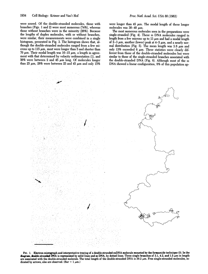
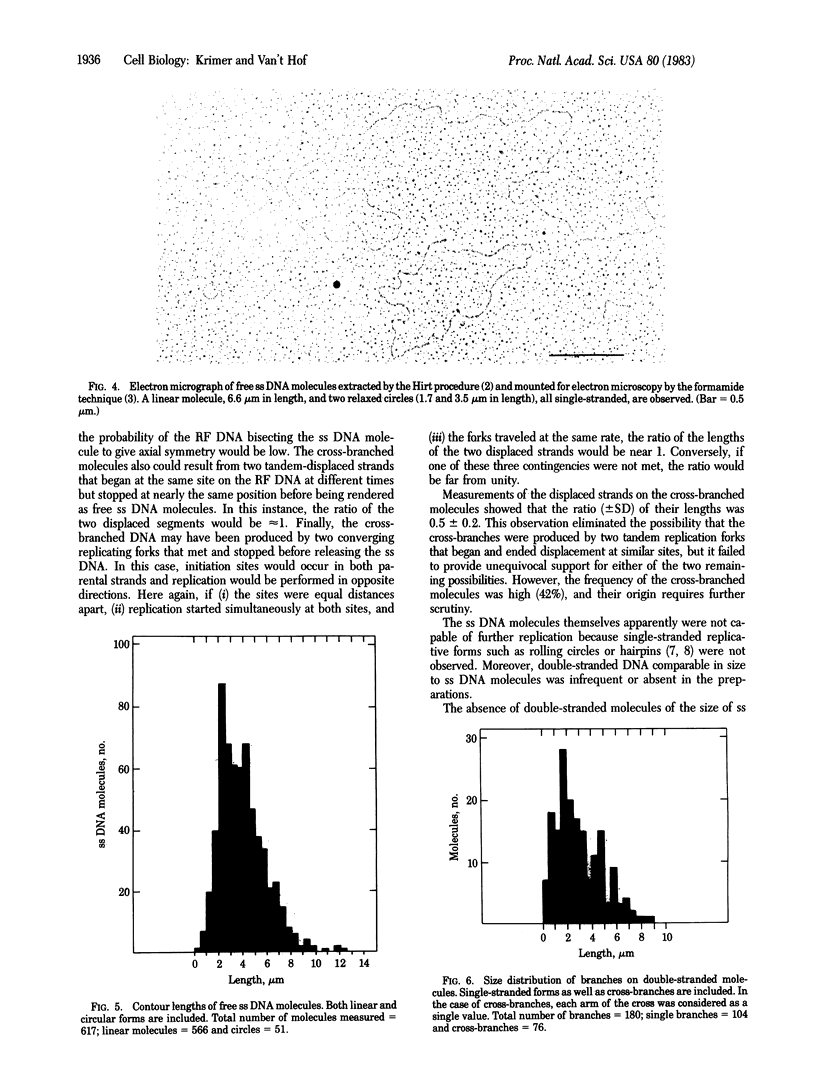
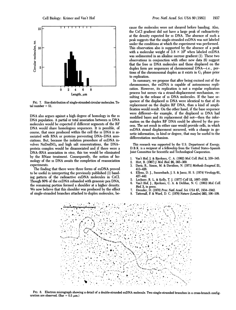
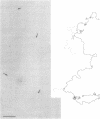
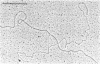
In cultured pea roots there is extrachromosomal DNA associated with cells that differentiate from the G2 phase of the cell cycle that is absent from those that differentiate from the G1 phase. We examined this extrachromosomal DNA by electron microscopy and found that it consisted of three types: (i) double-stranded linear molecules with single-stranded branches (74%), (ii) double-stranded molecules without branches (26%), and (iii) free single-stranded molecules. The double-stranded molecules with or without branches were similar in length, having a modal length of 10-15 μm. The free single-stranded molecules were shorter and had a mean length of 3.8 μm. The length of the branches attached to the duplex molecules was only slightly less than that of the free form. The duplex molecules with branches were interpreted as configurations reflecting an ongoing strand-displacement process that results in free single-stranded molecules. Finally, measurements on duplex molecules with multiple branches suggested that the extrachromosomal DNA may exist in the form of tandemly repeated sequences.
Keywords: DNA replication, single-stranded DNA, electron microscopy
Full text
PDF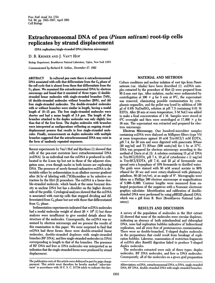
Images in this article

Selected References
These references are in PubMed. This may not be the complete list of references from this article.
- Dressler D. The rolling circle for phiX DNA replication. II. Synthesis of single-stranded circles. Proc Natl Acad Sci U S A. 1970 Dec;67(4):1934–1942. doi: 10.1073/pnas.67.4.1934. [DOI] [PMC free article] [PubMed] [Google Scholar]
- Ellens D. J., Sussenbach J. S., Jansz H. S. Studies on the mechanism of replication of adenovirus DNA. III. Electron microscopy of replicating DNA. Virology. 1974 Oct;61(2):427–442. doi: 10.1016/0042-6822(74)90279-7. [DOI] [PubMed] [Google Scholar]
- Hirt B. Selective extraction of polyoma DNA from infected mouse cell cultures. J Mol Biol. 1967 Jun 14;26(2):365–369. doi: 10.1016/0022-2836(67)90307-5. [DOI] [PubMed] [Google Scholar]
- Lechner R. L., Kelly T. J., Jr The structure of replicating adenovirus 2 DNA molecules. Cell. 1977 Dec;12(4):1007–1020. doi: 10.1016/0092-8674(77)90165-9. [DOI] [PubMed] [Google Scholar]
- Tattersall P., Ward D. C. Rolling hairpin model for replication of parvovirus and linear chromosomal DNA. Nature. 1976 Sep 9;263(5573):106–109. doi: 10.1038/263106a0. [DOI] [PubMed] [Google Scholar]
- Van't Hof J., Bjerknes C. A. Cells of pea (Pisum sativum) that differentiate from G2 phase have extrachromosomal DNA. Mol Cell Biol. 1982 Apr;2(4):339–345. doi: 10.1128/mcb.2.4.339. [DOI] [PMC free article] [PubMed] [Google Scholar]