Out of all superficial fungal infection in children below 15 years, tinea capitis is most common. Ninety-seven patients of this age group with superficial fungal infections were registered in out patient department (OPD) at SMS Hospital, Jaipur during August 2009-July 2010. Eighty patients (82.24%) out of them had tinea capitis, whereas 17 (17.76%) patients had other fungal infections, e.g. tinea cruris, tinea corporis, onychomycosis, etc., Tinea capitis being most common fungal infection, it is very important to diagnose it correctly and rapidly in a busy out patient department. Also seborrheic type of tinea capitis is difficult to differentiate from seborrhea capitis (which is not as uncommon as thought of). In our OPD, out of total 919 children with skin diseases who came between August 2009 and July 2010, 39 (4.24%) had seborrheic capitis.
We have found that examination of ear and other body parts is very important to differentiate between tinea capitis and seborrhea capitis. Out of 323 cases with classical presentation of tinea capitis, involvement of ear and other parts of body was seen in 185 patients (57.27%). In 97 (30.03%) patients, ear alone was involved; in 58 (17.95%) patients, ear and other body sites; and in 30 (9.28%) patients, other body sites only were involved.
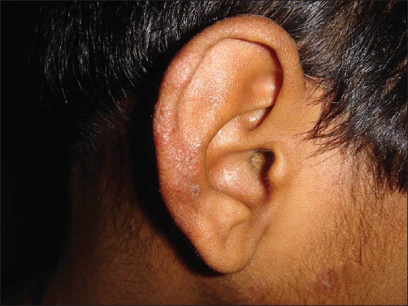
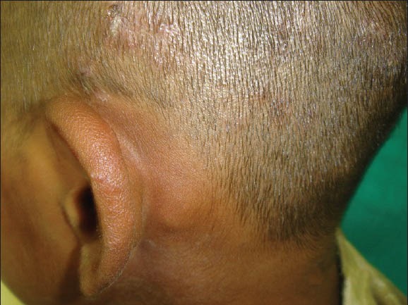
On ear, lesions were erythematous papules [Figure 1], scaling or well- to ill-defined plaques over helix, antihelix, and retroauricular region (away from retroauricular fold) in tinea capitis [Figure 2]. Patients with seborrheic capitis showed erythema, scaling, or fissuring in retroauricular fold and in other seborrheic sites. We have highlighted this finding as “Ear sign.”[1,2]
Figure 1.

Erythematous papules over external ear in a patient with tinea capitis
Figure 2.

Annular plaque over helix in patient with tinea capitis along with lymphadenopathy
Ear sign can be defined as presence of erythematous papules, scaling, or well- to ill-defined papules and plaques over helix, antihelix, and retroauricular region (away from retroauricular fold) in case of tinea capitis, whereas erythema, scaling, or fissuring in retroauricular fold in case of seborrhea capitis. Other body sites should also be examined and history of involvement of seborrheic sites in the past should also be taken. This simple finding can be very helpful for dermatologists and medical practitioners, especially to internists, pediatricians, and to postgraduates if highlighted properly. The sensitivity of ear sign was 50-60% and specificity was approaching near 100% when correlated with clinical response.[1]
Footnotes
Source of Support: Nil
Conflict of Interest: Nil
REFERENCES
- 1.Agarwal U, Sitaraman S, Panse GG, Bhola K, Besarwal RK. Useful sign to diagnose tinea capitis-‘ear sign’. Indian J Pediatr. 2012;79:679–80. doi: 10.1007/s12098-011-0571-1. [DOI] [PubMed] [Google Scholar]
- 2.Agarwal US, Shukla SR. Fungal infections. In: Agarwal US, Shukla SR, editors. Illustrated Textbook of Clinical Pediatric Dermatology. 1st ed. New Delhi: PEEPEE; 2011. pp. 37–8. [Google Scholar]


