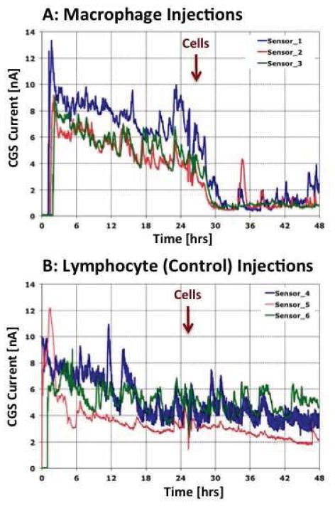
Figure 5. Impact of macrophage or lymphocytes injections at glucose sensor implantation sites on sensor function and CGM in vivo.
Presented in Figure 5 are the results of in vivo injection of MQ (Figure 5a) or spleen derived lymphocytes (Figure 5b) at the glucose sensor implantation in our murine model of CGM. For these studies MQ or lymphocytes (2.4x10^6 cells/30 ul) were injected in a volume of 30 ul at tip of sensor and CGM evaluated for the subsequent 48 hrs. Results presented in Figure 5 represents sensor response of triplicates (i.e. for each cell type 3 sensors were implanted in 3 separate mice) for MQ (Figure 5a) or lymphocyte (Figure 5b) injections. Sensor output for each sensor is represented as a separate solid line. Timing of cell injections is noted with the vertical red arrow.

