Abstract
The efficacy of conventional radiation therapy for gastric cancer is controversial. In this study, we evaluated the in vitro and in vivo effects of continuous low-dose-rate irradiation by I-125 seeds on different histological types of gastric cancer cell lines. Three human gastric cancer cell lines (MKN74, MKN45, and NUGC4) were treated with or without continuous low-dose irradiation by I-125 seeds in vitro and in vivo. Cell viability, apoptosis, caspase-3 assay, and cell-cycle distribution were examined in vitro. Body weight and tumor volumes of BALB/c nude mice bearing MKN74, MKN45, and NUGC4 gastric cancer xenografts were measured, and in vivo cell proliferation and apoptosis assays were performed by Ki67 and TUNEL staining, respectively. Continuous low-dose-rate irradiation by I-125 seeds reduced cell viability and induced cell apoptosis through the activation of caspase-3, and led to the accumulation of cells in the G2/M phase in vitro. It also suppressed the growth of gastric cancer xenografts in nude mice, while inhibiting cell proliferation and inducing apoptosis as demonstrated by Ki67 and TUNEL staining. Therefore, our data suggest that continuous low-dose-rate irradiation by I-125 seeds could be a promising new option for gastric cancer treatment, regardless of histological origin.
Keywords: I-125 seed irradiation, gastric cancer, apoptosis, cell cycle, caspase
Introduction
Gastric cancer is the fourth most frequent malignancy and the second leading cause of cancer-related mortality in the world.1 Although the incidence of gastric cancer has been decreasing, it remains a common malignancy worldwide, especially in Asia.2,3 Endoscopic submucosal dissection has recently emerged as a common treatment for early-stage IA gastric cancer,4,5 while surgical resection remains a standard therapeutic approach for early gastric cancer. However, for stages III and IV advanced gastric cancers, surgical treatment alone is not regarded as the definitive standard treatment. This is because the 5-y survival rates of stages III and IV patients were reported to be less than 50% even when a curative operation was performed.6,7 The high rate of relapse after surgical treatment makes it important to consider adjuvant treatment for patients with advanced gastric cancer. Several studies of chemotherapy for advanced gastric cancer were reported, but adjuvant chemotherapy has not resulted in higher survival rates than surgical treatment alone. As a result, chemoradiotherapy is being evaluated as an alternate treatment for gastric cancer.
Several studies, including the clinical trial INT0116,8 have reported that postoperative chemoradiotherapy could be a powerful treatment for controlling tumor progression in advanced gastric cancer.9-11 This led to the inclusion of radiotherapy as a standard treatment for patients with a high risk of recurrence12-14 in the National Comprehensive Cancer Network (NCCN) guidelines on gastric cancer treatment. Therefore, in the USA and Europe, postoperative chemoradiotherapy has becoming a standard treatment for advanced gastric cancer. However, there are some drawbacks of radiation therapy for gastric cancer. These include the difficulty of establishing the area to irradiate because of peristaltic movement, complications affecting the surrounding organs, risk of perforation and ulceration by high-dose radiation, and ineffectiveness against adenocarcinoma with low radiosensitivity. Therefore, in Japan, radiation therapy has not been established as a standard treatment for advanced gastric cancer. Indications for radiation therapy are limited, and it is performed only as a palliative therapy.15-17 Because of this, more effective and safer therapeutic strategies for advanced or unresectable gastric cancer are expected.
In recent years, I-125 seed implantation providing continuous low-dose-rate irradiation has been widely used to treat prostate cancer and other kinds of tumors in several Asian countries because of little trauma, strong effect, and fewer complications,18-21 while in Japan, I-125 seed implantation therapy is provided only for prostate cancer. In this study, we investigate the effect of continuous low-dose-rate irradiation by I-125 seeds on several types of gastric cancer in vitro and in vivo to determine its potential as a novel therapeutic strategy for advanced and unresectable gastric cancer.
Results
I-125 seeds reduced cell viability and induced cell apoptosis in vitro
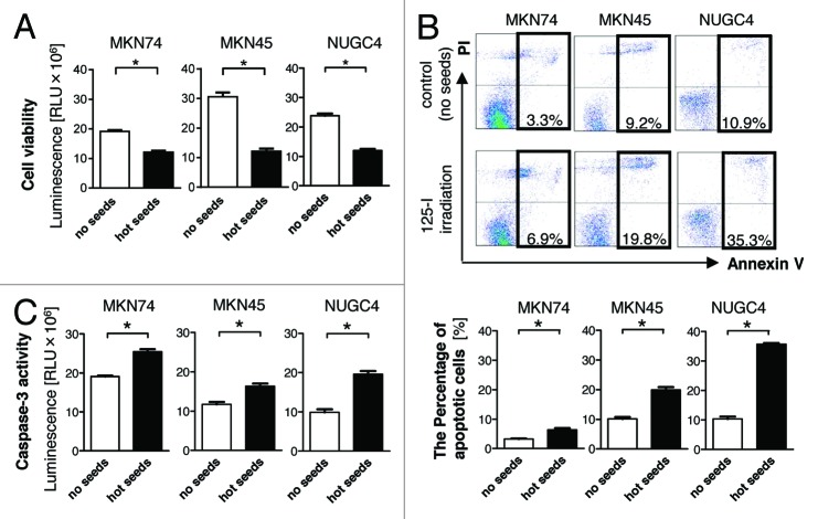
To determine the direct effects of I-125 seeds in gastric cancer cell lines, three gastric cancer cell lines (MKN74, MKN45, and NUGC4) were assayed after treatment with (2–3 Gy) or without (0 Gy, no seeds as a control) irradiation. Cell viability was significantly lower than in the control group in each of the three gastric cancer cell lines following irradiation (P < 0.05) (Fig. 1A). To analyze the induction of apoptosis by irradiation, double staining of cells with annexin V-FITC and propidium iodide (PI) was performed. Annexin V-positive/PI-negative cell staining was considered to denote early apoptosis, while annexin V/PI-double-positive cell staining was considered to denote late apoptosis (Fig. 1B). Figure 1B shows the total apoptosis rate (annexin V-positive rate) in all cell lines. The total apoptosis rate induced by irradiation was significantly increased in each of the three gastric cancer cell lines as compared with the control (P < 0.05). Recent studies have identified caspases, including caspase-3, as important mediators of apoptosis induced by various apoptotic stimuli.22 The activity of caspase-3 was significantly increased in all irradiated gastric cancer cell lines (P < 0.05) (Fig. 1C), thus confirming that I-125 seed irradiation caused apoptosis in gastric cancer cells through the activation of caspase-3.
Figure 1. Cell viability of the three gastric cancer cell lines (MKN74, MKN45, and NUGC4) was assessed following incubation for 96 h with or without irradiation to maintain constant cell culture conditions. The viability of each of the 3 cell lines was significantly lower than that of the control group (*P < 0.05) (A). Apoptosis was determined by flow cytometry and the total apoptosis rate was calculated. The total apoptosis rate induced by irradiation was significantly increased in each of the three gastric cancer cell lines as compared with the control (*P < 0.05) (representative data was shown in [B]). Caspase-3 assay was performed and the activity of caspase-3 was increased significantly in all 3 irradiated gastric cancer cell lines (P < 0.05) (C). Experiments were performed at least 3 times.
I-125 seeds reduced cell-cycle arrest in vitro
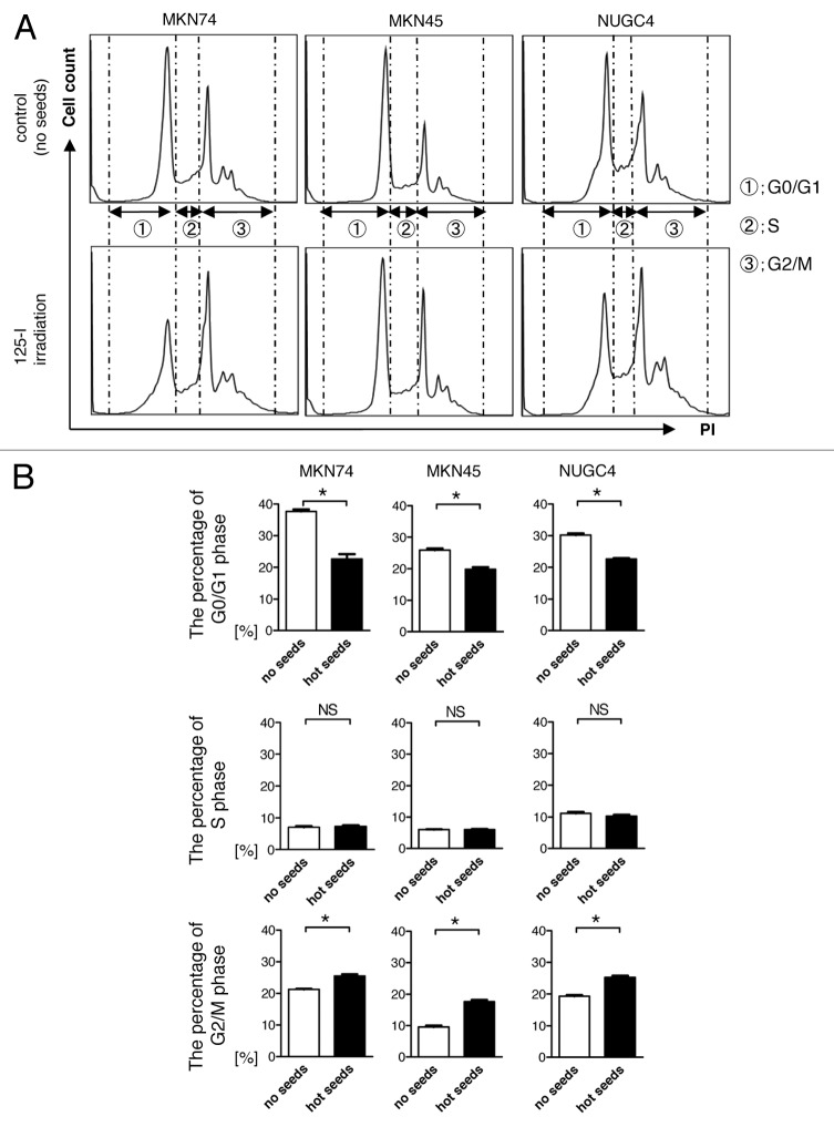
The results of the flow cytometry cell-cycle assay (Fig. 2A and B) indicated that the continuous low-dose-rate irradiation by I-125 seeds induced a lower percentage of G0/G1, and higher percentage of G2/M phase cell-cycle arrest in all three cell lines compared with the control, and the differences were statistically significant (P < 0.05). These results suggest that continuous low-dose-rate irradiation by I-125 seeds may enhance radiosensitivity by inducing accumulation of cells in the more radiosensitive G2/M phase.
Figure 2. Cell-cycle assay was performed on each of the three gastric cancer cell lines (MKN74, MKN45, and NUGC4) following incubation for 96 h with or without irradiation to maintain constant cell culture conditions (representative data was shown in [A]). Three cell-cycle segments are shown in order from left to right: G1/G0 phase, S phase, G2/M phase, and the percentage of cells in each phase is shown in (B). Continuous low-dose-rate irradiation by I-125 seeds induced a lower percentage of G0/G1, and higher percentage of G2/M phase cell-cycle arrest compared with the control for all 3 gastric cancer cell lines (P < 0.05). Experiments were performed at least 3 times.
I-125 seeds inhibited tumor growth of gastric cancer in vivo
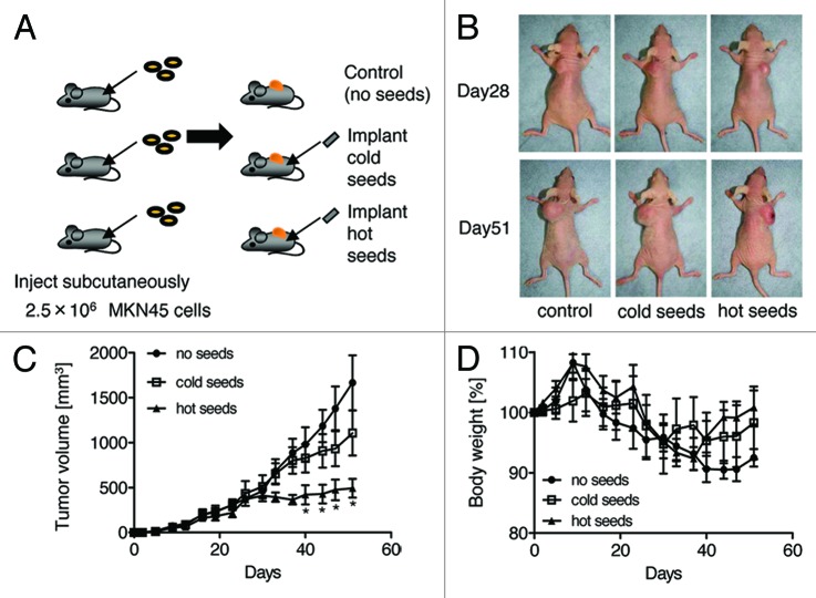
Tumor xenografts consisting of transplanted human gastric cancer cell lines MKN45 (derived from poorly differentiated adenocarcinoma) or NUGC4 (derived from signet-ring cell carcinoma) were used to evaluate the antitumor effects of I-125 seeds in vivo (Fig. 3A, data not shown for NUGC4). When tumors of both cell lines reached around 400 mm3 at day 28, cold and hot seeds were implanted. Tumors which I-125 seeds were implanted were smaller rather than cold seeds or the control at day 52 (Fig. 3B). There were no significant changes in tumor volumes during the first 2 weeks after seed implantation, but after that, I-125 irradiated tumors were significantly smaller than the non-irradiated tumors (P < 0.05) (Fig. 3C). This indicated that I-125 seeds significantly inhibited tumor growth during the 3- to 4-week treatment. As shown in Figure 3D, the body weights of mice were not affected by the I-125 irradiation. Besides, none of the mice died during the treatment, and no obvious radiation-induced damage was observed in vital organs (data not shown). These results underscore the safety of I-125 seed treatment.
Figure 3. Protocol for animal experiments using the MKN45 cell line (A). 2.5 × 10⁶ cells in 0.2 ml PBS were injected subcutaneously into the dorsa of each mouse. When tumors reached around 400 mm3 at about 3–4 weeks, I-125 seeds or cold seeds were implanted into each 5 mice per group via a needle. The untreated mice served as the no seed control group. The tumor’s macroscopic appearance was imaged on the day of implantation before the mouse was sacrificed (representative data was shown in [B]). Tumor size was measured once every 4 d. There were no significant changes in tumor volume during the first 2 weeks after seed implantation, but after that, I-125-irradiated tumors were much smaller than the others, and significant differences in tumor volumes were observed between the I-125 seed implanted group and the other 2 groups (P < 0.05) (C). The body weight of the animals was also measured every 4 d and mortality was monitored daily, but there were no significant differences between the 3 groups (D).
I-125 seeds inhibited cell proliferation and induced apoptosis in vivo
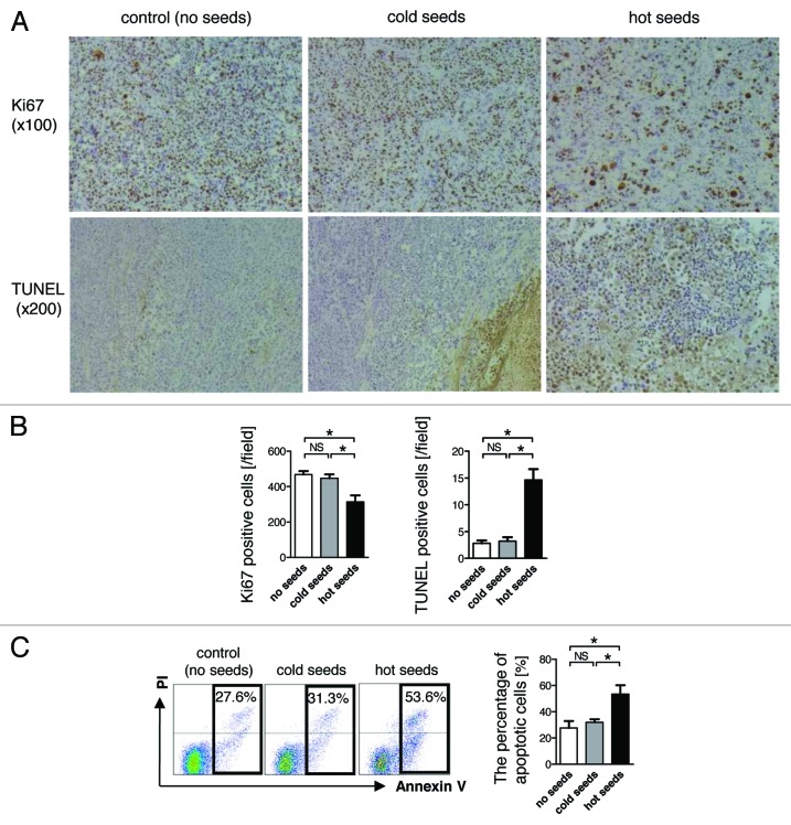
To quantitatively compare the proliferation and apoptotic rates, MKN45 and NUGC4 xenograft tumor sections were taken from mice in the control, cold seed, and I-125 seed implanted groups, and immunostained for Ki67 and TUNEL. In addition, before immunostaining, cells were isolated as described, stained with annexin V-FITC and PI, and analyzed using a flow cytometer to clarify the induction of apoptosis. There were clearly fewer Ki67-positive cells in the I-125 seed implanted group than in the control and cold seed implanted groups (Fig. 4A and B). In contrast to the proliferation rate, the average number of TUNEL-positive apoptotic cells in the I-125 seed implanted group was significantly increased compared with the control and cold seed implanted groups (P < 0.05). The apoptosis rate (annexin V-positive staining as determined by flow cytometry) in the I-125 implanted group was significantly increased over that of the control and cold seed implanted groups (P < 0.05) (Fig. 4C). These results suggested that I-125 seeds inhibited cell proliferation and induced apoptosis in MKN45 and NUGC4 xenografts.
Figure 4. Histological experiments with MKN45 xenografts. Tumor sections were immunostained for Ki67 and a TUNEL assay was performed. Ki67 and TUNEL immunostaining are shown at 100× and 200× magnification (representative data was shown in [A]). Ki67 and TUNEL-positive cells were quantified in 20 randomly selected, high-power fields in each tissue section. The average number of Ki67-positive cells in the I-125 implanted group was clearly less than those in the control and cold seed implanted groups. In contrast to the proliferation rate, the average number of TUNEL-positive apoptotic cells in the I-125 implanted group was significantly increased over those in the control and cold seed implanted groups (*P < 0.05) (B). Tumor tissue samples were isolated and stained with annexin V-FITC and PI, then analyzed using a flow cytometer to clarify the induction of apoptosis. The apoptosis rate in the I-125 implanted group was significantly increased compared with those of the control and cold seed implanted groups (P < 0.05) (representative data was shown in [C]).
Discussion
Gastric cancer remains a major cause of death in the world. Radiation therapy has recently started to play an important role in the treatment of advanced gastric cancer. However, the adverse effects of conventional external beam radiation therapy on surrounding organs pose a major problem. With recent technological advances in irradiation, e.g., intensity-modulated radiation therapy, irradiation is localized to the restricted area as far as possible. However, adverse effects remain a problem. In this study, we examined the effectiveness of I-125 seed irradiation therapy to address this problem. As described above, I-125 seeds serve as a localized radiation source with an irradiation range of <2 cm. If the seeds can provide the same therapeutic effects for gastric cancer in a more localized irradiated region than conventional external beam radiation therapy, then they may serve as a new radiation therapy, which reduces the adverse effects of radiation on the surrounding organs.
According to a recent report, continuous low-dose-rate irradiation by I-125 seeds plays an important role in apoptosis induction and cell-cycle arrest. However, this remains controversial18,23 and, moreover, this was reported in only one histological type of gastric cancer or cancer of other organs24-27 Undifferentiated cancer cells are generally more sensitive to radiation. Thus, radiation effects may vary with cancer histology. Therefore, three histologically different gastric cancer cell lines (well to moderately differentiated adenocarcinoma, poorly differentiated adenocarcinoma, and signet-ring cell carcinoma) were tested in vitro and in vivo in our study.
Our results demonstrated that I-125 irradiation reduced cell viability and activated caspase-3 to induce apoptosis in all histological types. Apoptosis induction rates tended to be higher for poorly differentiated cells, although no significant difference was noted.
Apoptosis is a specific form of cell death characterized by several morphological and biochemical events.28,29 Apoptosis plays an important role in a wide variety of biological processes including immune system and homeostatic system development.30 Atypical cells that survive by inhibiting apoptosis are expected to contribute to tumor progression and oncogenesis, and cancer cells often gain a selective growth advantage by blocking apoptosis. Therefore, we hypothesized that induction of apoptotic cell death must be an important mechanism in the anticancer properties of I-125 irradiation.
Cell-cycle analysis demonstrated that I-125 irradiation significantly decreased cells in the G0/G1 phase and increased cells in the G2/M phase. It is well recognized that the radiation sensitivity of cells is highest in the G2/M phase.31,32 Our results also demonstrated that I-125 irradiation inhibited the G2 to M phase transition during the cell cycle, delaying cell division through the accumulation of cells in the G2/M phase to enhance cell radiosensitivity in all histological types of gastric cancer in vitro. Furthermore, I-125 irradiation impairs the cell’s ability to repair the damage, thereby promoting cell apoptosis, which is consistent with our data.
A subsequent study using a subcutaneous implantation model of gastric cancer cells demonstrated that tumor growth was significantly suppressed only in the I-125 seed implanted group. Subsequent Ki67 and TUNEL staining demonstrated that I-125 irradiation significantly suppressed the proliferation of cancer and induced apoptosis in the residual tumors within the non-necrotic regions. These results suggest that the I-125 irradiation-induced cell death was caused by both necrosis and apoptosis in all three histological types of gastric cancer in vivo.
Thus, I-125 irradiation caused radiation-induced cell death in tumors, but did not significantly damage subcutaneous tissue and intra-abdominal organs in this experiment. Over the course of the study, the body weights of the mice did not significantly differ and all the mice survived, suggesting that I-125 irradiation is safe with few complications. Sugawara et al.33 conducted a study of patients with I-125 seeds that had migrated to a site other than the prostate among 267 patients who underwent brachytherapy for prostate cancer. The I-125 seeds leaked directly into the abdominal and pelvic cavities and migrated to the lungs, gastrointestinal tract, and kidneys through the bloodstream. However, none of the patients suffered serious complications, demonstrating that I-125 irradiation therapy is very safe. The late adverse effects of radiation and administration methods for hollow organs should be further investigated.
In conclusion, I-125 seed irradiation exerts anti-tumor effects by inducing apoptosis and suppressing proliferation in histologically varied gastric cancers (adenocarcinoma and signet-ring cell carcinoma). Thus, I-125 irradiation can serve as a novel radiation therapy for gastric cancer, with minimal adverse effects on the surrounding organ.
Materials and Methods
Cell culture
Three gastric cancer cell lines (MKN74, MKN45, and NUGC4), kindly provided by RIKEN BRC Cell Bank through the National Bio-Resource Project of the Ministry of Education, Culture, Sports, Science and Technology, Japan, were used in this study. MKN74 was derived from differentiated adenocarcinoma, MKN45 from poorly differentiated adenocarcinoma, and NUGC4 from signet-ring cell carcinoma. Cells were cultured in a RPMI 1640 (Invitrogen, Life Technologies Corp.) medium supplemented with 10% heat-inactivated fetal bovine serum (FBS), 100 U/ml penicillin, and 100 μg/ml streptomycin (Invitrogen, Life Technologies Corp.) at 37 °C in a 95%/5% humidified mixture of atmospheric air and CO2.
I-125 seed irradiation model in vitro
Model 6711 I-125 seeds were kindly provided by GE Healthcare Medi-Physics, Inc.. The seeds were 0.97 mm in diameter, 4.55 mm long, with a surface activity of 15.3 MBq, a half-life of 59.4 d, and average energy of 27.4–35.5 Kev. We used our in-house in vitro I-125 seed irradiation model as described previously with minor modifications.34,35 Parafilm® (Pechiney Plastic Packaging Company) was laid on the bottom of a 6-cm diameter cell culture dish. Eight I-125 seeds were evenly embedded within recesses (4.55 mm × 0.97 mm) around a 35-mm diameter circumference, with one I-125 seed placed in the center of the 6-cm dish, to obtain a relatively homogeneous dose distribution at the surface of the cell culture dish. A 6-cm culture dish was placed on the in-house I-125 irradiation model during the experiment. From each cell line (MKN74, MKN45, and NUGC4), 1 × 10⁵ cells were seeded into separate dishes, and incubated for 96 h under constant cell culture conditions. The culture dishes were rotated clockwise at specific time intervals to guarantee even irradiation of the cells. The cultured cells were divided into two groups: control group (0 Gy, without the embedded seeds), and I-125 seed irradiated group (described as hot seeds).
Preparation of cells for further experiments
Cells from the control and I-125 seed irradiated groups were digested with trypsin and gently washed with a serum-containing medium followed by a phosphate-buffered saline (PBS) wash. After that, cells were centrifuged at 190 × g for 5 min. The supernatant was discarded, and the cells were resuspended for counting.
Viability assay
Cell viability was assessed with the CellTiter-Glo® assay (Promega).36 Cells were added to a 96-well plate (opaque-walled multi-well plates) at 2 × 10⁵ cells per well in 100 μL media, and control wells containing only the medium were prepared to measure background luminescence. One hundred microliters of CellTiter-Glo® reagent was added to each well and the contents were mixed for 2 min on an orbital shaker to induce cell lysis. The plate was incubated at room temperature for 10 min. Data were then recorded using a plate-reading fluorometer Infinite® F200 PRO (Tecan Group Ltd.). Experiments were performed at least three times to ensure reproducibility.
Apoptosis analysis by flow cytometry
Cell concentrations were adjusted to 2 × 10⁵ cells/ml for apoptosis analysis by flow cytometry. Cell suspensions were centrifuged at 190 × g for 5 min, and supernatants were discarded. Apoptosis assay was performed using the MEBCYTO® Apoptosis Kit (AnnexinV-FITC Kit) (Medical and Biological Laboratories Co, Ltd.)37 for flow cytometry as described by the manufacturer. Cells were resuspended in 85 μL of binding buffer, followed by the addition of 10 μL of annexin V-FITC and 5 μL of propidium iodide, mixed well and incubated at room temperature (20–25 °C) for 15 min in the dark. After 100 μL of binding buffer was added, stained cells were analyzed using a flow cytometer (FACS Canto II; Becton Dickinson) and the data were analyzed using FlowJo software (Tree Star Inc.). Experiments were performed at least 3 times to ensure reproducibility.
Caspase-3 activity assay
Caspase-3 activity was assessed with the Caspase-Glo® 3/7 assay (Promega).38 Cells were added to a 96-well plate (opaque-walled multi-well plates) at 2 × 10⁵ cells per well in 100 μL media, and control wells containing only the medium were prepared to measure background luminescence. One hundred microliters of Caspase-Glo® reagent was added to each well and contents were mixed for 2 min on an orbital shaker to induce cell lysis. The plate was incubated at room temperature for 30 min. Data were then recorded using a plate-reading fluorometer Infinite® F200 PRO (TECAN Group Ltd.). Experiments were performed at least three times to ensure reproducibility.
Cell-cycle analysis by flow cytometry
Cell-cycle assay was performed using the Cell Cycle Phase Determination Kit® (Cayman Chemical Company)39 for flow cytometry according to the manufacturer’s instructions. The cell concentration was adjusted to 1 × 10⁶ cells/ml for cell-cycle analysis by flow cytometry. Cell suspensions were centrifuged at 190 × g for 5 min, and supernatants were discarded. Cells were washed with an assay buffer twice, and then fixed and permeabilized with fixation solution at −20 °C overnight. The fixed cells were centrifuged at 500 × g for 5 min, and the fixation solution was discarded. The cell pellet was suspended in 0.5 ml staining solution (200 μL RNase A with 200 μL PI, 10 ml assay buffer), and incubated for 30 min at room temperature in the dark. The DNA content was determined by flow cytometry (FACS Canto II, Becton Dickinson) and the data were analyzed using FlowJo software (Tree Star Inc.). Experiments were performed at least three times to ensure reproducibility.
Animal experiments
Female BALB/c nude mice, 35–42 d old and weighing 17–20 g, were purchased from CLEA Japan, Inc. Mice were maintained under specific pathogen-free conditions in the Animal Care Facility of Keio University School of Medicine. All experiments were approved by the regional animal study committee and were performed according to institutional guidelines and home office regulations. Animals were anesthetized via inhalation of diethyl ether and 2.5 × 10⁶ MKN45, or NUGC4 cells in 0.2 ml PBS was injected subcutaneously into the dorsa of each mouse. When tumors reached around 400 mm3 at about 4 weeks, the mice were randomly assigned to 3 groups (n = 5/group). The visible mass in mice from two of the groups was punctured by the 18-gauge needles of the Mick-applicator through which I-125 seeds or cold seeds were implanted. The remaining group served as the non-implanted control group. Tumor size was measured once every 4 d, and expressed as tumor volume using the formula: tumor volume (mm3) = (major axis) × (minor axis) × (height) × 0.52. The body weight of the animals was measured once every 4 d and mortality was monitored daily. After the treatment, all mice were sacrificed and weighed, and tumors were harvested and weighed.
Isolation of tumor cells from tumor xenografts
Tumor cells were isolated from tumors as described previously40,41 with minor modifications. Briefly, tumors were cut into fragments 2–3 mm in width, and incubated in a RPMI 1640 medium containing 10% FBS, collagenase type I (300 U/ml; invitrogen, Life Technologies Corp.) and DNase I (50 U/ml; Sigma-Aldrich Corp.) at 37 °C for 90 min. Thereafter, the digested fragments were teased through a steel mesh and single-cell suspensions were resuspended in 40% Percoll (Biochrome) and layered over 75% Percoll prior to centrifugation at 500 × g for 45 min. Interphase tumor cells were stained with the MEBCYTO® Apoptosis Kit (AnnexinV-FITC Kit) (Medical and Biological Laboratories Co, Ltd.) for flow cytometry analysis.
Ki67 and TUNEL staining of tumor samples
Tumor samples were fixed in PBS containing 10% neutral-buffered formalin. For the detection of apoptotic cells, tissue sections were subjected to a TUNEL assay using the In Situ Cell Death Detection Kit (Roche) according to the manufacturer’s instructions. Cell proliferation was assessed by quantitative morphometric analysis of proliferating cell nuclear antigen (Ki67) expression. Tissue sections were deparaffinized with xylene, rehydrated with graded ethanol, and fixed in 4% paraformaldehyde. The tissue sections were incubated in an EDTA pH 9.0 buffer solution at 95 °C for 20 min and 0.3% H2O2 for 3–5 min. The slides were washed three times in PBS and incubated for 60 min at room temperature with a mouse monoclonal Ki67 antibody (Thermo Fisher Scientific, Inc.) at a 1:100 dilution. After that, slides were washed three times in Tris-buffered saline (TBS), and incubated for 30 min at room temperature with a peroxidase-conjugated anti-mouse IgG polyclonal antibody (Nichirei Bioscience Inc.). The Ki67 stain was visualized with a DAB substrate system in which nuclei with DNA fragmentation were stained brown. Ki67- or TUNEL-positive cells were quantified in 20 randomly selected, high-power fields (200×) of each tissue section.
Statistical analysis
Statistical analysis was performed with GraphPad Prism software (version 4.0; GraphPad Software, Inc.). Results were expressed as mean ± standard error of the mean. Groups of data were analyzed by ANOVA followed by the Tukey post hoc test. Differences were considered to be statistically significant when the P value was less than 0.05.
Disclosure of Potential Conflicts of Interest
No potential conflicts of interest were disclosed.
Footnotes
Previously published online: www.landesbioscience.com/journals/cbt/article/26610
References
- 1.Herszényi L, Tulassay Z. Epidemiology of gastrointestinal and liver tumors. Eur Rev Med Pharmacol Sci. 2010;14:249–58. [PubMed] [Google Scholar]
- 2.Roder DM. The epidemiology of gastric cancer. Gastric Cancer. 2002;5(Suppl 1):5–11. doi: 10.1007/s10120-002-0203-6. [DOI] [PubMed] [Google Scholar]
- 3.Terry MB, Gaudet MM, Gammon MD. The epidemiology of gastric cancer. Semin Radiat Oncol. 2002;12:111–27. doi: 10.1053/srao.30814. [DOI] [PubMed] [Google Scholar]
- 4.Hoteya S, Yahagi N, Iizuka T, Kikuchi D, Kawano K, Noguchi T, Mizuno H, Hashimoto M. [Endoscopic resection for early gastric cancers by EMR/ESD] Gan To Kagaku Ryoho. 2007;34:16–20. [PubMed] [Google Scholar]
- 5.Hoteya S, Yamashita S, Kikuchi D, Nakamura M, Fujimoto A, Matsui A, Nishida N, Mitani T, Kuroki Y, Iizuka T, et al. Endoscopic submucosal dissection for submucosal invasive gastric cancer and curability criteria. Dig Endosc. 2011;23:30–6. doi: 10.1111/j.1443-1661.2010.01040.x. [DOI] [PubMed] [Google Scholar]
- 6.Hundahl SA, Phillips JL, Menck HR. The National Cancer Data Base Report on poor survival of U.S. gastric carcinoma patients treated with gastrectomy: Fifth Edition American Joint Committee on Cancer staging, proximal disease, and the “different disease” hypothesis. Cancer. 2000;88: 921-932. [PubMed] [Google Scholar]
- 7.Landry J, Tepper JE, Wood WC, Moulton EO, Koerner F, Sullinger J. Patterns of failure following curative resection of gastric carcinoma. Int J Radiat Oncol Biol Phys. 1990;19:1357–62. doi: 10.1016/0360-3016(90)90344-J. [DOI] [PubMed] [Google Scholar]
- 8.Macdonald JS, Smalley SR, Benedetti J, Hundahl SA, Estes NC, Stemmermann GN, Haller DG, Ajani JA, Gunderson LL, Jessup JM, et al. Chemoradiotherapy after surgery compared with surgery alone for adenocarcinoma of the stomach or gastroesophageal junction. N Engl J Med. 2001;345:725–30. doi: 10.1056/NEJMoa010187. [DOI] [PubMed] [Google Scholar]
- 9.Ajani JA, Winter K, Okawara GS, Donohue JH, Pisters PW, Crane CH, Greskovich JF, Anne PR, Bradley JD, Willett C, et al. Phase II trial of preoperative chemoradiation in patients with localized gastric adenocarcinoma (RTOG 9904): quality of combined modality therapy and pathologic response. J Clin Oncol. 2006;24:3953–8. doi: 10.1200/JCO.2006.06.4840. [DOI] [PubMed] [Google Scholar]
- 10.Saikawa Y, Kubota T, Kumagai K, Nakamura R, Kumai K, Shigematsu N, Kubo A, Kitajima M, Kitagawa Y. Phase II study of chemoradiotherapy with S-1 and low-dose cisplatin for inoperable advanced gastric cancer. Int J Radiat Oncol Biol Phys. 2008;71:173–9. doi: 10.1016/j.ijrobp.2007.09.010. [DOI] [PubMed] [Google Scholar]
- 11.Takahashi T, Saikawa Y, Kubota T, Akiba Y, Shigematsu N, Yoshida M, Otani Y, Kumai K, Hibi T, Kitajima M. Histological complete response in a case of advanced gastric cancer treated by chemotherapy with S-1 plus low-dose cisplatin and radiation. Jpn J Clin Oncol. 2003;33:584–8. doi: 10.1093/jjco/hyg110. [DOI] [PubMed] [Google Scholar]
- 12.National Comprehensive Cancer Network NCCN practice guidelines for upper gastrointestinal carcinomas. Oncology (Williston Park) 1998;12(11A):179–223. [Williston Park] [PubMed] [Google Scholar]
- 13.Smalley SR, Gunderson L, Tepper J, Martenson JA, Jr., Minsky B, Willett C, Rich T. Gastric surgical adjuvant radiotherapy consensus report: rationale and treatment implementation. Int J Radiat Oncol Biol Phys. 2002;52:283–93. doi: 10.1016/S0360-3016(01)02646-3. [DOI] [PubMed] [Google Scholar]
- 14.Zhang ZX, Gu XZ, Yin WB, Huang GJ, Zhang DW, Zhang RG. Randomized clinical trial on the combination of preoperative irradiation and surgery in the treatment of adenocarcinoma of gastric cardia (AGC)--report on 370 patients. Int J Radiat Oncol Biol Phys. 1998;42:929–34. doi: 10.1016/S0360-3016(98)00280-6. [DOI] [PubMed] [Google Scholar]
- 15.Yoshikawa T, Tsuburaya A, Hirabayashi N, Yoshida K, Nagata N, Kodera Y, Takahashi N, Oba K, Kimura M, Ishikura S, et al. A phase I study of palliative chemoradiation therapy with paclitaxel and cisplatin for local symptoms due to an unresectable primary advanced or locally recurrent gastric adenocarcinoma. Cancer Chemother Pharmacol. 2009;64:1071–7. doi: 10.1007/s00280-009-0963-3. [DOI] [PubMed] [Google Scholar]
- 16.Hashimoto K, Mayahara H, Takashima A, Nakajima TE, Kato K, Hamaguchi T, Ito Y, Yamada Y, Kagami Y, Itami J, et al. Palliative radiation therapy for hemorrhage of unresectable gastric cancer: a single institute experience. J Cancer Res Clin Oncol. 2009;135:1117–23. doi: 10.1007/s00432-009-0553-0. [DOI] [PubMed] [Google Scholar]
- 17.Asakura H, Hashimoto T, Harada H, Mizumoto M, Furutani K, Hasuike N, Matsuoka M, Ono H, Boku N, Nishimura T. Palliative radiotherapy for bleeding from advanced gastric cancer: is a schedule of 30 Gy in 10 fractions adequate? J Cancer Res Clin Oncol. 2011;137:125–30. doi: 10.1007/s00432-010-0866-z. [DOI] [PubMed] [Google Scholar]
- 18.Zhuang HQ, Wang JJ, Liao AY, Wang JD, Zhao Y. The biological effect of 125I seed continuous low dose rate irradiation in CL187 cells. J Exp Clin Cancer Res. 2009;28:12. doi: 10.1186/1756-9966-28-12. [DOI] [PMC free article] [PubMed] [Google Scholar]
- 19.Yu Y, Anderson LL, Li Z, Mellenberg DE, Nath R, Schell MC, Waterman FM, Wu A, Blasko JC. Permanent prostate seed implant brachytherapy: report of the American Association of Physicists in Medicine Task Group No. 64. Med Phys. 1999;26:2054–76. doi: 10.1118/1.598721. [DOI] [PubMed] [Google Scholar]
- 20.Wang JJ, Yuan HS, Li JN, Jiang WJ, Jiang YL, Tian SQ. Interstitial permanent implantation of 125I seeds as salvage therapy for re-recurrent rectal carcinoma. Int J Colorectal Dis. 2009;24:391–9. doi: 10.1007/s00384-008-0628-4. [DOI] [PubMed] [Google Scholar]
- 21.Joyce F, Burcharth F, Holm HH, Strøyer I. Ultrasonically guided percutaneous implantation of iodine-125 seeds in pancreatic carcinoma. Int J Radiat Oncol Biol Phys. 1990;19:1049–52. doi: 10.1016/0360-3016(90)90032-F. [DOI] [PubMed] [Google Scholar]
- 22.Debatin KM. Apoptosis pathways in cancer and cancer therapy. Cancer Immunol Immunother. 2004;53:153–9. doi: 10.1007/s00262-003-0474-8. [DOI] [PMC free article] [PubMed] [Google Scholar]
- 23.Wang J, Wang J, Liao A, Zhuang H, Zhao Y. The direct biologic effects of radioactive 125I seeds on pancreatic cancer cells PANC-1, at continuous low-dose rates. Cancer Biother Radiopharm. 2009;24:409–16. doi: 10.1089/cbr.2008.0563. [DOI] [PubMed] [Google Scholar]
- 24.Qiu H, Yashiro M, Shinto O, Matsuzaki T, Hirakawa K. DNA methyltransferase inhibitor 5-aza-CdR enhances the radiosensitivity of gastric cancer cells. Cancer Sci. 2009;100:181–8. doi: 10.1111/j.1349-7006.2008.01004.x. [DOI] [PMC free article] [PubMed] [Google Scholar]
- 25.Marples B, Wouters BG, Collis SJ, Chalmers AJ, Joiner MC. Low-dose hyper-radiosensitivity: a consequence of ineffective cell cycle arrest of radiation-damaged G2-phase cells. Radiat Res. 2004;161:247–55. doi: 10.1667/RR3130. [DOI] [PubMed] [Google Scholar]
- 26.Carlsson J, Håkansson E, Eriksson V, Grawe J, Wester K, Grusell E, Montelius A, Lundqvist H. Early effects of low dose-rate radiation on cultured tumor cells. Cancer Biother Radiopharm. 2003;18:663–70. doi: 10.1089/108497803322287754. [DOI] [PubMed] [Google Scholar]
- 27.Mirzaie-Joniani H, Eriksson D, Johansson A, Löfroth PO, Johansson L, Ahlström KR, Stigbrand T. Apoptosis in HeLa Hep2 cells is induced by low-dose, low-dose-rate radiation. Radiat Res. 2002;158:634–40. doi: 10.1667/0033-7587(2002)158[0634:AIHHCI]2.0.CO;2. [DOI] [PubMed] [Google Scholar]
- 28.Hengartner MO. The biochemistry of apoptosis. Nature. 2000;407:770–6. doi: 10.1038/35037710. [DOI] [PubMed] [Google Scholar]
- 29.Saraste A, Pulkki K. Morphologic and biochemical hallmarks of apoptosis. Cardiovasc Res. 2000;45:528–37. doi: 10.1016/S0008-6363(99)00384-3. [DOI] [PubMed] [Google Scholar]
- 30.Meier P, Finch A, Evan G. Apoptosis in development. Nature. 2000;407:796–801. doi: 10.1038/35037734. [DOI] [PubMed] [Google Scholar]
- 31.Collis SJ, Schwaninger JM, Ntambi AJ, Keller TW, Nelson WG, Dillehay LE, Deweese TL. Evasion of early cellular response mechanisms following low level radiation-induced DNA damage. J Biol Chem. 2004;279:49624–32. doi: 10.1074/jbc.M409600200. [DOI] [PubMed] [Google Scholar]
- 32.Nakamura H, Yasui Y, Saito N, Tachibana A, Komatsu K, Ishizaki K. DNA repair defect in AT cells and their hypersensitivity to low-dose-rate radiation. Radiat Res. 2006;165:277–82. doi: 10.1667/RR3519.1. [DOI] [PubMed] [Google Scholar]
- 33.Sugawara A, Nakashima J, Kunieda E, Nagata H, Mizuno R, Seki S, Shiraishi Y, Kouta R, Oya M, Shigematsu N. Incidence of seed migration to the chest, abdomen, and pelvis after transperineal interstitial prostate brachytherapy with loose (125)I seeds. Radiat Oncol. 2011;6:130. doi: 10.1186/1748-717X-6-130. [DOI] [PMC free article] [PubMed] [Google Scholar]
- 34.Ma JX, Jin ZD, Si PR, Liu Y, Lu Z, Wu HY, Pan X, Wang LW, Gong YF, Gao J, et al. Continuous and low-energy 125I seed irradiation changes DNA methyltransferases expression patterns and inhibits pancreatic cancer tumor growth. J Exp Clin Cancer Res. 2011;30:35. doi: 10.1186/1756-9966-30-35. [DOI] [PMC free article] [PubMed] [Google Scholar]
- 35.Chen H, Bao Y, Yu L, Jia R, Cheng W, Shao C. Comparison of cellular damage response to low-dose-rate 125I seed irradiation and high-dose-rate gamma irradiation in human lung cancer cells. Brachytherapy. 2012;11:149–56. doi: 10.1016/j.brachy.2011.05.002. [DOI] [PubMed] [Google Scholar]
- 36.Crouch SP, Kozlowski R, Slater KJ, Fletcher J. The use of ATP bioluminescence as a measure of cell proliferation and cytotoxicity. J Immunol Methods. 1993;160:81–8. doi: 10.1016/0022-1759(93)90011-U. [DOI] [PubMed] [Google Scholar]
- 37.Ohara M, Hayashi T, Kusunoki Y, Nakachi K, Fujiwara T, Komatsuzawa H, Sugai M. Cytolethal distending toxin induces caspase-dependent and -independent cell death in MOLT-4 cells. Infect Immun. 2008;76:4783–91. doi: 10.1128/IAI.01612-07. [DOI] [PMC free article] [PubMed] [Google Scholar]
- 38.Nicholson DW, Thornberry NA. Caspases: killer proteases. Trends Biochem Sci. 1997;22:299–306. doi: 10.1016/S0968-0004(97)01085-2. [DOI] [PubMed] [Google Scholar]
- 39.Sulić S, Panić L, Dikić I, Volarević S. Deregulation of cell growth and malignant transformation. Croat Med J. 2005;46:622–38. [PubMed] [Google Scholar]
- 40.Kim M, Miyamoto S, Yasui Y, Oyama T, Murakami A, Tanaka T. Zerumbone, a tropical ginger sesquiterpene, inhibits colon and lung carcinogenesis in mice. Int J Cancer. 2009;124:264–71. doi: 10.1002/ijc.23923. [DOI] [PubMed] [Google Scholar]
- 41.Ju SA, Park SM, Lee YS, Bae JH, Yu R, An WG, Suh JH, Kim BS. Administration of 6-gingerol greatly enhances the number of tumor-infiltrating lymphocytes in murine tumors. Int J Cancer. 2012;130:2618–28. doi: 10.1002/ijc.26316. [DOI] [PubMed] [Google Scholar]


