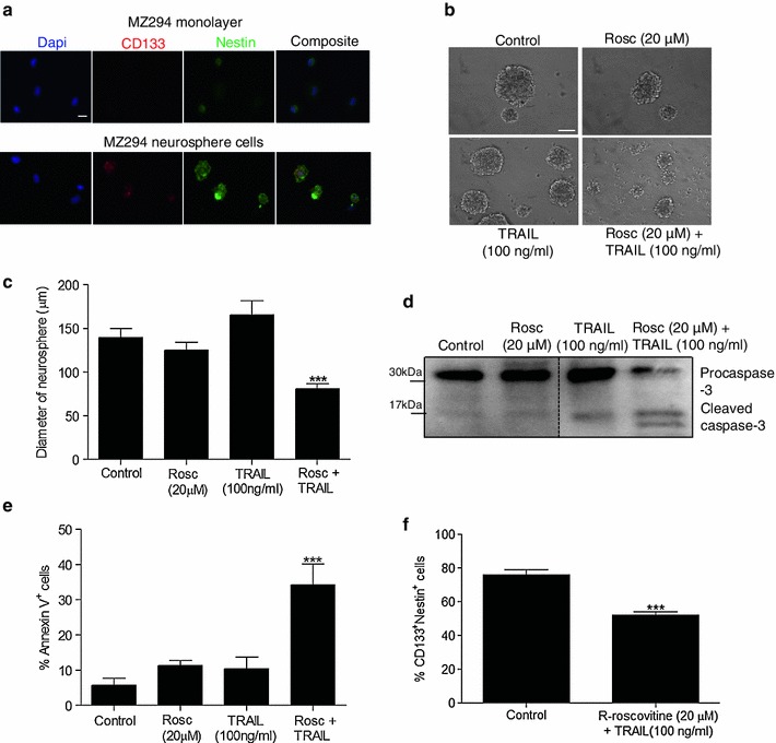
Fig. 4.

Treatment with R-roscovitine + TRAIL induces apoptosis in a 3D tumour model. MZ-294 neurospheres were generated from MZ-294 monolayer cells cultured in serum-free neurosphere-forming medium. a MZ-294 monolayer cells and MZ-294 neurospheres were dissociated and cytospun onto slides and then stained with the stem cell markers CD133, nestin and the nuclear marker Dapi (scale bar = 20 μM). b MZ-294 neurospheres were treated with R-roscovitine (20 μM), TRAIL (100 ng/ml), R-roscovitine (20 μM) + TRAIL (100 ng/ml) for 48 h. Brightfield images of the neurospheres were taken post treatment (scale bar = 50 μM). c The diameters of the neurospheres were measured following treatment with R-roscovitine (20 μM), TRAIL (100 ng/ml), R-roscovitine (20 μM) + TRAIL (100 ng/ml) for 48 h using an Eclipse TE 300 inverted microscope. Data are expressed as mean ± SEM, ***p < 0.001; n = 40–50 neurospheres. d Following treatment, neurospheres were harvested for western blot analysis of the expression of pro-apoptotic proteins procaspase-3 and cleaved caspase-3. e The percentage of apoptotic neurosphere cells was determined by Annexin staining using flow cytometry following treatment. Data are expressed as mean ± SEM, ***p < 0.001; data are from three independent experiments. f The percentage of neurosphere cells expressing CD133 and Nestin was assessed following treatment with R-roscovitine (20 μM) + TRAIL (100 ng/ml) for 48 h
