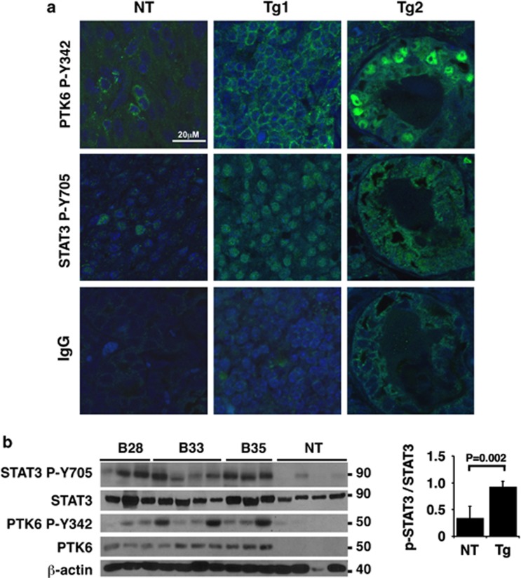
Figure 5.
Active PTK6 and STAT3 are expressed in mouse mammary gland tumors. (a) Mammary gland tumors that developed in nontransgenic (NT) and PTK6 transgenic lines (Tg) were analyzed for the expression of active PTK6 and active STAT3 using immunofluorescence. Morphologically similar areas of NT and Tg tumors are shown. P-PTK6 and P-STAT3 signals were low and sporadic in NT tumors. Membrane-associated active PTK6 (P-Y342) correlated with active nuclear STAT3 (P-Y705) in tumors from PTK6 transgenic mice (Tg1). Active PTK6 could be found at the membrane (Tg1) or sometimes within the nucleus (Tg2). The tumor in Tg1 was an adenocarcinoma composed of small glandular structures with small lumens consistent with an acinar pattern, and the nuclear-activated PTK6 appeared in the acinar cells. Background staining was monitored using immunoglobulin G (IgG) as a control. The scale bar represents 20 μm. (b) Immunoblotting was performed with total cell lysates prepared from tumors isolated from multiple B28, B33 and B35 animals, as well as tumors that developed in nontransgenic control mice. Each lane represents a unique tumor sample from an individual mouse. STAT3 activation was consistently observed in tumor samples from PTK6 transgenic mice. For quantitation, P-STAT3 levels were normalized to total STAT3 levels in nontransgenic and transgenic mice (right panel). Immunoblotting for PTK6 was performed using an antibody specific for the human protein expressed by the transgene.

