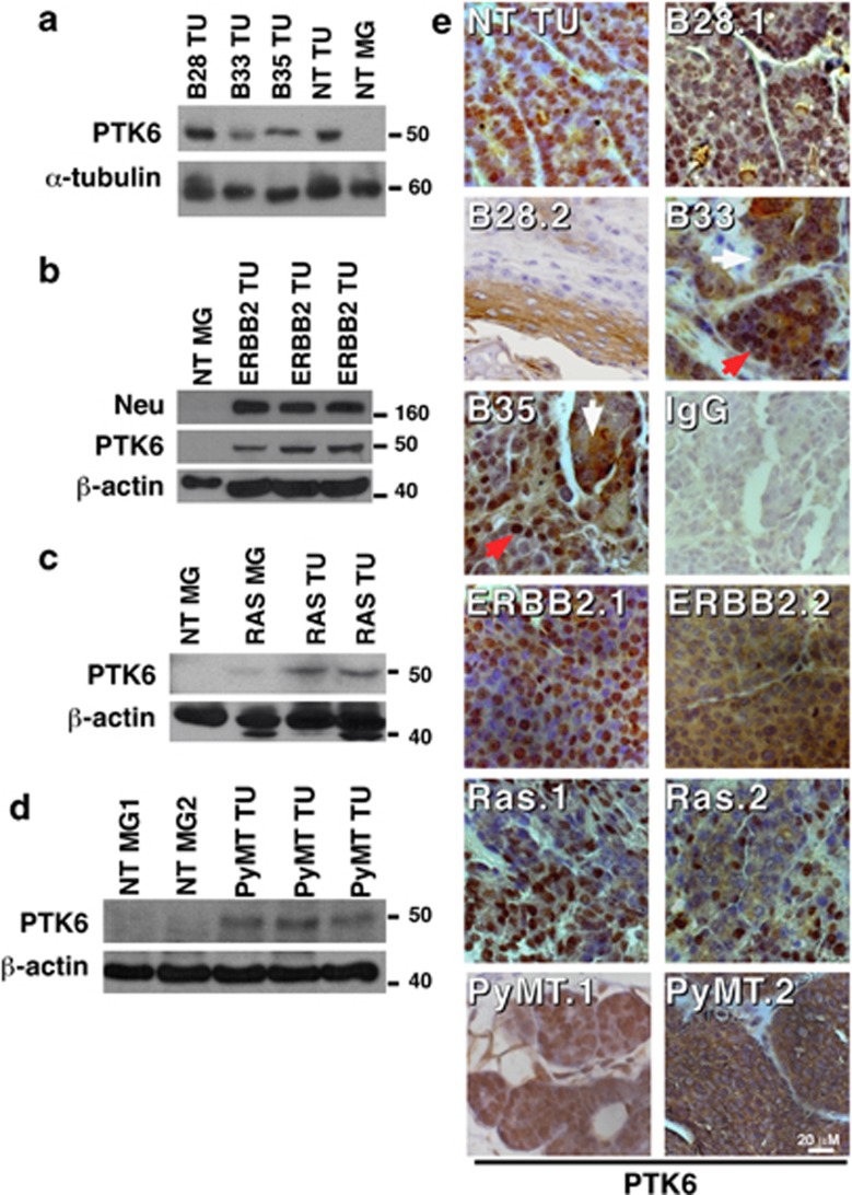
Figure 6.
Endogenous mouse PTK6 is induced in mammary gland tumors of different origins. (a–d) Mouse PTK6 protein expression was detected using immunoblotting, and an antibody specific for mouse PTK6 that does not cross-react with the human PTK6 encoded by the transgene. Nontransgenic mammary gland was used as negative control. Expression of α-tubulin or β-actin was examined as loading controls. Induction of the endogenous PTK6 protein was detected in all mouse mammary gland tumors examined, including tumors that formed in nontransgenic control mice (NT TU), MMTV-PTK6 (a), MMTV-ERBB2 (b), MMTV-Ha-Ras (c) MMTV-PyMT (d) transgenic animals. (e) Immunohistochemistry as used to examine endogenous PTK6 expression in mouse tumors of different origins. PTK6 is predominately nuclear in more differentiated acinar cells (NT, PyMT1) and in areas of tumors from different transgenic strains (B28.1, ERBB2.1 and Ras.1), but it can also be cytoplasmic as in ERBB2.2, Ras.2 and PyMT.2. Variation in PTK6 intracellular localization is sometimes observed in adjacent regions of the same tumors, as indicated by red (nuclear) and white (cytoplasmic/membrane) arrows in B33 and B35. Besides the neoplastic epithelial cells, endogenous PTK6 was also detected in the cytoplasm of cuboidal shaped epithelial cells and in metaplastic keratin producing squamous epithelial cells lining the ductules (B28.2). Sections were stained with normal rabbit IgG as a negative control (IgG).

