Abstract
M-type (KCNQ2/3) potassium channels are suppressed by activation of Gq/11-coupled receptors, thereby increasing neuronal excitability. We show here that rat KCNQ2 can bind directly to the multivalent A-kinase-anchoring protein AKAP150. Peptides that block AKAP150 binding to the KCNQ2 channel complex antagonize the muscarinic inhibition of the currents. A mutant form of AKAP150, AKAP(ΔA), which is unable to bind protein kinase C (PKC), also attenuates the agonist-induced current suppression. Analysis of recombinant KCNQ2 channels suggests that targeting of PKC through association with AKAP150 is important for the inhibition. Phosphorylation of KCNQ2 channels was increased by muscarinic stimulation; this was prevented either by coexpression with AKAP(ΔA) or pretreatment with PKC inhibitors that compete with diacylglycerol. These inhibitors also reduced muscarinic inhibition of M-current. Our data indicate that AKAP150-bound PKC participates in receptor-induced inhibition of the M-current.
The M-current is a low-threshold, slowly activating potassium current that exerts negative control over neuronal excitability. Activation of Gq/11-coupled receptors suppresses the M-current, creating a slow excitatory postsynaptic potential, enhancing excitability and reducing spike-frequency adaptation1,2. The M-type K+ channel is a promising therapeutic target, as the channel blocker linopirdine acts as a cognition enhancer3,4, and the channel activator retigabine functions as an anticonvulsant5,6. M-type channels are heteromeric complexes of certain KCNQ-family potassium channel subunits (KCNQ2–5)7–10. KCNQ2 and KCNQ3 were the first members of this family identified as M-channel forming subunits7. The KCNQ3 subunit is a core component that co-assembles with KCNQ2, KCNQ4 and KCNQ5 to form functional M-type channels10.
Although the subunits that form M-type K+ channels have been identified, the molecular details of the signaling pathways that lead to suppression of the M-currents upon receptor stimulation have not yet been fully defined2,11. We know that inhibition results from activation of G proteins of the Gq/11 family, with the α-subunit as the active moiety12,13, and that a ‘diffusible’ messenger is involved. That is, the receptor/G protein complex can be physically remote from the channel14,15. Thus, closure most likely results from some product of phospholipase C activity. M-type channels can be closed by raising intracellular calcium16, and there is evidence that this might be a ‘second messenger’ for bradykinin17 and nucleotides18, but not for acetylcholine17–20. Like many other ion channels, the activity of KCNQ/M channels is regulated by membrane phosphatidylinositol-4,5-bisphosphate (PIP2)21, such that recovery from muscarinic receptor–mediated inhibition requires resynthesis of PIP2, but whether the process of inhibition itself results directly from PIP2 breakdown is unclear.
It is also apparent that phosphorylation processes are involved in the tonic regulation of basal M-currents in neurons2, although the identity of the kinases and phosphatases that mediate this process are not entirely clear. Two candidate enzymes are PKC and calcineurin (a calcium/calmodulin-dependent protein phosphatase), as pharmacological stimulation of either enzyme can inhibit the M-current22,23. However, inhibitors of PKC (staurosporine, H-7 and pseudosubstrate) or intracellular perfusion of the auto-inhibitory peptide of calcineurin did not prevent transmitter-induced current inhibition9,11,19,22.
The types of channel regulation described above may involve macro-molecular signaling complexes that precisely control the phosphorylation status of the M-current24,25. Typical examples are the A-kinase anchoring proteins (AKAPs), a functionally defined group of proteins that bind to protein kinase A (PKA) and other second messenger regulated enzymes; several AKAPs are known to target these enzymes to sub-membrane sites close to channels26,27. For example, the protein yotiao associates with protein phosphatase-1, PKA and the NR1A subunit of NMDA receptors to modulate receptor activity28. Another example is AKAP150, a rodent ortholog of human AKAP79. This protein is a scaffold protein for PKA, calmodulin, PKC and calcineurin, and coordinates these enzymes at postsynaptic sites in neurons29–31.
In the present study, we show that AKAP150 and its anchored pool of PKC have an essential facilitatory role in the receptor-induced suppression of M-currents from rat superior cervical ganglion (SCG) neurons. Biochemical evidence and mapping studies showed that AKAP150 binds directly to an intracellular region of the KCNQ2 channel. We used whole-cell current recording techniques to show that the interaction between KCNQ2 and AKAP150 is important for signal transduction in Chinese hamster ovary (CHO) cells. We also report that a dominant-interfering AKAP150 form, which lacks PKC binding sites, reduced receptor-induced M-current suppression in neurons. Certain aspects of this effect could be replicated by the application of certain PKC inhibitors. Phosphorylation of KCNQ2 channels expressed in CHO cells was increased by muscarinic stimulation and attenuated by either coexpression of the mutant AKAP or by treatment with certain PKC inhibitors. Thus, we propose that an anchored pool of PKC functions as a signaling component for M-current regulation.
RESULTS
Interaction between KCNQ2 and AKAP150
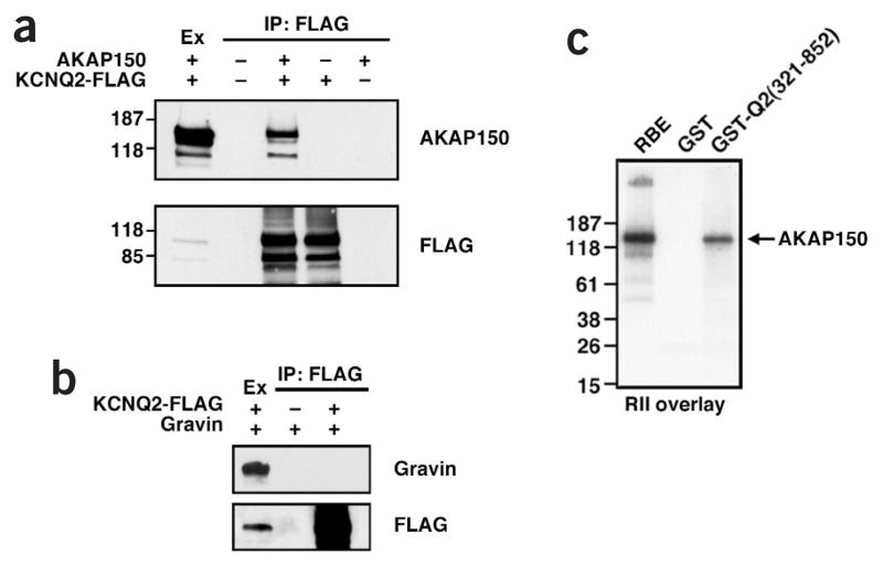
The neuronal anchoring protein AKAP150 organizes the subcellular location of candidate molecules proposed to modulate M-currents, including PKC, calcineurin, PIP2 and calmodulin21–23,31,32. Therefore, we tested whether AKAP150 interacts with KCNQ2 channels. FLAG-tagged KCNQ2 subunits were coexpressed with AKAP150 in HEK293 cells. The channel complex was immunoprecipitated with the FLAG antibody and co-purification of AKAP150 was assessed by western blot (Fig. 1a). AKAP150 was detected in the KCNQ2 precipitates but not in control conditions (Fig. 1a). Additional control experiments showed that gravin, another multivalent AKAP, did not copurify with KCNQ2 channels by immunoprecipitation (Fig. 1b). To further define the specificity of this interaction, we used a glutathione-S-transferase (GST) fusion protein containing the C-terminal tail of the KCNQ2 subunit to screen rat brain extracts for AKAP interaction. Although there were several AKAPs in the starting material, only AKAP150 interacted with the KCNQ channel fragment as assessed by the PKA type II regulatory subunit (RII) overlay procedure28 (Fig. 1c).
Figure 1.
KCNQ2 can form an AKAP150 signaling complex. (a) AKAP150 interacts with the KCNQ2 channel in cells. HEK293 cells were transfected with AKAP150 and FLAG-tagged KCNQ2. Cell extracts (Ex) were immunoprecipitated with anti-FLAG antibody and co-purification of AKAP150 with KCNQ2 was determined by Western blot analysis using a specific AKAP150 polyclonal antibody (upper panel). The presence of KCNQ2 was assessed by a FLAG western blot (lower panel). (b) Gravin does not associate with KCNQ2 channels. HEK293 cells were transfected with FLAG-tagged KCNQ2 and gravin. Cell extract and immunoprecipitates were probed with gravin (upper panel) or FLAG antibody (lower panel). (c) AKAP150 is the major AKAP that binds KCNQ2. Whole rat brain extract (RBE) was incubated with GST or GST-Q2(321-852) beads. AKAPs were detected by 32P-labeled RII in the solid-phase overlay assay. AKAP150 is indicated.
Binding regions within KCNQ2 and AKAP150
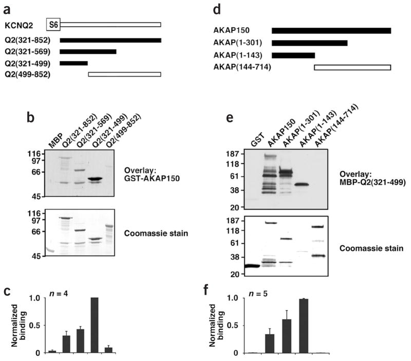
In vitro mapping experiments were performed to define the interacting surfaces on the KCNQ2 subunit and AKAP150 (Fig. 2). A GST–AKAP150 fusion protein was used as a probe to screen a family of C-terminal fragments of the KCNQ2 subunit that were fused to the maltose binding protein (MBP) (Fig. 2a–c). AKAP binding was detected with a polyclonal antibody against the anchoring protein (Fig. 2b). Fusion proteins containing residues 321–499 of the KCNQ2 channel bound to AKAP150, with the most intensive signal from the Q2(321–499) fragment (Fig. 2b,c). A GST fusion construct encompassing this fragment of the KCNQ2 subunit could also pull down AKAP150 from rat brain extract (data not shown). These data indicate that a region of about 200 amino acids, which is adjacent to the S6 transmembrane domain of KCNQ2, interacts with AKAP150.
Figure 2.
Mapping the reciprocal binding domains of KCNQ2 and AKAP150. (a) Schematic diagram showing the MBP–KCNQ2 protein fragments used in the mapping studies. The first and the last amino acids of each fragment are indicated. Fragments that interact with AKAP150 are highlighted. (b) The four KCNQ2 protein fragments were separated by SDS-PAGE and electrotransferred to PDVF membranes. Membranes were overlaid with recombinant GST–AKAP150 and binding was detected using an anti-AKAP150 antibody in far-western blot experiments (upper panel). Note the intense signal with MBP–Q2(321–499). Coomassie blue stain of the membrane indicates approximately equal loading of AKAP150 fragments (lower panel). (c) Histogram showing relative AKAP150 binding normalized to the corresponding strength of the stained KCNQ2 fragments (bottom). Data are presented as mean ± s.e.m. from four independent experiments. (d) Mapping the KCNQ2 binding domain for AKAP150. A schematic diagram depicts the protein fragments used to define the KCNQ2 binding site on AKAP150. (e) AKAP150 fusion proteins were separated by SDS–PAGE and electrotransferred to membrane. MBP–Q2(321–499) was overlaid onto the membrane and far-western blot experiments were performed with an anti-MBP antibody (upper panel). Coomassie stain indicates approximately equal loading of AKAP150 protein fragments (lower panel). (f) The amalgamation of five independent experiments is shown (bottom).
In reciprocal experiments, we mapped the site on AKAP150 that binds to the channel. A family of recombinant GST–AKAP150 fragments were assayed for solid-phase binding to the minimal interacting region of the KCNQ2 channel (residues 321–499). Binding was assessed by immunoblot using an antibody against the maltose-binding protein (MBP) moiety (Fig. 2d–f). The channel binding site was mapped to the N-terminal region between residues 1 and 143 of AKAP150. Attempts to narrow down the binding sites resulted in the detection of multiple binding sites scattered in this region, mapping to the regions between residues 31–110 and 113–143 (Supplementary Fig. 1 online).
Regulation of the KCNQ2 channel by AKAP150
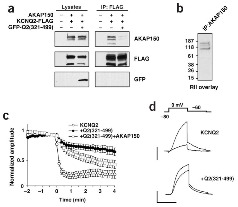
To test the involvement of the AKAP150 signaling complex on agonist-dependent channel inhibition, we attempted to displace AKAP150 from the KCNQ2 channel complex. To this end, we used the minimal binding region of KCNQ2, Q2(321–499), as a soluble antagonist of the AKAP150–channel interaction. Control experiments confirmed that expression of green fluorescent protein (GFP)-tagged Q2(321-499) could displace AKAP150 from KCNQ2 in cells (Fig. 3a). Next, we used CHO hm1 cells, which stably express the human M1 muscarinic acetylcholine receptor (mAChR), as a model system to study muscarinic regulation of KCNQ channels8,33. We found that this cell line expresses an endogenous Chinese hamster ortholog of AKAP150 (Fig. 3b). Therefore, we measured changes in the KCNQ currents by whole-cell patch clamp recording techniques upon manipulation of AKAP150 association. We applied 10 μM acetylcholine to cells coexpressing KCNQ2 and GFP–Q2(321-499). Expression of GFP–Q2(321–499) reduced the channel inhibition by acetylcholine, and this effect was partially reversed by expressing exogenous AKAP150 (Fig. 3c,d). These results suggest that AKAP150 and the associated signaling components are involved in KCNQ2 current inhibition.
Figure 3.
Role of AKAP150 in receptor-mediated KCNQ2 channel regulation. (a) Q2(321–499) fragment can displace AKAP150 from the KCNQ2 channel inside cells. HEK293 cells were transfected with combinations of FLAG-tagged KCNQ2, AKAP150 or GFP–Q2(321–499) as indicated above. Cell lysates were immunoprecipitated with anti-FLAG antibody, followed by western blot with the indicated antibodies (right). (b) Endogenous AKAP150 is found in CHO cells. CHO cell lysates were immunoprecipitated with anti-AKAP150 antibody. AKAP150 was detected by interaction with 32P-labeled RII in a solid phase overlay assay. (c) Agonist-induced current suppression is attenuated by expressing GFP–Q2(321–499). Averaged time courses show KCNQ2 current change by acetylcholine (ACh). ACh (10 μM) was applied at t = 0 for 20 s. CHO cells were transfected with KCNQ2 (○, n = 10), KCNQ2 + GFP–Q2(321–499) (●, n = 11) or KCNQ2 + GFP–Q2(321–499) + AKAP150 (□, n = 13). (d) Representative current traces for panel c showing control and 1 min after application of ACh. Scale bar, 1 nA and 500 ms.
Region A of AKAP150 required for inhibition
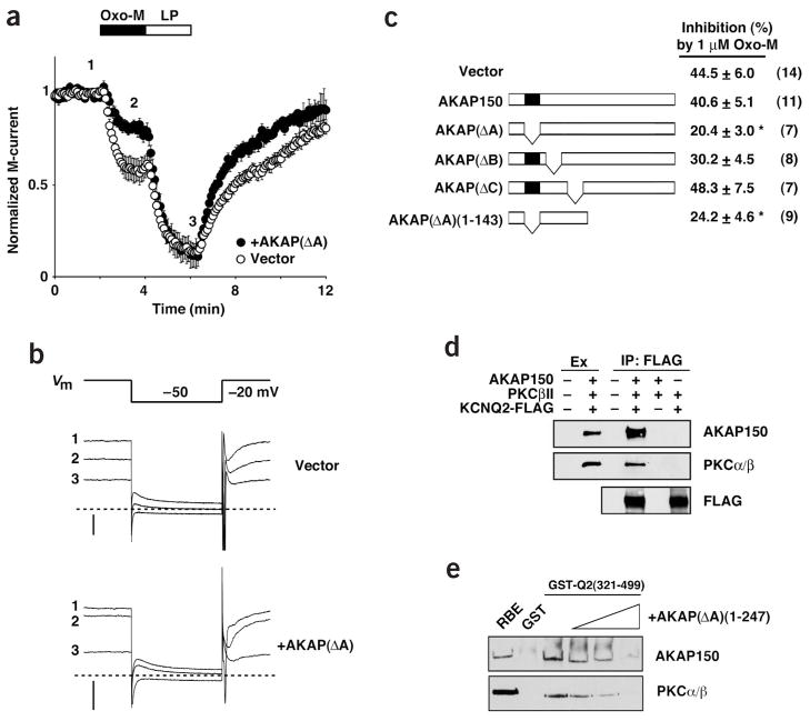
To examine the role of AKAP150 in mediating the inhibition of the M-current in rat SCG neurons, we identified relevant domains within AKAP150. First, we established the presence of AKAP150 in these neurons by immunoblotting (data not shown). Electrophysiological techniques were used to measure the effect of overexpressing wild-type AKAP150 or various deletion mutants on M-current inhibition by the mAChR agonist oxotremorine-M (Oxo-M; Fig. 4a–c). Oxo-M inhibited the M-currents in both control SCG neurons and in wild-type AKAP150-expressing cells, but this inhibition was strongly reduced in cells expressing a mutant AKAP150 lacking residues 31–51, called the A-site and designated AKAP(ΔA) (Fig. 4b–d). In contrast, the amount of M-current blocked by linopirdine, a KCNQ channel inhibitor3,4, was unchanged (Fig. 4a,b). A similar inhibitory effect was shown by the shortest truncation, AKAP(ΔA) (1–143) (Fig. 4c). In contrast, the dominant-negative effect on mAChR-induced blockade was not seen in cells expressing other deletion mutants of AKAP150, such as those lacking conserved basic regions B and C34 (Fig. 4c).
Figure 4.
Loss of PKC from channel complex attenuates the agonist-induced inhibition of the M-currents in SCG neurons. (a) Averaged time courses of normalized IK(M) amplitude during sequential application of Oxo-M (1 μM) and linopirdine (LP, 15 μM) observed from control vector-injected (○, n = 14) or AKAP(ΔA)-overexpressing (●, n = 7) SGC neurons. M-currents were monitored by 500-ms hyperpolarizing steps to −50 mV from a holding potential of −20 mV at 5-s intervals. Data shown as mean ± s.e.m. (b) Pulse protocol (top) and representative current traces from control vector (upper traces) and AKAP(ΔA) (lower traces) transfected SGC neurons at the points 1, 2 and 3 indicated in panel a. Dashed lines show the zero current level. Scale bar, 200 pA. (c) Schematic diagram of the AKAP150 deletion mutants expressed in neurons (left). The amalgamated data of the Oxo-M (1 μM) induced M-current inhibition in the presence of the indicated constructs (right). Values show mean ± s.e.m. The number of experiments is shown in parentheses. *P < 0.05 (compared to control). (d) AKAP150, PKC and KCNQ2 form a trimeric complex in cells. HEK293 cells were transfected with combinations of FLAG-tagged KCNQ2, AKAP150 and PKCβII as indicated at the top of the panel. Cell extracts (Ex) were immunoprecipitated with FLAG antibody. Resultant precipitates were subjected to immunoblotting with antibodies against AKAP150, PKCα/β and FLAG. Both AKAP150 and PKC were co-immunoprecipitated with KCNQ2. PKC in the precipitate is only detected in the presence of AKAP150. (e) Competition with AKAP(ΔA)(1–247) and AKAP150 signaling complex. GST–Q2(321–499) beads were incubated with rat brain lysate in the presence of increasing concentration of purified recombinant AKAP(ΔA)(1-247) (0, 1, 5, 10 μg). The amount of AKAP150 and PKCα/β was analyzed by immunoblotting. Note that the anti-AKAP150 antibody used in this assay recognizes the C terminus of AKAP150, which is lacking in AKAP(ΔA)(1-247).
The A site of AKAP150 has been previously identified as the PKC binding site30. Therefore, we examined whether KCNQ2 channels form a trimeric complex including AKAP150 and PKC. We performed immunoprecipitation using anti-FLAG antibody from HEK293 cell extract expressing KCNQ2-FLAG, AKAP150 and PKCβII. As expected, PKCβII precipitated with KCNQ2. Precipitation of PKCβII could be detected only in the presence of AKAP150, suggesting that association of the kinase with the channel was through their mutual interaction with the anchoring protein. Pulldown experiments confirmed that GST-AKAP150, but not GST-AKAP(ΔA) resin, is associated with PKC from rat brain extract (data not shown). The dominant-negative phenotype of AKAP(ΔA) suggests that this mutant forms an M-channel complex that is deficient in PKC binding. To test this idea, GST–Q2(321–499) beads were incubated with brain lysate in the presence of increasing concentrations of purified AKAP(ΔA) (1–247), which lacks the epitope for the antibody used in this assay. After extensive washing, the level of AKAP150 and PKC associated with the GST–Q2(321–499) was assessed by immunoblot (Fig. 4e). As the concentration of AKAP(ΔA) increased, retention of both AKAP150 and PKC decreased (Fig. 4e). This competition assay also provided a rough estimate for the dissociation constant (Kd) value for AKAP150 and GST-Q2(321–499): 14.1 ± 5.7 nM (n = 3). Together, our data suggest that expression of AKAP(ΔA) exerts a dominant-negative effect through the reduction of PKC in the M-channel complexes.
PKC involved in inhibition at low dose of Oxo-M
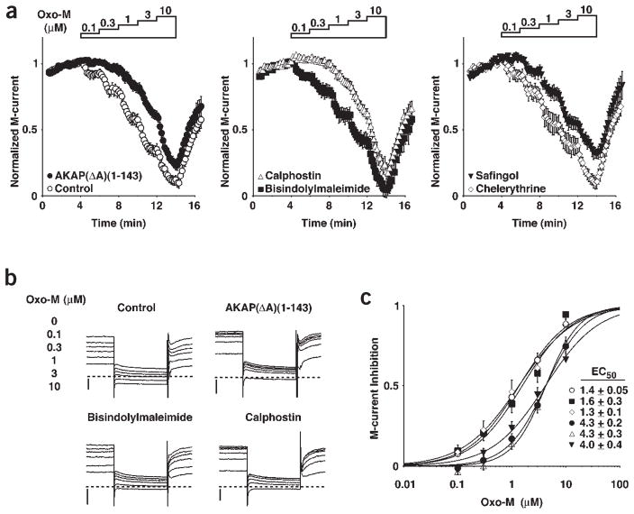
The data above suggested that PKC might be involved in mAChR-induced M-current inhibition. According to previous reports, however, some PKC inhibitors such as staurosporine have no effect on M-current suppression after agonist stimulation9,11,19. We suspect that the reason for the insensitivity of M-current suppression to such inhibitors is that PKC is associated with AKAP150 at its catalytic core35; accessibility of the catalytic site to such agents would then be restricted. To test this hypothesis, we pretreated SCG neurons with several inhibitors that act on different regions of the PKC enzyme and assessed M-current inhibition. Bisindolylmaleimide I (structurally similar to staurosporine) and chelerythrine act on the catalytic domain of PKC36,37, and we found that they had no effect on current suppression (Fig. 5a,c). In contrast, calphostin C and safingol, which act on the diacylglycerol binding site38,39, reduced the sensitivity to Oxo-M (Fig. 5a,b). This effect is observed as a shift in the dose–response curve to the right (Fig. 5c). Interestingly, the dose–response curves from calphostin-C-treated cells and AKAP(ΔA) (1–143)-overexpressing cells are almost identical (Fig. 5c). Moreover, addition of calphostin C to AKAP(ΔA)-expressing cells did not show a further shift (data not shown). These data strongly support the idea that an anchored pool of PKC is involved in M-current inhibition, especially at low doses of agonists.
Figure 5.
Effect of PKC inhibitors on the M-current inhibition. (a) Averaged time course of normalized IK(M) amplitude during cumulatively increasing concentrations of Oxo-M as indicated, for control SCG neurons (○, n = 20), neurons expressing AKAP(ΔA)(1–143) (●, n = 12), neurons pretreated by 100 nM bisindolylmaleimide I (■, n = 12), 100 nM calphostin C (△, n = 17), 1.2 μM chelerythrine (◇, n = 10) or 10 μM safingol (▽, n = 9). Each point represents the mean ± s.e.m. (b) Representative IK(M) traces from control, AKAP(ΔA)(1–143) expressing neurons and PKC inhibitor-treated neurons during application of Oxo-M at the indicated concentrations. Scale bar, 200 pA. (c) Concentration dependence of fractional M-current inhibition by Oxo-M deduced from panel a. Symbols are the same as in panel a. Curves show the best fit with the Hill equation with parameters shown (inset).
KCNQ2 phosphorylation correlates with the inhibition
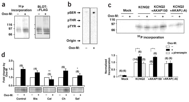
So far, our data indicate that AKAP150 anchoring of PKC promotes M-current inhibition. To test whether phosphorylation of the KCNQ channel correlates with the mAChR-induced inhibition, we metabolically labeled CHO hm1 cells with 32P-orthophosphate followed by Oxo-M stimulation. Incorporation of 32P-phosphate into the KCNQ2 channel was significantly potentiated upon stimulation (Fig. 6a,c). Channel phosphorylation is blocked by addition of an mAChR inhibitor, pirenzepine, confirming that this phosphorylation is induced by receptor activation (Fig. 6c, lower panel). Notably, KCNQ2 phosphorylation occurred predominantly on serine residues (Fig. 6b), which suggests the involvement of a serine/threonine kinase in this process. To determine whether anchored PKC is necessary for phosphorylation of the channel, CHO hm1 cells were co-transfected with either wild-type AKAP150 or mutant AKAP(ΔA), along with KCNQ2. There was no potentiation of mAChR-induced KCNQ2 phosphorylation in cells expressing AKAP(ΔA), whereas cells expressing wild-type AKAP150 behaved in a similar manner as control cells (Fig. 6c). This is further supported by our pharmacological experiments in which pre-treatment with the PKC inhibitors calphostin C and safingol abolished the Oxo-M-induced phosphorylation of KCNQ2 (Fig. 6d). In contrast, but in agreement with our current recordings (Fig. 5c), pretreatment with bisindolylmaleimide and chelerythrine had no effect (Fig. 6d). Together, these data suggest that PKC-dependent phosphorylation of KCNQ2 is associated with receptor-induced inhibition.
Figure 6.
PKC phosphorylation of KCNQ2 in Oxo-M treated cells. (a) Oxo-M induces phosphorylation of the KCNQ2 channel. CHO hm1 cells were transfected with FLAG-tagged KCNQ2 for 48 h and subsequently prelabeled for 4 h with [32P] orthophosphate followed by 3 min stimulation with Oxo-M (10 μM). Cells were lysed and immunoprecipitated with the anti-FLAG antibody. Precipitated material was separated by 6% SDS–PAGE and KCNQ2 phosphorylation was assessed by autoradiography. Control FLAG western blots confirmed that the primary phosphoprotein in immunoprecipitates was KCNQ2, and there was an equal amount of precipitate in both conditions. (b) KCNQ2 is phosphorylated on serine residues. The Oxo-M-treated (right) and untreated (left) KCNQ2 channels were subjected to hydrolysis in 6 M HCl. The resulting amino acids were separated by thin-layer electrophoresis. The migration of phosphoserine (pSER), phosphothreonine (pTHR) and phosphotyrosine (pTYR) standards are indicated with arrows. (c) Effect of AKAP150 on KCNQ2 channel phosphorylation. Basal and Oxo-M-induced phosphorylation of FLAG-tagged KCNQ2 channels were examined under various conditions. KCNQ2 was immunoprecipitated and separated by 6% SDS–PAGE. Incorporation of [32P] into KCNQ2 was assessed by autoradiography (top). Pooled data are summarized in the histogram (bottom). All data are normalized to the control unstimulated KCNQ2 channels. Control pirenzepine-treated cells (1 μM) are shown (hatched bar). Values show mean ± s.e.m. (n = 3–14, *P < 0.05). (d) PKC is required for Oxo-M stimulated KCNQ2 phosphorylation. FLAG-tagged KCNQ2 transfected cells were pretreated with PKC inhibitors as described in Methods. The levels of KCNQ2 channel phosphorylation in immunoprecipitates were assessed by phosphorimager analysis (bottom). Changes in phosphorylation for each inhibition are expressed as fold change over those before stimulating with Oxo-M (n = 4–6, *P < 0.05 and **P < 0.01 compared to control Oxo-M stimulated KCNQ2 channel).
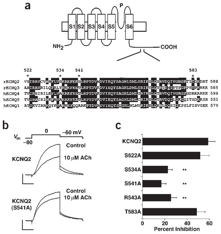
Finally, we searched KCNQ subfamily sequences for important residues that may be required for receptor-induced inhibition of channel activity. We found several PKC phosphorylation sites corresponding to the consensus sequence (S/T-X-R/K) throughout the KCNQ channel subfamily. For example, Ser217, located in the cytosolic loop between the S4 and S5 segments and Ser541, in the C-terminal tail of KCNQ2 protein, are conserved in most of the family members (Fig. 7a). To test whether these residues regulate the KCNQ2 current, we expressed wild-type or the alanine-substituted mutants in CHO hm1 cells. The S217A mutant gave a reduced current expression (43 ± 5% of wild-type current; n = 6), suggesting that the mutation disturbed the normal structure of the channel; we did not analyze this mutant further. Expression of the S541A mutant resulted in similar current density (74.3 ± 6 pA/pF; n = 15) to that of the wild-type channel (72.1 ± 8.4 pA/pF; n = 10). In contrast, cells expressing KCNQ2 (S541A) channels showed only small current inhibition by acetylcholine (Fig. 7b). Disruption of the PKC consensus sequence for Ser541 by changing the Arg543 residue to alanine also resulted in less agonist-induced current inhibition (Fig. 7c). Ser541 is located within the highly conserved region in the C-terminal tail of KCNQ family members (Fig. 7a). To clarify the role of this domain, we made alanine substitutions of potential PKC phosphorylation sites throughout this region. We identified a second, non-conserved PKC site, Ser534, which reduced agonist-induced current inhibition (Fig. 7a). We attempted to examine a Ser534/Ser541 double alanine substituted mutant; however, this mutant did not express as expected and was not further analyzed. These results suggest that Ser534 and 541 are key sites for PKC phosphorylation, although we have not ruled out the possibility that other PKC sites are involved in this process.
Figure 7.
Effects of mutations of potential PKC-dependent phosphorylation sites of KCNQ2 channels on receptor-induced suppression in CHO hm1 cells. (a) Structure of the KCNQ channel (top) indicating the transmembrane domains (S1–S6), pore region (P) and the conserved region at the C-terminal tail focused in this study. ClustalW alignment (bottom) of the conserved region among KCNQ subfamily members from rat and human counterparts. Potential PKC-dependent phosphorylation sites in KCNQ2 are indicated (*). Identical residues are marked with a black background. (b) The current inhibition induced by acetylcholine (10 μM) measured in CHO hm1 cells transfected with the wild-type KCNQ2 or the mutant KCNQ2(S541A). The currents were elicited by 500-ms steps to 0 mV from a holding potential of −80 mV then to −60 mV. Scale bars, 400 pA and 200 ms. (c) Effect of a series of point mutations on the current inhibition of indicated KCNQ2 channels (n = 11–18) induced by 10 μM ACh. **P < 0.01 compared to wild-type KCNQ2 channels.
DISCUSSION
Our main finding is that association with AKAP150 promotes the PKC-induced serine phosphorylation of KCNQ2, which in turn facilitates the inhibition of M-type K+ channels induced by M1 mAChR stimulation. Presumably, the anchoring protein serves as an adapter that brings kinase and substrate together to permit the rapid and preferential phosphorylation of the KCNQ2 channel. This is supported by our biochemical evidence that AKAP150 can interact simultaneously with both KCNQ2 and PKC. Similar roles have been proposed for PKA anchoring to glutamate receptors and calcium channels via AKAPs27,40–43, but this is the first demonstration of anchoring-dependent PKC modulation of an ion channel.
The interference with agonist-induced suppression was accomplished either by disrupting the signaling complex with GFP–Q2(321–499) or by forming a PKC-deficient complex using AKAP(ΔA). These observations support the idea that the KCNQ2–AKAP150 complex is required for normal KCNQ channel regulation by muscarinic receptor stimulation, and that anchored PKC is involved in this process. This is further supported by the effects of PKC inhibitors that act at the diacylglycerol binding site. Results from experiments involving KCNQ channels with mutations in potential PKC-dependent phosphorylation sites support the theory that this phosphorylation facilitates inhibition of channel activity. While we have used SCG neurons and the heterologous expression system for our experiments, the KCNQ2 and KCNQ3 channels have been reported to form a signaling complex with AKAP79 in the human brain44. We speculate that M-current inhibition in the central nervous system may also be governed by a similar mechanism. AKAP79 and KCNQ2 seem to be expressed in overlapping but distinct patterns in certain brain areas44,45. Hence, the involvement of AKAP79 may vary in different parts of the brain. It is important to note that the effect of AKAP-facilitated, PKC-mediated KCNQ phosphorylation is to sensitize M-type channels to mAChR-induced inhibition, so variable association with AKAP79 might result in variable channel sensitivity to inhibition.
Previous experiments have been inconclusive with regard to the role of PKC activation in receptor-mediated M-current inhibition2,11. In particular, inhibitors directed at the PKC catalytic site are ineffective in preventing mAChR-mediated inhibition9,15,19. Our results suggest that insensitivity to such blockers results from limited accessibility of the catalytic domain to such agents due to the PKC–AKAP150 interaction. In addition, it has been reported that AKAP79 inhibits PKC even in the presence of diacylglycerol35. We speculate that the interaction between these proteins modifies drug sensitivities and enzyme activity, which leads to different interpretations of pharmacological experiments. It has also been reported that the partial suppression of the M-current by low concentrations of muscarine antagonizes the response to phorbol esters15. Our hypothesis explains this observation, in that receptor-activated PKC phosphorylates M-type channels at the same site, thereby reducing the sensitivity to phorbol esters.
In essence, our results suggest that the role of AKAP-bound PKC is to prime M-type channels for inhibition by increasing the sensitivity to mAChR stimulation about threefold. Thus, responses to low concentrations of agonist are strongly inhibited both by overexpressing mutant AKAP lacking the PKC binding site or by inhibiting the DAG site on PKC. On the other hand, sufficiently high concentrations of agonist are still capable of producing profound inhibition, implying that an additional pathway is capable of mediating the inhibition of unprimed channels after strong receptor stimulation. The availability of an alternative pathway is supported by the fact that the KCNQ1 channel is also inhibited by mAChR stimulation8 but does not possess the critical PKC phosphorylation site corresponding to S541 in KCNQ2, which is conserved in the neural members of the KCNQ family, KCNQ2–5. Furthermore, KCNQ1 does not form a macromolecular complex with AKAP79, but instead forms yotiao-containing complexes46. Thus, it is likely that the KCNQ1 channel uses an alternative pathway for agonist-induced inhibition, and that this is also available to other members of this family following strong receptor stimulation.
One alternative pathway that has been suggested to mediate M-channel inhibition following activation of bradykinin B2 or nucleotide P2Y receptors involves the IP3-induced release of Ca2+ upon phospholipase C activation17,18. Ca2+ reportedly closes M-type K+ channels when applied directly through inside-out membrane patches, either by activating calcineurin or by some other mechanism16,22. Ca2+-activated molecules such as calcineurin and calmodulin would likely be contained in AKAP150 complexes, yet available evidence indicates that mAChRs do not use the IP3/Ca2+ path for M-current suppression9,17–20. This difference between mAChR and BK2 receptors appears to derive from the fact that BK2 receptors form a spatially restricted complex with the IP3 receptor, thus amplifying the Ca2+ signal, whereas mAChRs do not47. Another, rather ubiquitous regulator of these channels is the membrane phospholipid PIP2. Re-synthesis of PIP2 is required for recovery from muscarinic suppression of M-current21. Interestingly, AKAP150 associates with PIP2 through its conserved basic regions, A, B and C, and this interaction is responsible for the membrane localization of AKAP15034. In this context, it would clearly be useful to know whether the regulation of KCNQ channels by PIP2 is affected by AKAP150 and anchored PKC-mediated phosphorylation. Finally, our present results emphasize the significance of signaling complexes in the regulation of signal transduction from G-protein coupled receptors to downstream ion channels.
METHODS
Antibodies
The following primary antibodies were used for immunoblotting and immunoprecipitation: goat polyclonal antibody to AKAP150 (C-20 from Santa Cruz Biotechnology), rabbit polyclonal antibody to AKAP150 (VO88), mouse monoclonal PKCα/β (BD Transduction Laboratories), mouse monoclonal MBP Ab-2 (clone R29 from NeoMarkers) mouse monoclonal GST (Upstate Biotechnology) and mouse monoclonal FLAG M2 (Sigma).
Expression constructs
cDNAs for KCNQ2, KCNQ3 and AKAP150 were obtained, using RT-PCR, with LA-Taq DNA polymerase (Takara Shuzo) or Platinum Pfx DNA polymerase (Invitrogen), from rat brain RNA according to the sequences in GenBank (accession numbers AF087453, AF087454 and U67136, respectively). Mutagenesis was conducted using recombinant PCR. cDNA of KCNQ clones was inserted into pZeoSV (Invitrogen). cDNA of AKAP clones was inserted into pTracer-SV40 (Invitrogen). For construction of GST fusion proteins of AKAP150, the deletions were PCR-amplified and sub-cloned into pGEX vectors. For MBP fusion proteins of KCNQ2, C-terminal tails of KCNQ2 were PCR-amplified and subcloned into pMAL-c. For GFP–Q2(321–499), the corresponding KCNQ2 fragment was subcloned into pEGFP-C2. All PCR-derived constructs were verified by sequencing.
Cell cultures and cDNA delivery
Sympathetic neurons were isolated from 2–3 week-old rats and cultured as described previously48. All experiments were approved by the institutional committee at Kanazawa University. DNA plasmids were pressure-injected into the nucleus of SCG neurons as described48. HEK293 cells were grown in Dulbecco’s modified Eagle medium (DMEM) with 10% fetal calf serum. CHO hm1 cells were grown in alpha-modified Eagle’s medium (α-MEM) supplemented with 10% fetal calf serum. Cells were transfected one day after plating using LipofectAmine Plus (Invitrogen) or Fugene 6 (Roche Molecular Biochemicals). Cells were replated at low density on 35-mm culture dishes 24 h after transfection. Two days after transfection, co-transfected cells were visually selected by fluorescence of GFP. To evaluate the effect of PKC inhibitors, cells were incubated overnight with enzyme inhibitors in the following concentrations: 100 nM bisindolylmaleimide I, 100 nM calphostin C and 1.2 μM chelerythrine chloride (all from Calbiochem). Pretreatment with calphostin C was carried out under dim light in the incubator. For treatment with L-threo-dihydrosphingosine (safingol), ethanol stock solution of safingol (Sigma) was diluted into α-MEM containing 1 mM BSA, sonicated briefly, and then incubated at room temperature for 1 h to form complexes as described39. The cells were then treated with either 10 μM safingol or vehicle for 4–6 h before recording.
Electrophysiological measurements
Whole-cell recordings were performed on isolated cells using an Axopatch 200B patch-clamp amplifier (Axon Instruments). Signals were sampled at 2 kHz, filtered at 1 kHz, and acquired using pClamp software (version 7 and 8, Axon Instruments). The perforated patch method was used to measure macroscopic currents from SCG neurons as described previously14. Briefly, amphotericin B (0.1–0.2 mg/ml) was dissolved in the intracellular solution containing 130 mM potassium acetate, 15 mM KCl, 3 mM MgCl2, 6 mM NaCl and 10 mM HEPES (adjusted to pH 7.3 by NaOH). When filled with this internal solution, pipette resistance was 2–3 MΩ. Access resistances after permeabilization ranged from 7–17 MΩ. The external solution consisted of 120 mM NaCl, 6 mM KCl, 1.5 mM MgCl2, 2.5 mM CaCl2, 11 mM glucose and 10 mM HEPES (titrated to pH 7.4 with NaOH). Amplitudes of the M-currents were measured as deactivating currents during 500-ms test pulses to −50 mV from a holding potential of −20 mV. For CHO hm1 cells, currents were recorded with the whole-cell configuration of patch clamp technique as described49. Patch pipettes with resistance of 4–8 MΩ were filled with solution containing 90 mM potassium citrate, 20 mM KCl, 3 mM MgCl2, 1 mM CaCl2, 3 mM EGTA and 40 mM HEPES (pH 7.4). The external solution contained 140 mM NaCl, 5 mM KCl, 1 mM CaCl2, 2 mM MgCl2 and 10 mM HEPES (pH 7.4). Series resistance (90–95%) and whole-cell capacitance compensation were used. Linear leak and capacitance transient in CHO hm1 experiments were subtracted by P/4 protocol from a holding potential of −80 mV. Data are presented as mean ± s.e.m.
In vitro binding studies and immunoblotting
For immunoprecipitations of heterologous cells, HEK293 cells were harvested and lysed 48 h after transfection in 500 μl IP buffer containing 150 mM NaCl, 5 mM EDTA, 5 mM EGTA, 10 mM phosphate (pH 7.4) with 1% Triton X-100 and complete protease inhibitor cocktail (Roche Molecular Biochemicals). Supernatants were incubated with 20 μg/ml phosphatidylserine, 4 μg antibody and 20 μl of prewashed protein G–agarose beads. After overnight incubation at 4 °C, the immunoprecipitates were washed twice with IP buffer, twice in the same buffer with 650 mM NaCl, and twice in tris-buffered saline containing 0.1% Tween-20 (TTBS). Bound proteins were detected by immunoblotting.
RII overlay assays were conducted as described using [32P]-labeled recombinant murine RIIα28. Overlay assay for mapping studies was performed as previously described50. Briefly, purified recombinant fusion protein (1 μg) was separated by SDS-PAGE, transferred to polyvinylidine difluoride (PVDF) or nitro-cellulose membranes, renatured by incubating at 37 °C, and incubated 1 h at room temperature with 1 μg of recombinant probes in TTBS and 5% skim milk. The membrane was washed extensively with TTBS and subjected to immunoblotting using anti-AKAP150 antibody or anti-MBP antibody. For GST pulldown experiments, extracts of rat brain were prepared by Dounce homogenization in 5 ml HSE buffer containing 150 mM NaCl, 5 mM EDTA, 20 mM HEPES (pH 7.4) and complete protease inhibitor cocktail, and then centrifuged at 38,000g for 30 min. The pellet was resuspended in 5 ml HSE buffer containing 1% Triton X-100 and centrifuged for an additional 30 min at 38,000g for 30 min. The supernatants were incubated with gluthathione sepharose 4B (Amersham Pharmacia Biotech) charged with GST fusions (5 μg) for 18 h at 4 °C. The resin was washed twice with the IP buffer, twice with IP buffer + 650 mM NaCl, twice with TTBS and once with TE. Bound proteins were analyzed by immunoblotting. For competition, beads charged with GST–KCNQ2(321–499) or control GST (1 μg) were preincubated with increasing concentrations (0, 1, 5 or 10 μg) of Xa-cleaved and purified AKAP(ΔA)(1–247) in the hypotonic buffer for 4 h at 4 °C. The brain extract, prepared as above, was then added and incubated overnight at 4 °C. The presence of AKAP150 and PKC bound to the beads was tested by immunoblotting.
Phospholabeling of CHO hm1 cells
CHO hm1 cells were transfected with FLAG-tagged KCNQ2 and treated with PKC inhibitors as detailed above. 48 h after transfection, expressing cells were incubated for 4 h in phosphate-free medium containing 0.5 mCi/ml [32P]orthophosphate (Amersham Biosciences), some of which contain PKC inhibitors. Cells were then exposed to Oxo-M (10 μM) for 3 min and scraped into 700 μl of ice-cold TNE solution: 150 mM NaCl, 2 mM EDTA, 1% NP-40, 50 mM NaF, 1 mM Na3VO4, 1 mM DTT and complete protease inhibitor cocktail. Solubilization was carried out for 20 min at 4 °C with gentle rocking. The lysates were centrifuged and the supernatants were precleared with 30 μl of Protein G Sepharose 4 Fast flow (Amersham Biosciences). Supernatants were incubated for 2 h with 4 μg of M2 anti-FLAG antibody followed by addition of protein-G beads for 1 h at 4 °C. The immunoprecipitates were washed six times with TNE buffer. After 6% SDS–PAGE, radiolabeled KCNQ2 channels were detected by phosphorimager (BAS1000, Fujifilm) analysis. For the phosphoamino acid analysis, the transferred PDVF membrane was cleaved and incubated with 6 M HCl at 110 °C for 90 min followed by cellulose thin-layer electrophoresis.
Supplementary Material
Acknowledgments
The authors thank M. Okamura, T. Haga and T. I. Bonner for the gift of CHO hm1 cells, and T. Rafiq for help with ganglion cell cultures. This work was supported by grants to N.H. and H.H. from the Japanese Ministry of Education, Culture, Sports, Science and Technology, by grant PG7909913 to D.A.B. from the UK Medical Research Council and by National Institute of Health grant GM48231 for the support of J.D.S.
Footnotes
Note: Supplementary information is available on the Nature Neuroscience website.
COMPETING INTERESTS STATEMENT
The authors declare that they have no competing financial interests.
References
- 1.Brown DA. M-current. In: Narahashi T, editor. Ion Channels. Plenum; New York: 1988. pp. 55–94. [DOI] [PubMed] [Google Scholar]
- 2.Marrion NV. Control of M-current. Annu Rev Physiol. 1997;59:483–504. doi: 10.1146/annurev.physiol.59.1.483. [DOI] [PubMed] [Google Scholar]
- 3.Aiken SP, Lampe BJ, Murphy PA, Brown BS. Reduction of spike frequency adaptation and blockade of M-current in rat CA1 pyramidal neurones by linopirdine (DuP 996), a neurotransmitter release enhancer. Br J Pharmacol. 1995;115:1163–1168. doi: 10.1111/j.1476-5381.1995.tb15019.x. [DOI] [PMC free article] [PubMed] [Google Scholar]
- 4.Lamas JA, Selyanko AA, Brown DA. Effects of a cognition-enhancer, linopirdine (DuP 996), on M-type potassium currents (IK(M)) and some other voltage- and ligand-gated membrane currents in rat sympathetic neurons. Eur J Neurosci. 1997;9:605–616. doi: 10.1111/j.1460-9568.1997.tb01637.x. [DOI] [PubMed] [Google Scholar]
- 5.Rundfeldt C, Netzer R. The novel anticonvulsant retigabine activates M-currents in Chinese hamster ovary cells tranfected with human KCNQ2/3 subunits. Neurosci Lett. 2000;282:73–76. doi: 10.1016/s0304-3940(00)00866-1. [DOI] [PubMed] [Google Scholar]
- 6.Tatulian L, Delmas P, Abogadie FC, Brown DA. Activation of expressed KCNQ potassium currents and native neuronal M-type potassium currents by the anti-convulsant drug retigabine. J Neurosci. 2001;21:5535–5545. doi: 10.1523/JNEUROSCI.21-15-05535.2001. [DOI] [PMC free article] [PubMed] [Google Scholar]
- 7.Wang HS, et al. KCNQ2 and KCNQ3 potassium channel subunits: molecular correlates of the M-channel. Science. 1998;282:1890–1893. doi: 10.1126/science.282.5395.1890. [DOI] [PubMed] [Google Scholar]
- 8.Selyanko AA, et al. Inhibition of KCNQ1-4 potassium channels expressed in mammalian cells via M1 muscarinic acetylcholine receptors. J Physiol. 2000;522:349–355. doi: 10.1111/j.1469-7793.2000.t01-2-00349.x. [DOI] [PMC free article] [PubMed] [Google Scholar]
- 9.Shapiro MS, et al. Reconstitution of muscarinic modulation of the KCNQ2/KCNQ3 K+ channels that underlie the neuronal M-current. J Neurosci. 2000;20:1710–1721. doi: 10.1523/JNEUROSCI.20-05-01710.2000. [DOI] [PMC free article] [PubMed] [Google Scholar]
- 10.Jentsch TJ. Neuronal KCNQ potassium channels: physiology and role in disease. Nat Rev Neurosci. 2000;1:21–30. doi: 10.1038/35036198. [DOI] [PubMed] [Google Scholar]
- 11.Brown BS, Yu SP. Modulation and genetic identification of the M-channel. Prog Biophys Mol Biol. 2000;73:135–166. doi: 10.1016/s0079-6107(00)00004-3. [DOI] [PubMed] [Google Scholar]
- 12.Haley JE, et al. The alpha subunit of Gq contributes to muscarinic inhibition of the M-type potassium current in sympathetic neurons. J Neurosci. 1998;18:4521–4531. doi: 10.1523/JNEUROSCI.18-12-04521.1998. [DOI] [PMC free article] [PubMed] [Google Scholar]
- 13.Haley JE, et al. Bradykinin, but not muscarinic, inhibition of M-current in rat sympathetic ganglion neurons involves phospholipase C-beta 4. J Neurosci. 2000;20:RC105. doi: 10.1523/JNEUROSCI.20-21-j0003.2000. [DOI] [PMC free article] [PubMed] [Google Scholar]
- 14.Selyanko AA, Stansfeld CE, Brown DA. Closure of potassium M-channels by muscarinic acetylcholine-receptor stimulants requires a diffusible messenger. Proc R Soc Lond B Biol Sci. 1992;250:119–125. doi: 10.1098/rspb.1992.0139. [DOI] [PubMed] [Google Scholar]
- 15.Marrion NV. M-current suppression by agonist and phorbol ester in bullfrog sympathetic neurons. Pflugers Arch. 1994;426:296–303. doi: 10.1007/BF00374785. [DOI] [PubMed] [Google Scholar]
- 16.Selyanko AA, Brown DA. Intracellular calcium directly inhibits potassium M channels in excised membrane patches from rat sympathetic neurons. Neuron. 1996;16:151–162. doi: 10.1016/s0896-6273(00)80032-x. [DOI] [PubMed] [Google Scholar]
- 17.Cruzblanca H, Koh DS, Hille B. Bradykinin inhibits M current via phospholipase C and Ca2+ release from IP3-sensitive Ca2+ stores in rat sympathetic neurons. Proc Natl Acad Sci USA. 1998;95:7151–7156. doi: 10.1073/pnas.95.12.7151. [DOI] [PMC free article] [PubMed] [Google Scholar]
- 18.Bofill-Cardona E, et al. Two different signaling mechanisms involved in the excitation of rat sympathetic neurons by uridine nucleotides. Mol Pharmacol. 2000;57:1165–1172. [PubMed] [Google Scholar]
- 19.Robbins J, Marsh SJ, Brown DA. On the mechanism of M-current inhibition by muscarinic m1 receptors in DNA-transfected rodent neuroblastoma × glioma cells. J Physiol. 1993;469:153–178. doi: 10.1113/jphysiol.1993.sp019809. [DOI] [PMC free article] [PubMed] [Google Scholar]
- 20.del Rio E, et al. Muscarinic M1 receptors activate phosphoinositide turnover and Ca2+ mobilisation in rat sympathetic neurones, but this signalling pathway does not mediate M-current inhibition. J Physiol. 1999;520:101–111. doi: 10.1111/j.1469-7793.1999.00101.x. [DOI] [PMC free article] [PubMed] [Google Scholar]
- 21.Suh BC, Hille B. Recovery from muscarinic modulation of M-current channels requires phosphatidylinositol 4,5-bisphosphate synthesis. Neuron. 2002;35:507–520. doi: 10.1016/s0896-6273(02)00790-0. [DOI] [PubMed] [Google Scholar]
- 22.Marrion NV. Calcineurin regulates M channel modal gating in sympathetic neurons. Neuron. 1996;16:163–173. doi: 10.1016/s0896-6273(00)80033-1. [DOI] [PubMed] [Google Scholar]
- 23.Higashida H, Brown DA. Two polyphosphatidylinositide metabolites control two K+ currents in a neuronal cell. Nature. 1986;323:333–335. doi: 10.1038/323333a0. [DOI] [PubMed] [Google Scholar]
- 24.Pawson T, Scott JD. Signaling through scaffold, anchoring, and adaptor proteins. Science. 1997;278:2075–2080. doi: 10.1126/science.278.5346.2075. [DOI] [PubMed] [Google Scholar]
- 25.Sudol M, Hunter T. NeW wrinkles for an old domain. Cell. 2000;103:1001–1004. doi: 10.1016/s0092-8674(00)00203-8. [DOI] [PubMed] [Google Scholar]
- 26.Colledge M, Scott JD. AKAPs: from structure to function. Trends Cell Biol. 1999;9:216–221. doi: 10.1016/s0962-8924(99)01558-5. [DOI] [PubMed] [Google Scholar]
- 27.Fraser ID, Scott JD. Modulation of ion channels: a “current” view of AKAPs. Neuron. 1999;23:423–426. doi: 10.1016/s0896-6273(00)80795-3. [DOI] [PubMed] [Google Scholar]
- 28.Westphal RS, et al. Regulation of NMDA receptors by an associated phosphatase-kinase signaling complex. Science. 1999;285:93–96. doi: 10.1126/science.285.5424.93. [DOI] [PubMed] [Google Scholar]
- 29.Coghlan VM, et al. Association of protein kinase A and protein phosphatase 2B with a common anchoring protein. Science. 1995;267:108–111. doi: 10.1126/science.7528941. [DOI] [PubMed] [Google Scholar]
- 30.Klauck TM, et al. Coordination of three signaling enzymes by AKAP79, a mammalian scaffold protein. Science. 1996;271:1589–1592. doi: 10.1126/science.271.5255.1589. [DOI] [PubMed] [Google Scholar]
- 31.Colledge M, et al. Targeting of PKA to glutamate receptors through a MAGUK-AKAP complex. Neuron. 2000;27:107–119. doi: 10.1016/s0896-6273(00)00013-1. [DOI] [PubMed] [Google Scholar]
- 32.Wen H, Levitan IB. Calmodulin is an auxiliary subunit of KCNQ2/3 potassium channels. J Neurosci. 2002;22:7991–8001. doi: 10.1523/JNEUROSCI.22-18-07991.2002. [DOI] [PMC free article] [PubMed] [Google Scholar]
- 33.Dorje F, et al. Antagonist binding profiles of five cloned human muscarinic receptor subtypes. J Pharmacol Exp Ther. 1991;256:727–733. [PubMed] [Google Scholar]
- 34.Dell’Acqua ML, et al. Membrane-targeting sequences on AKAP79 bind phos-phatidylinositol-4, 5-bisphosphate. EMBO J. 1998;17:2246–2260. doi: 10.1093/emboj/17.8.2246. [DOI] [PMC free article] [PubMed] [Google Scholar]
- 35.Faux MC, et al. Mechanism of A-kinase-anchoring protein 79 (AKAP79) and protein kinase C interaction. Biochem J. 1999;343:443–452. [PMC free article] [PubMed] [Google Scholar]
- 36.Toullec D, et al. The bisindolylmaleimide GF 109203X is a potent and selective inhibitor of protein kinase C. J Biol Chem. 1991;266:15771–15781. [PubMed] [Google Scholar]
- 37.Herbert JM, Augereau JM, Gleye J, Maffrand JP. Chelerythrine is a potent and specific inhibitor of protein kinase C. Biochem Biophys Res Commun. 1990;172:993–999. doi: 10.1016/0006-291x(90)91544-3. [DOI] [PubMed] [Google Scholar]
- 38.Kobayashi E, Nakano H, Morimoto M, Tamaoki T. Calphostin C (UCN-1028C), a novel microbial compound, is a highly potent and specific inhibitor of protein kinase C. Biochem Biophys Res Commun. 1989;159:548–553. doi: 10.1016/0006-291x(89)90028-4. [DOI] [PubMed] [Google Scholar]
- 39.Sachs CW, Safa AR, Harrison SD, Fine RL. Partial inhibition of multidrug resistance by safingol is independent of modulation of P-glycoprotein substrate activities and correlated with inhibition of protein kinase C. J Biol Chem. 1995;270:26639–26648. doi: 10.1074/jbc.270.44.26639. [DOI] [PubMed] [Google Scholar]
- 40.Rosenmund C, et al. Anchoring of protein kinase A is required for modulation of AMPA/kainate receptors on hippocampal neurons. Nature. 1994;368:853–856. doi: 10.1038/368853a0. [DOI] [PubMed] [Google Scholar]
- 41.Tavalin SJ, et al. Regulation of GluR1 by the A-kinase anchoring protein 79 (AKAP79) signaling complex shares properties with long-term depression. J Neurosci. 2002;22:3044–3051. doi: 10.1523/JNEUROSCI.22-08-03044.2002. [DOI] [PMC free article] [PubMed] [Google Scholar]
- 42.Johnson BD, Scheuer T, Catterall WA. Voltage-dependent potentiation of L-type Ca2+ channels in skeletal muscle cells requires anchored cAMP-dependent protein kinase. Proc Natl Acad Sci USA. 1994;91:11492–11496. doi: 10.1073/pnas.91.24.11492. [DOI] [PMC free article] [PubMed] [Google Scholar]
- 43.Gao T, et al. cAMP-dependent regulation of cardiac L-type Ca2+ channels requires membrane targeting of PKA and phosphorylation of channel subunits. Neuron. 1997;19:185–196. doi: 10.1016/s0896-6273(00)80358-x. [DOI] [PubMed] [Google Scholar]
- 44.Cooper EC, et al. Colocalization and coassembly of two human brain M-type potassium channel subunits that are mutated in epilepsy. Proc Natl Acad Sci USA. 2000;97:4914–4919. doi: 10.1073/pnas.090092797. [DOI] [PMC free article] [PubMed] [Google Scholar]
- 45.Sik A, et al. Localization of the A kinase anchoring protein AKAP79 in the human hippocampus. Eur J Neurosci. 2000;12:1155–1164. doi: 10.1046/j.1460-9568.2000.00002.x. [DOI] [PubMed] [Google Scholar]
- 46.Marx SO, et al. Requirement of a macromolecular signaling complex for beta adrenergic receptor modulation of the KCNQ1-KCNE1 potassium channel. Science. 2002;295:496–499. doi: 10.1126/science.1066843. [DOI] [PubMed] [Google Scholar]
- 47.Delmas P, et al. Signaling microdomains define the specificity of receptor-mediated InsP(3) pathways in neurons. Neuron. 2002;34:209–220. doi: 10.1016/s0896-6273(02)00641-4. [DOI] [PubMed] [Google Scholar]
- 48.Delmas P, et al. On the role of endogenous G-protein beta gamma subunits in N-type Ca2+ current inhibition by neurotransmitters in rat sympathetic neurones. J Physiol. 1998;506:319–329. doi: 10.1111/j.1469-7793.1998.319bw.x. [DOI] [PMC free article] [PubMed] [Google Scholar]
- 49.Takahashi Y, et al. 12-Lipoxygenase overexpression in rodent NG108-15 cells enhances membrane excitability by inhibiting M-type K+ channels. J Physiol. 1999;521:567–574. doi: 10.1111/j.1469-7793.1999.00567.x. [DOI] [PMC free article] [PubMed] [Google Scholar]
- 50.Hoshi N, et al. KCR1, a membrane protein that facilitates functional expression of non-inactivating K+ currents associates with rat EAG voltage-dependent K+ channels. J Biol Chem. 1998;273:23080–23085. doi: 10.1074/jbc.273.36.23080. [DOI] [PubMed] [Google Scholar]
Associated Data
This section collects any data citations, data availability statements, or supplementary materials included in this article.


