ABSTRACT
Twenty-one limbs of bovine cadavers (42 digits) were exposed to interdigital cassetteless imaging plate using computed radiography. The radiographic findings included exostosis, a rough planta surface, osteolysis of the apex of the distal phalanx and widening of the laminar zone between the distal phalanx and the hoof wall. All these findings were confirmed by computed tomography. The hindlimbs (19 digits) showed more changes than the forelimbs (10 digits), particularly in the lateral distal phalanx. The cassetteless computed radiography technique is expected to be an easily applicable method for the distal phalanx rather than a conventional cassette-plate and/or the film-screen cassetteless methods.
Keywords: bovine, cassetteless, computed radiography, interdigital
Bovine lameness has major financial implications for farmers [1]. Lameness causes direct and indirect losses through decreased milk yield, decreased fertility and increased treatment costs [2]. Therefore, reducing the incidence of lameness will bring substantial benefits to farmers and improve animal production [3]. Surveys have revealed that most (90%) lameness problems involve claws, regardless of breed, use and stabling [7]. Early detection of abnormal changes allows for appropriate treatment and may prevent the lameness from becoming a chronic problem that could affect the welfare and productivity of the animal.
Conventional cassette-plate radiographs of bovine digits have been used for years [4, 6]. However, the conventional cassette-plate is too thick to insert in the interdigital space, resulting in superimposition of the lateral and medial digits in the lateral view. The cassetteless imaging plate (IP) is thin and can thus be easily inserted in the interdigital space, and subsequent imaging reveals the individual details of each medial and lateral digit unless they are superimposed. Computed radiography (CR) was developed by Fuji Film (Tokyo, Japan) and has been in use since the 1980s. The basic components of this system include an IP for acquiring the image, a device to read the IP, an analog to digital converter and a computer and software to process the digitized image. The first step with CR is image acquisition, and a standard X-ray exposure of the patient is made. A flexible IP containing photosensitive phosphors is used for image capture. Photosensitive phosphors have a complex crystalline structure containing halogens and activators. Following X-ray exposure, the flourohalide and activator act together to capture a latent image. Electrons in the crystalline phosphor are exitted to a higher energy level where they are trapped at various sites in the phosphor. Then, the latent image is processed by the reader [9]. The present study was designed to evaluate the interdigital cassetteless technique using CR.
Twenty-one limbs (42 digits) of Holstein and Japanese Black cows (10 forelimbs and 11 hindlimbs) of different ages were randomly obtained from animals slaughtered at the local abattoir in Obihiro, Hokkaido, Japan. Cassetteless CR was performed with a 70 kVp, 2.0 mAs radiography unit (Hitachi Sirius 125A, Hitachi Ltd., Tokyo, Japan) with a 70 cm focal film distance. IPs (25.2 × 30.3 cm, 100 g, Fuji film, Tokyo, Japan) were taken out from the imaging cassette (Fuji IP Cassette Type CC, Fuji Film) used in CR and covered by a commercially available hard black plastic bag (Heiko, Tokyo, Japan). The appearance of IP is shown in Fig. 1. Dorsopalmar/dorsoplantar, interdigital, medial and lateral radiographs were visualized using a CR system (FCR CAPSULA-2V, Fuji Film). Computed tomography (CT) was performed to confirm the radiographic findings (Asteion Super 4, Toshiba, Tokyo, Japan), and sagittal images of the digits were reconstructed. The acquisition settings were 135 kV and 250 mA with a 1.0 mm slice thickness. The Virtual Place Advance workstation (AZE Ltd., Tokyo, Japan) was used to view images and select optimal windows for structures of different densities.
Fig. 1.
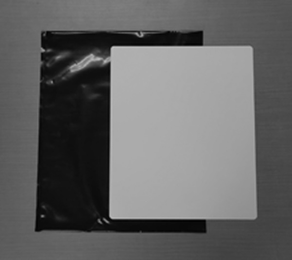
The appearance of cassetteless method of a imaging plate and a commercially available hard black plastic bag.
Twenty-five radiographic abnormal findings of 42 digits (21 limbs, lateral and medial) and 29 CT findings from 42 digits were obtained. Most of the radiographic findings were consistent with the CT findings, except for the enthesophytes of the extensor digiti minimi insertion (4 digits), because interdigital technique is sufficient for the distal phalanx, but not for the medial phalanx. This is an anatomical limitation of the extensor digiti minimi insertion, which locates proximal to extensor process of distal phalanx. The radiographic findings of the distal phalanx included exostosis (Figs. 2, 3), a rough planta surface (Fig. 3), osteolysis of the apex of the distal phalanx (Fig. 4) and widening of the laminar zone in the dorsal surface between the distal phalanx and the hoof wall (Fig. 5). The cassetteless technique is considered as an advantage over the conventional cassette-plate because of the thin imaging plate, which can be easily inserted in the interdigital space. Furthermore, film-screen radiography is often overexposed/underexposed when taken at a farm site because of improper imaging techniques, which in turn affects imaging quality of the radiograph. Therefore, the film-screen method has not been widely used at farm animal clinics. However, CR is not dependent on the imaging technique, because the technology compensates for improper exposure and focal film distance. Improved contrast resolution and computer digital reformatting make it a valuable alternative technique to the film-screen method. Furthermore, the CR does not require a dark room and thus facilitates X-ray examinations at the farm animal clinic. While, there is a disadvantage of cassetteless technique. IP is not covered by hard cassette and therefore may be fragile. Handling IP should be careful, especially in the field.
Fig. 2.
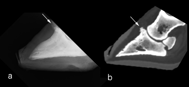
Interdigital computed radiography (a) showing exostosis at the level of the distal phalanx extensor process (arrow) and a sagittal computed tomography (b, WW: 609, WL: 348).
Fig. 3.
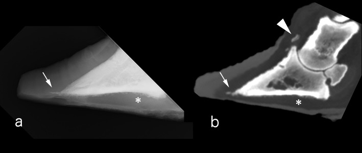
Interdigital computed radiography (a) showing exostosis at the apex of the distal phalanx (arrow), a rough planta surface (*) and a sagittal computed tomography (b, WW: 600, WL: 380). Enthesophyte of the extensor digiti minimi insertion (arrowhead) was not visualized on the radiograph. This is the anatomical limitation of interdigital technique.
Fig. 4.
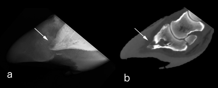
Interdigital computed radiography (a) showing osteolysis of the apex of the distal phalanx (arrow) and a sagittal computed tomography (b, WW: 1600, WL: 300).
Fig. 5.
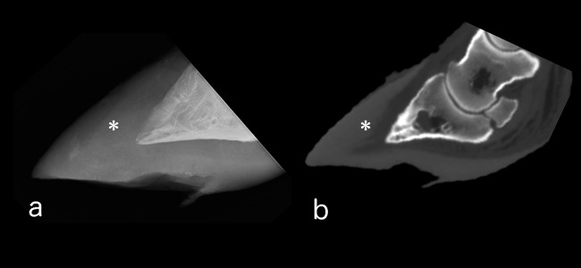
Interdigital computed radiography (a) showing widening of the laminar zone in the dorsal surface between the distal phalanx and the hoof wall (*) and a sagittal computed tomography (b, WW: 1600, WL: 377).
In our study, the hindlimbs (19 digits) showed more CT changes than the forelimbs (10 digits). The lateral digits (17 digits) were more affected than the medial digits (12 digits), particularly in the hindlimbs. These results were consistent with a previous report [4]. Higher incidence of disorders in the hindlimb than in the forelimb, because of greater exposure of the hind hooves to a dirty environment; furthermore, the hindlimbs are more susceptible to disorders, because of the size and weight of the udder [4, 6]. The lateral digits showed a higher prevalence of severe radiographic changes than the medial digits, particularly in the hindlimbs. The causes of overload and predisposition of the lateral hind hooves to disease are poorly understood. Even after trimming, the cow’s weight is not evenly distributed between the hindlimb hooves; thus, the lateral hooves carry considerably more weight than the medial hooves [8]. The lateral hooves of the hindlimbs may present particular anatomical characteristics that predispose them to chronic overload and disease [5].
We believe that the cassetteless CR technique may be an easily applicable method for imaging of the distal phalanx compared with the conventional cassette-plate and/or film-screen cassetteless method. A clinical trial should be the next step.
REFERENCES
- 1.Alban L. 1995. Lameness in Danish dairy cows: frequency and possible risk factors. Prev. Vet. Med. 22: 213–225. doi: 10.1016/0167-5877(94)00411-B [DOI] [Google Scholar]
- 2.Enting H., Kooij D., Dijkhuizen A. A., Huirne R. M., Noordhuizen-Stassen E. N. 1997. Economic losses due to clinical lameness in dairy cattle. Livest. Prod. Sci. 49: 259–267. doi: 10.1016/S0301-6226(97)00051-1 [DOI] [Google Scholar]
- 3.Ettema J. F., Ostergaard S. 2006. Economic decision making on prevention and control of clinical lameness in Danish dairy herds. Livest. Prod. Sci. 102: 92–106. doi: 10.1016/j.livprodsci.2005.11.021 [DOI] [Google Scholar]
- 4.Kofler J. 1999. Clinical study of toe ulcer and necrosis of the apex of the distal phalanx in 53 cattle. Vet. J. 157: 139–147. doi: 10.1053/tvjl.1998.0290 [DOI] [PubMed] [Google Scholar]
- 5.Muggli E., Sauter-Louis C., Braun U., Nuss K. 2011. Length asymmetry of the bovine digits. Vet. J. 188: 295–300. doi: 10.1016/j.tvjl.2010.05.016 [DOI] [PubMed] [Google Scholar]
- 6.Sogstad Å. M., Fjeldaas T., Østerås O. 2005. Lameness and claw lesions of the Norwegian red dairy cattle house in free stalls in relation to environment, parity and stage of lactation. Acta Vet. Scand. 46: 203–217. doi: 10.1186/1751-0147-46-203 [DOI] [PMC free article] [PubMed] [Google Scholar]
- 7.Starke A., Hepplemann M., Beyerbach M., Rehage J. 2007. Septic arthritis of the distal interphalangeal joint in cattle: comparison of digital amputation and joint resection by solar approach. Vet. Surg. 36: 350–359. doi: 10.1111/j.1532-950X.2007.00257.x [DOI] [PubMed] [Google Scholar]
- 8.van der Tol P. P., van der Beek S. S., Metz J. H., Noordhuizen-Stassen E. N., Back W., Braam C. R., Weijs W. A. 2004. The effect of preventive trimming on weight bearing and force balance on the claws of dairy cattle. J. Dairy Sci. 87: 1732–1738. doi: 10.3168/jds.S0022-0302(04)73327-5 [DOI] [PubMed] [Google Scholar]
- 9.Widmer W. R. 2008. Acquisition hardware for digital imaging. Vet. Radiol. Ultrasound 49, S2–8. doi: 10.1111/j.1740-8261.2007.00326.x [DOI] [PubMed] [Google Scholar]


