



Abstract
Purpose of review
Despite apparent blood pressure (BP) control and renin–angiotensin system (RAS) blockade, the chronic kidney disease (CKD) outcomes have been suboptimal. Accordingly, this review is addressed to renal microvascular and autoregulatory impairments that underlie the enhanced dynamic glomerular BP transmission in CKD progression.
Recent findings
Clinical data suggest that failure to achieve adequate 24-h BP control is likely contributing to the suboptimal outcomes in CKD. Whereas evidence continues to accumulate regarding the importance of preglomerular autoregulatory impairment to the dynamic glomerular BP transmission, emerging data indicate that nitric oxide-mediated efferent vasodilation may play an important role in mitigating the consequences of glomerular hypertension. By contrast, the vasoconstrictor effects of angiotensin II are expected to potentially reduce glomerular barotrauma and possibly enhance ischemic injury. When adequate BP measurement methods are used, the evidence for BP-independent injury initiating mechanisms is considerably weaker and the renoprotection by RAS blockade largely parallels its antihypertensive effectiveness.
Summary
Adequate 24-h BP control presently offers the most feasible intervention for reducing glomerular BP transmission and improving suboptimal outcomes in CKD. Investigations addressed to improving myogenic autoregulation and/or enhancing nitric oxide-mediated efferent dilation in addition to the more downstream mediators may provide additional future therapeutic targets.
Keywords: angiotensin II, autoregulation, chronic kidney disease, hypertension, nitric oxide
INTRODUCTION
The mechanisms responsible for the seemingly autonomous progressive glomerulosclerosis and nephron loss in chronic kidney disease (CKD) have been investigated and debated for several decades. On the basis of micropuncture studies in the rat remnant kidney model of 5/6 renal ablation, the concept was initially formulated by Brenner and colleagues that ‘glomerular hyperfiltration, an adaptation seen in response to a reduction in functional nephron number’ irrespective of the cause, was in fact maladaptive and eventually led to the progressive glomerulosclerosis of the initially normal remnant glomeruli [1,2]. It was concluded that ‘the elevated single nephron glomerular filtration rate (SNGFR) common to these pathophysiologic conditions is usually caused by increases in the glomerular capillary plasma flow rate (QA) and mean glomerular capillary hydraulic pressure (PGC), which in turn are due to adaptive reductions in preglomerular and postglomerular arteriolar resistances’ and that ‘systemic hypertension is not required for glomerular capillary hyperfiltration and hypertension’. Similar hyperfiltration, increased PGC and glomerulosclerosis were described in experimental diabetes [1]. The increases in PGC were ascribed to the greater dilation of the afferent than efferent arteriole. This relative efferent vasoconstriction was attributed to the tonic vasoconstrictor effects of angiotensin II (Ang II), because angiotensin-converting enzyme (ACE) inhibitors were shown to dilate the efferent arteriole, reduce PGC and ameliorate glomerulosclerosis [1].
Observations from our laboratory have provided an alternative interpretation of these hemodynamic data. As discussed in greater detail elsewhere [3], we have questioned that significant glomerular hypertension is necessary for hyperfiltration, which can be achieved in normotensive states by coordinated increases in glomerular filtration surface area (hypertrophy) and increases in single nephron plasma flow through proportionate afferent and efferent vasodilation without significant PGC increases [4,5]. In fact, increases in PGC may not even be very effective in increasing SNGFR due to the inverse relationship between PGC and the ultrafiltration coefficient Kf [6,7]. Instead, we have attributed the glomerular hypertension in these models to represent the enhanced glomerular transmission of the coexistent systemic hypertension, through a dilated and poorly autoregulating preglomerular vasculature [3-5]. Accordingly, we have ascribed the protection provided by renin–angiotensin system (RAS) blockade to its antihypertensive effects and suggested that the contribution of Ang II-mediated efferent constriction to glomerular hypertension has likely been greatly overestimated during micropuncture studies due to renin release and efferent constriction triggered by anesthesia, surgery and neurohormonal activation [3,8].
Over the years, other investigators have assigned a primary importance to RAS-mediated as well as other nonhemodynamic injury pathways in CKD progression (Fig. 1) [9-12,13■,14]. However, interpretations are confounded by the interactions between blood pressure (BP)-dependent and BP-independent pathways, the limitations of the BP measurement methodologies employed and a lack of clear discrimination between initiating mechanisms and the somewhat more nonspecific downstream responses to injury including fibrosis. Nevertheless, such a separation is consistent with current management guidelines which recommend BP control and RAS blockade as separate interventions addressed to these two pathways for slowing CKD progression [15,16]. In any event, the following discussion is primarily addressed to angiotensin-dependent and independent alterations in afferent and efferent arteriolar function in CKD and their potential interactions with BP-dependent injury mechanisms in the pathogenesis of CKD progression. The increasingly recognized role of nitric oxide and endothelial dysfunction in such interactions and CKD progression will also be addressed.
FIGURE 1.
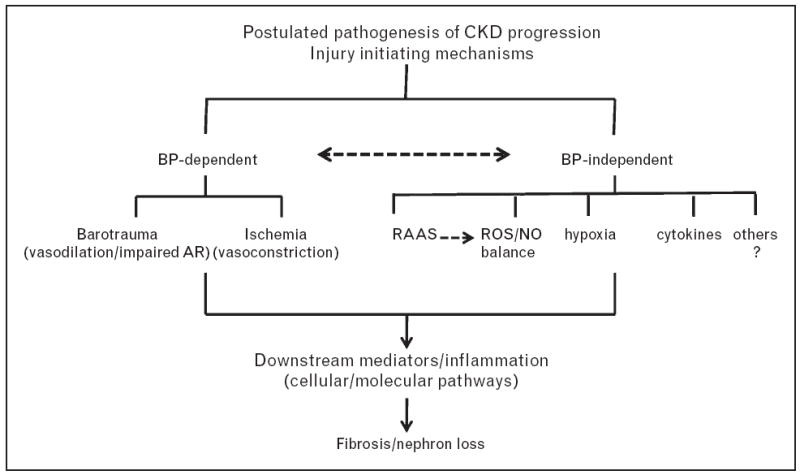
An overview of the pathogenesis of CKD progression. Although there is considerable interaction between BP-dependent and BP-independent initiating mechanisms, BP-dependent mechanisms predominate in hypertensive CKD states. BP-independent mechanisms may modulate hypertensive injury and also contribute to CKD progression in normotensive states. AR, autoregulation; BP, blood pressure; CKD, chronic kidney disease; NO, nitric oxide; RAAS, renin–angiotensin–aldosterone system; ROS, reactive oxygen species.
RENAL MICROVASCULAR FUNCTION AND PATHOPHYSIOLOGY OF HYPERTENSIVE RENAL DAMAGE
Hypertension is second only to diabetes as a primary cause of end-stage renal disease (ESRD). Nevertheless, despite the large population risk, the most striking feature of the relationship between hypertension and renal damage is the very small individual risk (< 0.5%) in those with essential hypertension. By contrast, substantial evidence indicates that hypertension plays a major role in the progression of most forms of CKD including diabetic nephropathy. These large differences in individual risk are reflected in differences in the quantitative relationships between BP and renal damage, as well as differences in morphologic patterns of injury [8]. Such differences are best explained when considered in the context of preglomerular autoregulatory function in essential hypertension and CKD states.
Hypertensive renal damage in essential hypertension
Normally, increases in BP – episodic or sustained – result in proportionate autoregulatory vasoconstriction of the preglomerular resistance vessels, primarily the afferent arteriole, such that renal blood flow (RBF), GFR and PGC are maintained relatively constant (Fig. 2a) [8]. The vast majority of patients with primary essential hypertension have preserved autoregulation. The glomerular capillaries are protected from barotrauma and significant proteinuria is not seen. However, the preglomerular vasculature exposed to the hypertension develops the slowly progressive vascular pathology of benign nephrosclerosis [8,17]. The ischemic glomerulosclerosis and nephron loss that occur over time are usually not sufficient to result in ESRD. However, a subset of genetically susceptible individuals, largely African-Americans, seems to exhibit a more accelerated course [18-22]. Histologic studies in such individuals have suggested a focal loss of autoregulation and barotraumas-mediated focal segmental glomerulosclerosis in addition to ischemic glomerulosclerosis [21,23]. The term benign decompensated nephrosclerosis has been used to describe such pathology [24]. The responsible mechanisms and their relationship to recently identified genetic susceptibility variants in African-Americans have notyet been defined. By contrast, if the hypertension becomes very severe and exceeds a certain critical threshold, malignant nephrosclerosis develops [8,17]. Severe barotrauma and acute disruptive injury to the renal vasculature and glomeruli result. Proteinuria, hematuria and rapid loss of renal function ensue.
FIGURE 2.
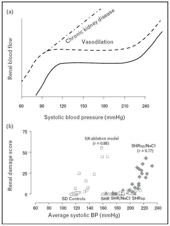
(a) Summary of renal autoregulatory patterns obtained in rats with intact kidneys and normal autoregulation, vasodilated vascular bed and normal autoregulation (i.e. uninephrectomy) and vasodilated vascular bed and impaired autoregulation (i.e. 5/6 renal ablation model of CKD). Reproduced with permission from [49]. (b) Compilation of data obtained in our laboratory which demonstrates that the relationship between renal injury and average SBP parallels that of renal autoregulatory function in experimental rodent models of hypertension. Minimal injury is seen in SHR with intact autoregulation despite severe hypertension. Injury in the salt-sensitive SHRsp administered a high NaCl diet occurs at BPs that exceed the autoregulatory capacity, whereas the 5/6 renal ablation model, with impaired autoregulation, exhibits a much lower BP threshold for hypertensive injury than normal or SHR kidneys. CKD, chronic kidney disease; SHR, spontaneously hypertensive rat; SHRsp, stroke-prone spontaneously hypertensive rat. Reproduced with permission from [27].
Hypertensive renal damage in chronic kidney disease
In contrast to the slowly progressive ischemic pathogenesis of nephron loss in benign nephrosclerosis, the preglomerular vasodilation present in CKD allows a greater fractional transmission of systemic BP to the glomeruli with resultant barotrauma [8]. Vasodilation alone, with preserved autoregulation as after uninephrectomy, only modestly increases the susceptibility to hypertensive injury. But, with greater reduction in functional renal mass, autoregulation is impaired, and even modest increases in BP – episodic or sustained – are transmitted to the glomerular capillaries. In the absence of severe hypertension, vascular pathology may not be prominent and the predominant lesion is that of glomerulosclerosis. These differences in the quantitative relationships between BP and renal damage with intact vs. impaired autoregulation are illustrated in Fig. 2b using models of genetic hypertension and CKD.
MECHANISMS UNDERLYING RENAL AUTOREGULATION
Renal autoregulation is believed to be mediated by the combined and interacting contributions of two mechanisms: a faster myogenic and a slower tubuloglomerular feedback (TGF) system (Fig. 3) [8,25-27]. Recently, additional slower mechanisms have been postulated [26]. Although the myogenic and TGF mechanisms are thought to act in concert to insulate the renal excretory function from BP fluctuations in the regulation of salt and water homeostasis [27,28], the myogenic response may be primarily responsible for protecting against hypertensive injury [8,27]. The rapid activation kinetics of the afferent arteriolar myogenic response and its potential triggering by the systolic (peak) rather than mean BP are consistent with this protective function [8,27,29,30], given that SBP may have the greatest potential for target organ damage. Pressure-induced vascular wall stretch, depolarization, activation of voltage-gated Ca2+ channels, Ca2+ entry and vasoconstriction are involved, but the determinants of afferent arteriolar myogenic reactivity remain largely undefined [31]. Unique roles of 20-hydroxyeicosatetraenoic acid, epithelial sodium channel, Connexin 40, transient receptor potential channels, protein kinase C and inward rectifier K channels (KIR) have all been implicated [32-38]. Recent investigations have implicated reactive oxygen species (ROS) and superoxide in myogenic vasoconstriction [39,40■]. However, ROS have also been implicated in impairment of myogenic responses [40■,41-43], suggesting involvement of separate ROS species. However, definitive evidence that alteration in any of these pathways is involved in CKD is lacking. For example, blockade of KIR markedly blunts the afferent arteriolar myogenic response, suggesting a critical role of this channel [32]. However, unpublished findings from our laboratory indicate that KIR currents are not altered in myocytes isolated from afferent arterioles obtained from remnant rat kidneys.
FIGURE 3.
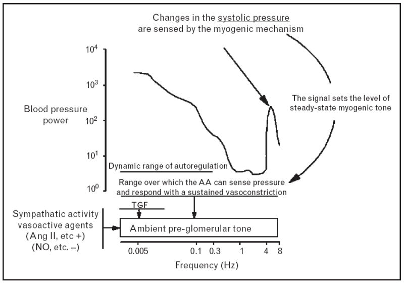
Overview of the mechanisms regulating renal blood flow (RBF) autoregulation and glomerular BP transmission. Changes in the oscillating SBP are sensed and responded to by the myogenic mechanism. The ambient preglomerular tone is also influenced by neurohumoral agents, such as nitric oxide and angiotensin II. The dynamic autoregulation of RBF occurs at frequencies below the oscillating SBP (~6 Hz in rat) and includes a faster myogenic (~0.25 Hz) and a slower tubuloglomerular feedback (~0.04 Hz) component. BP, blood pressure; TGF, tubuloglomerular feedback. Reproduced with permission from [27].
BLOOD PRESSURE LABILITY, RENAL AUTOREGULATION AND DYNAMICS OF GLOMERULAR BLOOD PRESSURE TRANSMISSION
Blood pressure is fundamentally labile, and the glomerular transmission of the continuously oscillating systemic pressures in real time must be a very dynamic process. Accordingly, the transmission of individual BP fluctuations is expected to depend on their rate (frequency) and the kinetics and magnitude of the autoregulatory response [8,27]. Of note, it is intact renal autoregulation that prevents the normally occurring BP fluctuations, even in normotensive states, from reaching the glomeruli. Conversely, with impaired autoregulation, glomerular pressure exposure may not be normalized even if normotension is achieved [44■]. Consistent with such interpretations, glomerulosclerosis in normotensive models of renal mass reduction has been shown to exhibit greater correlations with percentage SBP fluctuations above 150 mmHg than average SBP [45]. Given the exaggerated BP lability in hypertensive states, isolated BP (and PGC) measurements are likely to be more limited as indices of total glomerular BP exposure in CKD states. It is for such reasons that direct and continuous rather than isolated tail-cuff BP measurements have been recommended for the investigations of hypertensive renal damage in experimental animal models [46]. The likely round-the-clock nature of such glomerular BP transmission is particularly relevant in CKD patients as masked daytime and/or nocturnal hypertension and BP lability are both widely prevalent and more difficult to control, further emphasizing the limitations of isolated clinic BP measurements [44■,47].
RENAL AUTOREGULATION, ANTIHYPERTENSIVE AGENTS AND CHRONIC KIDNEY DISEASE PROGRESSION
Only limited data are available regarding the effects of specific antihypertensives on renal autoregulation and hypertensive injury. Predictably, calcium channel blockers (CCBs) further impair autoregulation and cause the slope of the relationship between radiotelemetrically measured BP and glomerulosclerosis in 5/6 ablated rats to become steeper, resulting in greater glomerulosclerosis for any given BP increase as compared to untreated or RAS blockade-treated animals (Fig. 4) [8,48,49]. By contrast, RAS blockade does not additionally alter renal autoregulation or the relationship between BP and glomerulosclerosis from that observed in untreated animals (Fig. 4) (vide infra).
FIGURE 4.
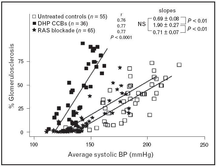
Compilation of data obtained in our laboratory which illustrates the quantitative relationships between BP and glomerulosclerosis (GS) in rats with 5/6 renal ablation who were left untreated or received either calcium channel blockers or RAS blockade. The deleterious effects of calcium channel blockers on GS as compared to untreated or RAS blockade treated rats are evident. BP, blood pressure; DHP CCBs, dihydropyridine calcium channel blockers; RAS, renin–angiotensin system. Reproduced with permission from [49].
ANGIOTENSIN II-INDUCED RENAL MICROVASCULAR DYSFUNCTION AND CHRONIC KIDNEY DISEASE PROGRESSION
Although Ang II plays a critical homeostatic role in BP regulation, it is the BP-independent deleterious effects of Ang II that have received the greatest emphasis in the context of CKD progression [9-12,13■,14]. Although widely accepted, the evidence in support of these effects in vivo is much less definitive than implied, as noted above and discussed at greater length elsewhere [8,48-53]. To briefly illustrate, the protection provided by RAS blockade is believed to be largely mediated by mechanisms independent of its antihypertensive effects. However, given the wide individual variability of renal damage in these models [45,48,51,52] and the fact that the BP-dependent component of renal damage is expected to be a function of the total chronic BP exposure, to truly demonstrate BP independence, the achieved renoprotection has to be shown to be independent of the continuously measured BP in the same animals in which such injury is assessed. In the only studies that have performed such correlations between the magnitude of renal injury and radiotelemetrically measured chronic BP load, the protection after RAS blockade has been found to be proportionate to the achieved BP reductions (Fig. 4) and similar to that obtained with other non-CCB antihypertensive regimens (i.e. triple therapy) [52]. However, a recent study utilizing intermittent BP radiotelemetry in a mouse renal ablation model of CKD did find RAS blockade to be more antiproteinuric as compared to hydralazine, but renal pathology data were not provided [54].
In any event, in addition to the much emphasized BP-independent deleterious mechanisms such as nonhemodynamic profibrotic signaling and efferent constriction, Ang II has also been postulated to impair renal autoregulatory mechanisms. For instance, in contrast to the lack of effect of RAS blockade on impaired autoregulation in the 5/6 renal ablation model at 3 weeks [48], losartan corrected the impaired TGF and altered hemodynamics observed at 7 days [55,56]. Additionally, Inscho and colleagues have reported that Ang II impairs autoregulation and thereby promotes hypertensive injury [57-60]. Other studies by these and other investigators have indicated that Ang II and salt are synergistic in their deleterious effects of renal autoregulation [41,58]. In any event, given this plethora of mechanisms by which Ang II promotes renal injury, exogenous Ang II infusion models of hypertension would be expected to result in rapid development of severe glomerular injury. Yet, remarkably, most studies have shown only minimal to modest injury after 2–6 weeks of Ang II hypertension [41,61,62].
Indeed, recent studies (unpublished data) from our laboratory using BP radiotelemetry to examine the quantitative relationships between Ang II hypertension and glomerular injury after 6 weeks have shown modest injury (6%) and a very flat relationship (slope 0.15 + 0.07). By contrast, the slope for comparably hypertensive rats (~180 mmHg systolic) with 3/4 renal ablation, impaired autoregulation and threefold greater glomerulosclerosis (20%) was 0.6 + 0.25 (P < 0.01). The explanation for these seemingly unexpected findings may reside in the fact that as noted previously [63], the deleterious consequences of impaired autoregulation in hypertensive glomerulosclerosis are only expected to occur with a vasodilated preglomerular vasculature. By contrast, persistent Ang II vasoconstriction that fails to respond to pressure changes (impaired autoregulation) is expected to reduce glomerular BP transmission but increase the potential for ischemic tubulointerstitial injury. Not surprisingly, given the facilitating effects of Ang II on myogenic vasoconstriction [35,64], other studies have found augmented myogenic vasoconstrictor responses in isolated perfused kidneys from Ang II-infused rats, described as ‘superautoregulation’ [65]. Consistent with such data, an enhanced myogenic resonance peak was observed by Just et al. [66] during dynamic autoregulation studies in the conscious dog. Similarly, our studies in conscious chronically instrumented rats have shown both an enhanced and faster myogenic response as well as RBF responses to spontaneous changes in BP that are consistent with the ‘superautoregulation’ description [67]. Recent data that Notch 3 gene deletion reduced vasoconstriction and BP responses to Ang II but nevertheless increased renal damage and mortality are consistent with the protective effects of such vasoconstriction [68■].
NITRIC OXIDE EFFECTS ON RENAL MICROVASCULATURE AND CHRONIC KIDNEY DISEASE PROGRESSION
Abundant evidence supports the deleterious impact of reduced nitric oxide generation/availability in CKD progression [69■]. Although multiple pathways are likely involved, the present discussion is confined to the hemodynamic effects of nitric oxide loss on hypertensive mechanisms of CKD progression. Whereas the severity of hypertension is expected to increase by nitric oxide loss, the adverse impact on glomerular hypertension is expected to be mitigated by the preglomerular vasoconstriction [70,71■]. Additionally, nitric oxide is an inhibitor of autoregulatory responses and accordingly nitric oxide loss should reduce dynamic glomerular BP transmission [72,73]. Thus, the large adverse impact of nitric oxide loss on hypertensive glomerulosclerosis in models such as 5/6 ablation is somewhat counter-intuitive [71■,74-76]. On the basis of studies in Sprague–Dawley rats from two different suppliers who exhibit contrasting susceptibilities to nitric oxide inhibition nephropathy, we have suggested that an underappreciated nitric oxide-mediated efferent vasodilation may serve a critical protective role in preventing/ameliorating glomerular hypertension [71■]. The rich distribution of both nitric oxide synthase (NOS) 1 and 3 in efferent arteriolar endothelial cells is consistent with such interpretations [77]. Elegant anatomical studies have also shown that these endothelial cells bulge into the lumen at the initial intraglomerular segment and may serve as shear stress sensors [78]. Such interpretations are consistent with the increase in filtration fraction seen after NOS inhibition [70,73]. Thus, nitric oxide release from these sites is ideally suited to mediate a protective decompression of glomerular hypertension. It is even possible that in chronic Ang II infusion models, AT2 receptor-mediated nitric oxide release and efferent vasodilation may limit renal injury [79]. These efferent vasodilatory effects of nitric oxide may be particularly important in diabetes and may account for the well known resistance to nephropathy of most rodent diabetic models [80,81]. A dramatic illustration of this phenomenon is provided by the striking conversion of the very nephropathy-resistant C57BL/6 mouse to a diabetic nephropathy-susceptible phenotype by the genetic deletion of endothelial NOS [81]. Genetic and/or acquired differences in the capacity to produce nitric oxide or resist its suppression may therefore play a significant role in determining susceptibility to hypertensive injury in human CKD, as has been shown in rodent strains.
ADDITIONAL BLOOD PRESSURE-INDEPENDENT MODULATORS OF HYPERTENSIVE RENAL INJURY
In addition to the severity of hypertension and the renal microvascular determinants of PGC, hypertensive glomerulosclerosis is likely to be significantly impacted by the local glomerular capillary susceptibility to barotrauma [63,82,83]. Glomerular capillary hypertrophy is a likely such independent risk factor for glomerulosclerosis. In addition to the expected increase in wall tension (Laplace Law: tension = pressure × radius), hypertrophy of glomerular capillaries results in a reduction of podocyte density due to its limited replication potential [82,83]. It has been proposed that the consequent loss of such structural support may limit the ability to maintain physical integrity and mechanical support during hypertensive stress. Such mechanisms likely contribute to progression in podocyte depletion models [82,83,84■,85■]. In this context, it has been suggested that the induction of an enhanced hypoxia inducible factor 1 alpha may provide BP-independent renoprotection in the 5/6 ablation model [55,86,87], although the effects of such induction regimens (i.e. cobalt and dimethyloxalyglycine) on BP and its potential glomerular transmission have not been rigorously examined. Similarly, differences in downstream signaling and injury responses to barotrauma (Fig. 1) are also likely to play important roles in individual differences in CKD progression, but despite much recent investigation have not yet been clearly established as intervention targets.
CONCLUSION
An enhanced susceptibility to develop hypertension and a reduced ability to protect against its glomerular transmission because of alterations in renal microvascular function are characteristic of CKD states and models. When significant hypertension is present, pressure-dependent mechanisms represent the predominant pathway for progressive glomerular injury and nephron loss. Given the recent data that masked daytime and/or nocturnal hypertension and BP lability are both more common and difficult to control in CKD patients, adequate 24-h BP control may be necessary to minimize glomerular BP transmission and barotrauma. When adequate BP measurement methodologies are used, the renoprotective effects of RAS blockade appear to be largely BP-dependent. Improving preglomerular autoregulatory function and enhancing efferent arteriolar vasodilatation in CKD states represent additional targets for investigation and potential interventions. Available data also suggest that BP-independent pathways may also impact CKD progression by the modulation of pressure-dependent injury mechanisms but may become quantitatively more important in normotensive states or when BP is adequately controlled.
KEY POINTS.
Renal autoregulation provides the primary protection against hypertensive injury.
Enhanced glomerular blood pressure (BP) transmission due to impaired renal autoregulation represents the predominant mechanism for chronic kidney disease (CKD) progression.
Adequate 24-h BP control may be necessary to minimize CKD progression.
The mechanism for impaired autoregulation in CKD remains unknown and an important target for future investigation and intervention.
Nitric oxide-mediated efferent arteriole dilation may be an additional important protective mechanism against glomerular hypertension.
Acknowledgments
The work was supported by National Institute of Diabetes and Digestive and Kidney Disease Grants DK-40426 (A.K.B.) and DK-61653 (K.A.G.), by a Merit Review (K.A.G.) and a Career Development Award 1IK2BX001285 (A.J.P.) from the Office of Research and Development of the Department of Veterans Affairs, as well as the Canadian Institutes of Health Research and the Alberta Heritage Foundation for Medical Research (R.L.).
Footnotes
Conflicts of interest
There are no conflicts of interest.
REFERENCES AND RECOMMENDED READING
Papers of particular interest, published within the annual period of review, have been highlighted as:
-
■
of special interest
-
■■
of outstanding interest
Additional references related to this topic can also be found in the Current World Literature section in this issue (pp. 127–128).
- 1.Brenner BM, Lawler EV, Mackenzie HS. The hyperfiltration theory: a paradigm shift in nephrology. Kidney Int. 1996;49:1774–1777. doi: 10.1038/ki.1996.265. [DOI] [PubMed] [Google Scholar]
- 2.Hostetter TH, Olson JL, Rennke HG, et al. Hyperfiltration in remnant nephrons: a potentially adverse response to renal ablation. Am J Physiol. 1981;241:F85–93. doi: 10.1152/ajprenal.1981.241.1.F85. [DOI] [PubMed] [Google Scholar]
- 3.Griffin KA, Kramer H, Bidani AK. Adverse renal consequences of obesity. Am J Physiol Renal Physiol. 2008;294:F685–F696. doi: 10.1152/ajprenal.00324.2007. [DOI] [PubMed] [Google Scholar]
- 4.Bidani AK, Mitchell KD, Schwartz MM, et al. Absence of glomerular injury or nephron loss in a normotensive rat remnant kidney model. Kidney Int. 1990;38:28–38. doi: 10.1038/ki.1990.163. [DOI] [PubMed] [Google Scholar]
- 5.Griffin KA, Picken MM, Churchill M, et al. Functional and structural correlates of glomerulosclerosis after renal mass reduction in the rat. J Am Soc Nephrol. 2000;11:497–506. doi: 10.1681/ASN.V113497. [DOI] [PubMed] [Google Scholar]
- 6.Arendshorst WJ, Gottschalk CW. Glomerular ultrafiltration dynamics: euvolemic and plasma volume-expanded rats. Am J Physiol. 1980;239:F171–F186. doi: 10.1152/ajprenal.1980.239.2.F171. [DOI] [PubMed] [Google Scholar]
- 7.Tucker BJ, Blantz RC. Effects of glomerular filtration dynamics on the glomerular permeability coefficient. Am J Physiol. 1981;240:F245–F254. doi: 10.1152/ajprenal.1981.240.3.F245. [DOI] [PubMed] [Google Scholar]
- 8.Bidani AK, Griffin KA, Williamson G, et al. Protective importance of the myogenic response in the renal circulation. Hypertension. 2009;54:393–398. doi: 10.1161/HYPERTENSIONAHA.109.133777. [DOI] [PMC free article] [PubMed] [Google Scholar]
- 9.Boor P, Ostendorf T, Floege J. Renal fibrosis: novel insights into mechanisms and therapeutic targets. Nat Rev Nephrol. 2010;6:643–656. doi: 10.1038/nrneph.2010.120. [DOI] [PubMed] [Google Scholar]
- 10.Harris RC, Neilson EG. Toward a unified theory of renal progression. Annu Rev Med. 2006;57:365–380. doi: 10.1146/annurev.med.57.121304.131342. [DOI] [PubMed] [Google Scholar]
- 11.Kagami S. Involvement of glomerular renin-angiotensin system (RAS) activation in the development and progression of glomerular injury. Clin Exp Nephrol. 2012;16:214–220. doi: 10.1007/s10157-011-0568-0. [DOI] [PMC free article] [PubMed] [Google Scholar]
- 12.Mimura I, Nangaku M. The suffocating kidney: tubulointerstitial hypoxia in end-stage renal disease. Nat Rev Nephrol. 2010;6:667–678. doi: 10.1038/nrneph.2010.124. [DOI] [PubMed] [Google Scholar]
- 13■.Ruster C, Wolf G. Angiotensin II as a morphogenic cytokine stimulating renal fibrogenesis. J Am Soc Nephrol. 2011;22:1189–1199. doi: 10.1681/ASN.2010040384. Summarizes the case for angiotensin II as an initiator of pro-fibrotic signaling. [DOI] [PubMed] [Google Scholar]
- 14.Zeisberg M, Neilson EG. Mechanisms of tubulointerstitial fibrosis. J Am Soc Nephrol. 2010;21:1819–1834. doi: 10.1681/ASN.2010080793. [DOI] [PubMed] [Google Scholar]
- 15.K/DOQI clinical practice guidelines on hypertension and antihypertensive agents in chronic kidney disease. Am J Kidney Dis. 2004;43(5 Suppl 1):S1–290. [PubMed] [Google Scholar]
- 16.Chobanian AV, Bakris GL, Black HR, et al. The Seventh Report of the Joint National Committee on Prevention, Detection, Evaluation, and Treatment of High Blood Pressure: the JNC 7 report. J Am Med Assoc. 2003;289:2560–2572. doi: 10.1001/jama.289.19.2560. [DOI] [PubMed] [Google Scholar]
- 17.Olson JL. Renal disease caused by hypertension. In: Jennette JC, Olson JL, Schwartz MM, Silva FG, editors. Heptinstall’s pathology of the kidney. 6. Philadelphia: Lippincott Williams & Wilkins; 2006. pp. 937–990. [Google Scholar]
- 18.Appel LJ, Wright JT, Jr, Greene T, et al. Intensive blood-pressure control in hypertensive chronic kidney disease. N Engl J Med. 2010;363:918–929. doi: 10.1056/NEJMoa0910975. [DOI] [PMC free article] [PubMed] [Google Scholar]
- 19.Appel LJ, Wright JT, Jr, Greene T, et al. Long-term effects of renin-angiotensin system-blocking therapy and a low blood pressure goal on progression of hypertensive chronic kidney disease in African Americans. Arch Intern Med. 2008;168:832–839. doi: 10.1001/archinte.168.8.832. [DOI] [PMC free article] [PubMed] [Google Scholar]
- 20.Genovese G, Friedman DJ, Ross MD, et al. Association of trypanolytic ApoL1 variants with kidney disease in African Americans. Science. 2010;329:841–845. doi: 10.1126/science.1193032. [DOI] [PMC free article] [PubMed] [Google Scholar]
- 21.Hill GS. Hypertensive nephrosclerosis. Curr Opin Nephrol Hypertens. 2008;17:266–270. doi: 10.1097/MNH.0b013e3282f88a1f. [DOI] [PubMed] [Google Scholar]
- 22.Kopp JB, Smith MW, Nelson GW, et al. MYH9 is a major-effect risk gene for focal segmental glomerulosclerosis. Nat Genet. 2008;40:1175–1184. doi: 10.1038/ng.226. [DOI] [PMC free article] [PubMed] [Google Scholar]
- 23.Marcantoni C, Ma LJ, Federspiel C, Fogo AB. Hypertensive nephrosclerosis in African Americans versus Caucasians. Kidney Int. 2002;62:172–180. doi: 10.1046/j.1523-1755.2002.00420.x. [DOI] [PubMed] [Google Scholar]
- 24.Bohle A, Ratschek M. The compensated and the decompensated form of benign nephrosclerosis. Pathol Res Pract. 1982;174:357–367. doi: 10.1016/S0344-0338(82)80017-4. [DOI] [PubMed] [Google Scholar]
- 25.Cupples WA, Braam B. Assessment of renal autoregulation. Am J Physiol Renal Physiol. 2007;292:F1105–F1123. doi: 10.1152/ajprenal.00194.2006. [DOI] [PubMed] [Google Scholar]
- 26.Just A. Mechanisms of renal blood flow autoregulation: dynamics and contributions. Am J Physiol Regul Integr Comp Physiol. 2007;292:R1–17. doi: 10.1152/ajpregu.00332.2006. [DOI] [PubMed] [Google Scholar]
- 27.Loutzenhiser R, Griffin K, Williamson G, Bidani A. Renal autoregulation: new perspectives regarding the protective and regulatory roles of the underlying mechanisms. Am J Physiol Regul Integr Comp Physiol. 2006;290:R1153–R1167. doi: 10.1152/ajpregu.00402.2005. [DOI] [PMC free article] [PubMed] [Google Scholar]
- 28.Thomson SC, Blantz RC. Glomerulotubular balance, tubuloglomerular feedback, and salt homeostasis. J Am Soc Nephrol. 2008;19:2272–2275. doi: 10.1681/ASN.2007121326. [DOI] [PubMed] [Google Scholar]
- 29.Chen J, Sgouralis I, Moore LC, et al. A mathematical model of the myogenic response to systolic pressure in the afferent arteriole. Am J Physiol Renal Physiol. 2011;300:F669–F681. doi: 10.1152/ajprenal.00382.2010. [DOI] [PMC free article] [PubMed] [Google Scholar]
- 30.Williamson GA, Loutzenhiser R, Wang X, et al. Systolic and mean blood pressures and afferent arteriolar myogenic response dynamics: a modeling approach. Am J Physiol Regul Integr Comp Physiol. 2008;295:R1502–R1511. doi: 10.1152/ajpregu.00490.2007. [DOI] [PubMed] [Google Scholar]
- 31.Davis MJ, Hill MA. Signaling mechanisms underlying the vascular myogenic response. Physiol Rev. 1999;79:387–423. doi: 10.1152/physrev.1999.79.2.387. [DOI] [PubMed] [Google Scholar]
- 32.Chilton L, Smirnov SV, Loutzenhiser K, et al. Segment-specific differences in the inward rectifier K(+) current along the renal interlobular artery. Cardiovasc Res. 2011;92:169–177. doi: 10.1093/cvr/cvr179. [DOI] [PubMed] [Google Scholar]
- 33.Drummond HA, Grifoni SC, Abu-Zaid A, et al. Renal inflammation and elevated blood pressure in a mouse model of reduced {beta}-ENaC. Am J Physiol Renal Physiol. 2011;301:F443–F449. doi: 10.1152/ajprenal.00694.2010. [DOI] [PMC free article] [PubMed] [Google Scholar]
- 34.Grifoni SC, Chiposi R, McKey SE, et al. Altered whole kidney blood flow autoregulation in a mouse model of reduced beta-ENaC. Am J Physiol Renal Physiol. 2010;298:F285–F292. doi: 10.1152/ajprenal.00496.2009. [DOI] [PMC free article] [PubMed] [Google Scholar]
- 35.Kirton CA, Loutzenhiser R. Alterations in basal protein kinase C activity modulate renal afferent arteriolar myogenic reactivity. Am J Physiol. 1998;275(2 Pt 2):H467–H475. doi: 10.1152/ajpheart.1998.275.2.H467. [DOI] [PubMed] [Google Scholar]
- 36.Roman RJ, Harder DR. Cellular and ionic signal transduction mechanisms for the mechanical activation of renal arterial vascular smooth muscle. J Am Soc Nephrol. 1993;4:986–996. doi: 10.1681/ASN.V44986. [DOI] [PubMed] [Google Scholar]
- 37.Sharif-Naeini R, Dedman A, Folgering JH, et al. TRP channels and mechanosensory transduction: insights into the arterial myogenic response. Pflugers Arch. 2008;456:529–540. doi: 10.1007/s00424-007-0432-y. [DOI] [PubMed] [Google Scholar]
- 38.Sorensen CM, Giese I, Braunstein TH, et al. Role of Connexin40 in the autoregulatory response of the afferent arteriole. Am J Physiol Renal Physiol. 2012;303:F855–F863. doi: 10.1152/ajprenal.00026.2012. [DOI] [PubMed] [Google Scholar]
- 39.Lai EY, Solis G, Luo Z, et al. p47(phox) is required for afferent arteriolar contractile responses to angiotensin II and perfusion pressure in mice. Hypertension. 2012;59:415–420. doi: 10.1161/HYPERTENSIONAHA.111.184291. [DOI] [PMC free article] [PubMed] [Google Scholar]
- 40■.Lai EY, Wellstein A, Welch WJ, Wilcox CS. Superoxide modulates myogenic contractions of mouse afferent arterioles. Hypertension. 2011;58:650–656. doi: 10.1161/HYPERTENSIONAHA.111.170472. Provides evidence for the role of reactive oxygen species in modulating the myogenic responses in isolated mouse afferent arterioles. [DOI] [PMC free article] [PubMed] [Google Scholar]
- 41.Saeed A, Dibona GF, Marcussen N, Guron G. High-NaCl intake impairs dynamic autoregulation of renal blood flow in ANG II-infused rats. Am J Physiol Regul Integr Comp Physiol. 2010;299:R1142–R1149. doi: 10.1152/ajpregu.00326.2010. [DOI] [PubMed] [Google Scholar]
- 42.Sharma K, Cook A, Smith M, et al. TGF-beta impairs renal autoregulation via generation of ROS. Am J Physiol Renal Physiol. 2005;288:F1069–F1077. doi: 10.1152/ajprenal.00345.2004. [DOI] [PubMed] [Google Scholar]
- 43.Vavrinec P, van Dokkum RP, Goris M, et al. Losartan protects mesenteric arteries from ROS-associated decrease in myogenic constriction following 5/6 nephrectomy. J Renin Angiotensin Aldosterone Syst. 2011;12:184–194. doi: 10.1177/1470320310391328. [DOI] [PubMed] [Google Scholar]
- 44■.Bidani AK, Griffin KA, Epstein M. Hypertension and chronic kidney disease progression: why the suboptimal outcomes? Am J Med. 2012;125:1057–1062. doi: 10.1016/j.amjmed.2012.04.008. Discusses the clinical evidence that indicates the potential importance of inadequate 24-h BP control in CKD progression. [DOI] [PMC free article] [PubMed] [Google Scholar]
- 45.Griffin KA, Picken MM, Bidani AK. Blood pressure lability and glomerulosclerosis after normotensive 5/6 renal mass reduction in the rat. Kidney Int. 2004;65:209–218. doi: 10.1111/j.1523-1755.2004.00356.x. [DOI] [PubMed] [Google Scholar]
- 46.Kurtz TW, Griffin KA, Bidani AK, et al. Recommendations for blood pressure measurement in humans and experimental animals. Part 2: blood pressure measurement in experimental animals: a statement for professionals from the subcommittee of professional and public education of the American Heart Association council on high blood pressure research. Hypertension. 2005;45:299–310. doi: 10.1161/01.HYP.0000150857.39919.cb. [DOI] [PubMed] [Google Scholar]
- 47.Pogue V, Rahman M, Lipkowitz M, et al. Disparate estimates of hypertension control from ambulatory and clinic blood pressure measurements in hypertensive kidney disease. Hypertension. 2009;53:20–27. doi: 10.1161/HYPERTENSIONAHA.108.115154. [DOI] [PubMed] [Google Scholar]
- 48.Griffin KA, Picken MM, Bidani AK. Deleterious effects of calcium channel blockade on pressure transmission and glomerular injury in rat remnant kidneys. J Clin Invest. 1995;96:793–800. doi: 10.1172/JCI118125. [DOI] [PMC free article] [PubMed] [Google Scholar]
- 49.Griffin KA, Bidani AK. Progression of renal disease: renoprotective specificity of renin-angiotensin system blockade. Clin J Am Soc Nephrol. 2006;1:1054–1065. doi: 10.2215/CJN.02231205. [DOI] [PubMed] [Google Scholar]
- 50.Bidani AK, Griffin KA. The benefits of renin-angiotensin blockade in hypertension are dependent on blood-pressure lowering. Nat Clin Pract Nephrol. 2006;2:542–543. doi: 10.1038/ncpneph0299. [DOI] [PubMed] [Google Scholar]
- 51.Bidani AK, Griffin KA, Bakris G, Picken MM. Lack of evidence of blood pressure-independent protection by renin-angiotensin system blockade after renal ablation. Kidney Int. 2000;57:1651–1661. doi: 10.1046/j.1523-1755.2000.00009.x. [DOI] [PubMed] [Google Scholar]
- 52.Griffin KA, Picken M, Bidani AK. Radiotelemetric BP monitoring, antihypertensives and glomeruloprotection in remnant kidney model. Kidney Int. 1994;46:1010–1018. doi: 10.1038/ki.1994.361. [DOI] [PubMed] [Google Scholar]
- 53.Kurtz TW. False claims of blood pressure-independent protection by blockade of the renin angiotensin aldosterone system? Hypertension. 2003;41:193–196. doi: 10.1161/01.hyp.0000049882.23078.eb. [DOI] [PubMed] [Google Scholar]
- 54.Leelahavanichkul A, Yan Q, Hu X, et al. Angiotensin II overcomes strain-dependent resistance of rapid CKD progression in a new remnant kidney mouse model. Kidney Int. 2010;78:1136–1153. doi: 10.1038/ki.2010.287. [DOI] [PMC free article] [PubMed] [Google Scholar]
- 55.Deng A, Arndt MA, Satriano J, et al. Renal protection in chronic kidney disease: hypoxia-inducible factor activation vs. angiotensin II blockade. Am J Physiol Renal Physiol. 2010;299:F1365–F1373. doi: 10.1152/ajprenal.00153.2010. [DOI] [PMC free article] [PubMed] [Google Scholar]
- 56.Singh P, Deng A, Blantz RC, Thomson SC. Unexpected effect of angiotensin AT1 receptor blockade on tubuloglomerular feedback in early subtotal nephrectomy. Am J Physiol Renal Physiol. 2009;296:F1158–F1165. doi: 10.1152/ajprenal.90722.2008. [DOI] [PMC free article] [PubMed] [Google Scholar]
- 57.Inscho EW, Lewis K. Dahl Memorial Lecture. Mysteries of renal autoregulation. Hypertension. 2009;53:299–306. doi: 10.1161/HYPERTENSIONAHA.108.119982. [DOI] [PMC free article] [PubMed] [Google Scholar]
- 58.Inscho EW, Cook AK, Clarke A, et al. P2X1 receptor-mediated vasoconstriction of afferent arterioles in angiotensin II-infused hypertensive rats fed a high-salt diet. Hypertension. 2011;57:780–787. doi: 10.1161/HYPERTENSIONAHA.110.168955. [DOI] [PMC free article] [PubMed] [Google Scholar]
- 59.Inscho EW, Cook AK, Murzynowski JB, Imig JD. Elevated arterial pressure impairs autoregulation independently of AT(1) receptor activation. J Hypertens. 2004;22:811–818. doi: 10.1097/00004872-200404000-00025. [DOI] [PubMed] [Google Scholar]
- 60.Inscho EW, Imig JD, Deichmann PC, Cook AK. Candesartan cilexetil protects against loss of autoregulatory efficiency in angiotensin II-infused rats. J Am Soc Nephrol. 1999;10(Suppl 11):S178–S183. [PubMed] [Google Scholar]
- 61.Guan Z, Fuller BS, Yamamoto T, et al. Pentosan polysulfate treatment preserves renal autoregulation in ANG II-infused hypertensive rats via normalization of P2X1 receptor activation. Am J Physiol Renal Physiol. 2010;298:F1276–F1284. doi: 10.1152/ajprenal.00743.2009. [DOI] [PMC free article] [PubMed] [Google Scholar]
- 62.Sasser JM, Moningka NC, Cunningham MW, Jr, et al. Asymmetric dimethylarginine in angiotensin II-induced hypertension. Am J Physiol Regul Integr Comp Physiol. 2010;298:R740–R746. doi: 10.1152/ajpregu.90875.2008. [DOI] [PMC free article] [PubMed] [Google Scholar]
- 63.Bidani AK, Griffin KA. Pathophysiology of hypertensive renal damage: implications for therapy. Hypertension. 2004;44:595–601. doi: 10.1161/01.HYP.0000145180.38707.84. [DOI] [PubMed] [Google Scholar]
- 64.Brands MW, Banes-Berceli AK, Inscho EW, et al. Interleukin 6 knockout prevents angiotensin II hypertension: role of renal vasoconstriction and janus kinase 2/signal transducer and activator of transcription 3 activation. Hypertension. 2010;56:879–884. doi: 10.1161/HYPERTENSIONAHA.110.158071. [DOI] [PMC free article] [PubMed] [Google Scholar]
- 65.Guan Z, Willgoss DA, Matthias A, et al. Facilitation of renal autoregulation by angiotensin II is mediated through modulation of nitric oxide. Acta Physiol Scand. 2003;179:189–201. doi: 10.1046/j.1365-201X.2003.01125.x. [DOI] [PubMed] [Google Scholar]
- 66.Just A, Ehmke H, Wittmann U, Kirchheim HR. Role of angiotensin II in dynamic renal blood flow autoregulation of the conscious dog. J Physiol. 2002;538(Pt 1):167–177. doi: 10.1113/jphysiol.2001.012593. [DOI] [PMC free article] [PubMed] [Google Scholar]
- 67.Bidani AK, Abu-Naser M, Abu-Amarah I, et al. Pressure-flow relationships and transfer functions in kidneys of angiotensin II infused conscious rats. J Am Soc Nephrol. 2004;15:208A. [Google Scholar]
- 68■.Boulos N, Helle F, Dussaule JC, et al. Notch3 is essential for regulation of the renal vascular tone. Hypertension. 2011;57:1176–1182. doi: 10.1161/HYPERTENSIONAHA.111.170746. Illustrates the potential importance of anigotensin II-induced afferent arteriole vasoconstriction in protecting against hypertensive injury. [DOI] [PubMed] [Google Scholar]
- 69■.Baylis C. Nitric oxide synthase derangements and hypertension in kidney disease. Curr Opin Nephrol Hypertens. 2012;21:1–6. doi: 10.1097/MNH.0b013e32834d54ca. Review discussing the accumulating evidence of the importance of loss of nitric oxide in CKD progression. [DOI] [PMC free article] [PubMed] [Google Scholar]
- 70.Baylis C, Qiu C. Importance of nitric oxide in the control of renal hemodynamics. Kidney Int. 1996;49:1727–1731. doi: 10.1038/ki.1996.256. [DOI] [PubMed] [Google Scholar]
- 71■.Griffin K, Polichnowski A, Licea-Vargas H, et al. Large BP-dependent and -independent differences in susceptibility to nephropathy after nitric oxide inhibition in Sprague-Dawley rats from two major suppliers. Am J Physiol Renal Physiol. 2012;302:F173–F182. doi: 10.1152/ajprenal.00070.2011. Proposes a role for the underemphasized efferent arteriolar nitric oxide generation as a protective mechanism against hypertensive glomerular injury. [DOI] [PMC free article] [PubMed] [Google Scholar]
- 72.Dautzenberg M, Keilhoff G, Just A. Modulation of the myogenic response in renal blood flow autoregulation by NO depends on endothelial nitric oxide synthase (eNOS), but not neuronal or inducible NOS. J Physiol. 2011;589(Pt 19):4731–4744. doi: 10.1113/jphysiol.2011.215897. [DOI] [PMC free article] [PubMed] [Google Scholar]
- 73.Wang X, Cupples WA. Interaction between nitric oxide and renal myogenic autoregulation in normotensive and hypertensive rats. Can J Physiol Pharmacol. 2001;79:238–245. [PubMed] [Google Scholar]
- 74.Erdely A, Wagner L, Muller V, et al. Protection of wistar furth rats from chronic renal disease is associated with maintained renal nitric oxide synthase. J Am Soc Nephrol. 2003;14:2526–2533. doi: 10.1097/01.asn.0000086476.48686.7d. [DOI] [PMC free article] [PubMed] [Google Scholar]
- 75.Fujihara CK, De Nucci G, Zatz R. Chronic nitric oxide synthase inhibition aggravates glomerular injury in rats with subtotal nephrectomy. J Am Soc Nephrol. 1995;5:1498–1507. doi: 10.1681/ASN.V571498. [DOI] [PubMed] [Google Scholar]
- 76.Kang DH, Nakagawa T, Feng L, Johnson RJ. Nitric oxide modulates vascular disease in the remnant kidney model. Am J Pathol. 2002;161:239–248. doi: 10.1016/S0002-9440(10)64175-2. [DOI] [PMC free article] [PubMed] [Google Scholar]
- 77.Bachmann S, Bosse HM, Mundel P. Topography of nitric oxide synthesis by localizing constitutive NO synthases in mammalian kidney. Am J Physiol. 1995;268(5 Pt 2):F885–F898. doi: 10.1152/ajprenal.1995.268.5.F885. [DOI] [PubMed] [Google Scholar]
- 78.Elger M, Sakai T, Kriz W. The vascular pole of the renal glomerulus of rat. Adv Anat Embryol Cell Biol. 1998;139:1–98. doi: 10.1007/978-3-642-80449-6. [DOI] [PubMed] [Google Scholar]
- 79.Griffin KA, Bidani AK. Angiotensin II type 2 receptor in chronic kidney disease: the good side of angiotensin II? Kidney Int. 2009;75:1006–1008. doi: 10.1038/ki.2009.59. [DOI] [PubMed] [Google Scholar]
- 80.Bidani AK, Picken M, Hacioglu R, et al. Spontaneously reduced blood pressure load in the rat streptozotocin-induced diabetes model: potential pathogenetic relevance. Am J Physiol Renal Physiol. 2007;292:F647–F654. doi: 10.1152/ajprenal.00017.2006. [DOI] [PMC free article] [PubMed] [Google Scholar]
- 81.Brosius FC, 3rd, Alpers CE, Bottinger EP, et al. Mouse models of diabetic nephropathy. J Am Soc Nephrol. 2009;20:2503–2512. doi: 10.1681/ASN.2009070721. [DOI] [PMC free article] [PubMed] [Google Scholar]
- 82.Kriz W, Elger M, Mundel P, Lemley KV. Structure-stabilizing forces in the glomerular tuft. J Am Soc Nephrol. 1995;5:1731–1739. doi: 10.1681/ASN.V5101731. [DOI] [PubMed] [Google Scholar]
- 83.Pavenstadt H, Kriz W, Kretzler M. Cell biology of the glomerular podocyte. Physiol Rev. 2003;83:253–307. doi: 10.1152/physrev.00020.2002. [DOI] [PubMed] [Google Scholar]
- 84■.Fukuda A, Wickman LT, Venkatareddy MP, et al. Angiotensin II-dependent persistent podocyte loss from destabilized glomeruli causes progression of end stage kidney disease. Kidney Int. 2012;81:40–55. doi: 10.1038/ki.2011.306. Demonstrates the potential deleteroius effects of podocyte loss to capillary wall instability and CKD progression. [DOI] [PMC free article] [PubMed] [Google Scholar]
- 85■.Sachs N, Claessen N, Aten J, et al. Blood pressure influences end-stage renal disease of Cd151 knockout mice. J Clin Invest. 2012;122:348–358. doi: 10.1172/JCI58878. Another demonstration of the potential deleteroius effects of podocyte loss to capillary wall instability and CKD progression. [DOI] [PMC free article] [PubMed] [Google Scholar]
- 86.Song YR, You SJ, Lee YM, et al. Activation of hypoxia-inducible factor attenuates renal injury in rat remnant kidney. Nephrol Dial Transplant. 2010;25:77–85. doi: 10.1093/ndt/gfp454. [DOI] [PubMed] [Google Scholar]
- 87.Tanaka T, Kojima I, Ohse T, et al. Cobalt promotes angiogenesis via hypoxia-inducible factor and protects tubulointerstitium in the remnant kidney model. Lab Invest. 2005;85:1292–1307. doi: 10.1038/labinvest.3700328. [DOI] [PubMed] [Google Scholar]