Abstract
Background
Interleukin-5 (IL-5) is thought to play a pivotal role in the pathogenesis of asthma. High levels of circulating IL-5 have been documented in acute asthma. However, serum IL-5 levels in mild to moderate asthmatics and the influence of regular use of inhaled glucocorticoids, is not known.
Methods
Fifty-six asthmatics and 56 age and sex matched controls were recruited prospectively from an outpatient department. Information on asthma severity and treatment was gathered by a questionnaire. Serum IL-5, total IgE and specific IgE levels were measured in a blinded fashion.
Results
There were 32 atopic and 24 non-atopic mild-to-moderate asthmatics. The median serum IL-5 levels in atopic asthmatics (9.5 pg/ml) and in non-atopic asthmatics (8.1 pg/ml) were significantly higher than in normal controls (4.4 pg/ml, both p < 0.003). However, median serum IL-5 levels in atopic and non-atopic asthmatics were not significantly different. The median serum IL-5 level was insignificantly higher in fourteen moderate persistent asthmatics (10.6 pg/ml) compared to forty-two mild persistent asthmatics (7.3 pg/ml) (p = 0.13). The median serum IL-5 levels in asthmatics using regular inhaled steroids (7.8 pg/ml) was not significantly different from those not using inhaled steroids (10.2 pg/ml). Furthermore, serum total IgE levels and eosinophil counts were not significantly different in those using versus those not using inhaled glucocorticoids.
Conclusion
Serum IL-5 levels are elevated in mild and moderate persistent atopic and non-atopic asthmatics. Regular use of inhaled glucocorticoids may not abrogate the systemic Th2 type of inflammatory response in mild-moderate persistent asthma.
Keywords: Interleukin-5, non-atopic asthma, atopic asthma, inhaled steroids
Background
The vast majority of patients with asthma exhibit mild or moderate states of clinical severity [1]. Interleukin-5 (IL-5) plays a pivotal role in the pathogenesis of airway inflammation in asthma [2-5]. Although activated CD3+ T cells are the primary source of IL-5 in atopic asthma, CD8 cells are thought to be the main source in non-atopic asthma [6]. A reduction in IL-5 induced eosinophilia upon subcutaneous administration of glucocorticoids in an animal model of asthma, has been documented by Wada and associates [7]. Furthermore, oral glucocorticoids have been shown to reduce elevated serum IL-5 levels in patients with acute asthma [8]. Visser and associates attempted to measure serum IL-5 levels in children on inhaled steroids [9]. However, the assay could not detect serum IL-5 levels in 45% of asthmatics. To our knowledge, the single report in the literature on serum IL-5 levels in adult asthmatics on inhaled glucocorticoids studied a small group of patients (14 atopic asthmatics) whose asthma severity was not graded [10]. Therefore, we set up this prospective cross sectional study to document serum IL-5 levels in an adult population of atopic and non-atopic asthmatics with mild and moderate persistent asthma, approximately half of these patients were receiving regular treatment with inhaled glucocorticoids.
Methods
Stable asthmatics attending the outpatient department of a university hospital between January 2001 and May 2002 were recruited in this prospective study. Fifty-six patients (15 males, 41 females, aged 18 – 55 years) with a clinical diagnosis of asthma according to the Expert Panel Report 2 [11] were studied. Exclusion criteria included pregnancy, a positive history of smoking and treatment with oral glucocorticoids, anti leukotreines, theophyllines or antihistamines within 4 weeks of the study. Patients receiving medication for any other condition were also excluded. The institutional research ethics committee approved the study protocol. After obtaining informed written consent, details of asthma symptoms and treatment, especially inhaled glucocorticoid usage, were collected using a pre-designed questionnaire.
Patients were classified as atopic if one or more specific IgE tests were positive to the common allergens prevalent locally. Furthermore, study patients were classified into one of three asthma subgroups: mild intermittent, mild persistent or moderate persistent using a modification of the clinical criteria proposed by the NIH [11] as peak flow variability data was not available in this cross-sectional study. Of the 56 patients, 31 had not received inhaled glucocorticoids at least 12 months prior to the study, of which, two patients reported infrequent usage of inhaled glucocorticoids two weeks prior to recruitment. Twenty-five patients reported regular use of inhaled glucocorticoids prior to recruitment to the study. Among the 25 regularly using inhaled glucocorticoids, 20 reported usage for at least 12 months, four for 9 months and one for 3 months. The type of inhaled glucocorticoids, was Fluticasone 500 microgram twice daily in 19 patients and Budosenide 400 microgram twice daily in six patients.
Healthy adults between the ages 18 and 55 years, matched for age and sex were recruited as normal controls. Exclusion criteria for normal controls were: history of childhood asthma, a family history of asthma, a febrile illness or chest infection within the previous four weeks, or episodes of cough and wheezing in the past 12 months. Finally, individuals with a serum total IgE value of ≥120 IU/L were excluded from the control group. Upon recruitment, a forearm venous blood was drawn from all study subjects for complete blood count and cytokine estimation.
Serum total IgE and specific IgE estimation
Blood samples for total IgE and IL-5 estimation were collected in plain vacutainers and the serum separated and stored at -80°C until assayed. Total serum IgE was measured by a Microparticle Enzyme Immunoassay (IMx Total IgE Assay, Abbott Laboratories, IL, USA). Serum-specific IgE to six common allergens (House dust mite-Dermatophagoides pteronyssinus and Dermatophagoides farinae, Bermuda grass, mesquite, cockroach and Aspergillus fumigatus) were measured using ImmunoCAP technology (Pharmacia Upjohn Diagnostics, Uppsala, Sweden). The complete blood count was documented using a Coulter counter.
Measurement of serum levels of interleukin-5
IL-5 was measured using a specific enzyme linked immunoassay (BioSource, Camarillo, CA). This assay has been calibrated against the WHO reference preparation 90/586 (NIBSC, Hertfordshire, UK). The IL-5 standard curve was constructed using serial dilutions (750,375,187.5,93.7, 46.8,23.4, 11.7, and 0 pg/ml) of standard IL-5. A curve-fitting software program was used to quantitate IL-5 concentrations. The minimum level of detection of IL-5 with this ELISA was 2 pg/ml. The intra-assay coeffieient of variation was 7.4% and the inter-assay coefficient of variation was 10%. All assays were performed by technical staff who were blinded to patients groupings.
Statistical analysis
Data were analyzed using computer software SPSS. All continuous variables not normally distributed, were compared using the Kruskal-Wallis test. The Mann-Whitney test was used for multiple pair-wise comparisons and Bonferroni correction applied for the final p value. Associations between duration of asthma and serum IL-5 were estimated using Spearman's rank correlation. A p value of < 0.05 was considered statistically significant.
Results
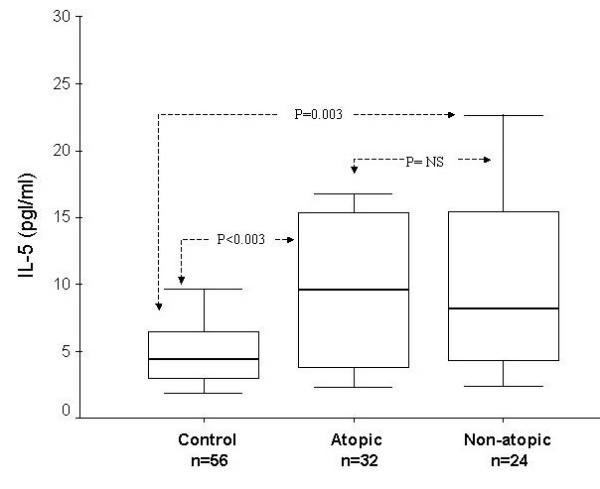
Table 1 shows the age, sex and pertinent laboratory values among study subjects. Of the fifty-six asthmatics, 32 were atopic and 24 non-atopic. Asthma severity was mild-intermittent in 4 patients, mild-persistent in 38 and moderate-persistent in 14. As there were only 4 mild-intermittent patients, they were combined with the mild-persistent group for statistical analysis. Of the 56 asthmatics, 25 reported regular use of inhaled glucocorticoids. Figure 1 shows the median and inter-quartile range (IQR) values of serum IL-5 levels in asthmatics and normal controls. The median serum IL-5 levels varied significantly between atopic, non-atopic asthmatics and controls (p < 0.001). The serum IL-5 levels (median, IQR) in atopic asthmatics (9.57 pg/ml, 3.74–15.64) and in non-atopic asthmatics (8.17 pg/ml, 4.21–15.47) were significantly higher compared to normal controls (4.4 pg/ml, 2.99–6.51) (both p = 0.003). However, the median serum IL-5 levels in atopic and non-atopic asthmatics were not significantly different.
Table 1.
Clinical & laboratory parameters in study subjects
| Normal Control (n = 56) | Asthmatics (n = 56) | P value | ||
| On Inhaled Steroids (n = 31) | Not on Inhaled Steroids (n = 25) | |||
| Age (years) | 27 (22–39) | 27 (23–38) | 28 (21–40) | 0.6* |
| Duration of asthma (years) | - | 5 (1–15) | 10 (4.5–14.5) | 0.18* |
| Atopic asthma | - | 16 | 16 | 0.42 † |
| Non-atopic asthma | - | 9 | 15 | |
| FEV1 (liter) | - | 2.3 (2–2.67) | 2 (1.7–2.5) | 0.2* |
| WBC count (× 109/L) | 6.5 (5.1–7.5) | 6.4 (5.1–8.3) | 6 (5.1–8.1) | 0.8* |
| Eosinophils % | 0.11(0.07–0.18) | 5 (2–9) | 6 (4–10) | 0.2* |
| Serum IgE (IU/L) | 26 (10–62) | 240 (54–595) | 200 (96–612) | 0.8* |
Data are expressed as medians with interquartile range in parenthesis * By Mann Whitney test between steroid and non-steroid users † By Fisher's exact test between steroid and non-steroid users
Figure 1.

Serum IL-5 levels according to the type of asthma. The box & whisker plot shows the median and interquartile values of serum IL-5 levels. Although the median serum Il-5 value in atopic and non-atopic groups were significantly higher compared to normal controls, there was no significant difference in the median serum IL-5 level between atopic and non-atopic groups
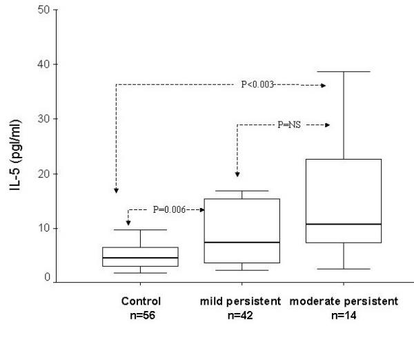
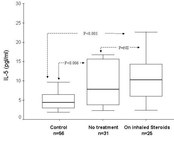
Figure 2 illustrates serum IL-5 levels in mild-persistent and moderate-persistent asthmatics and controls. Of the 42 mild persistent asthmatics, 22 were atopic and 20 non-atopic. In the moderate persistent group 10 were atopics and 4 were non-atopic. The median serum IL-5 level was insignificantly higher in fourteen moderate persistent asthmatics (10.61 pg/ml, 7.34 – 26.6) compared to forty-two mild persistent asthmatics (7.3 pg/ml, 3.56 – 15.41) (p = 0.13). Whereas median serum IL-5 levels in mild persistent asthmatics and moderate-persistent asthmatics were significantly higher than in controls (both p= 0.003). Figure 3 shows serum IL-5 levels in asthmatics using regular inhaled glucocorticoids, in those not using inhaled glucocorticoids and in normal controls. The median serum IL-5 levels in asthmatics using regular inhaled steroids (10.29 pg/ml, 4.96–14.55) was not significantly different compared to those not using inhaled steroids (7.81 pg/ml, 3.69–15.92). However, the serum IL-5 levels were significantly higher in asthmatics on inhaled steroids and not on inhaled steroids compared with controls (4.43 pg/ml, 2.99–6.51, p < 0.003 and < 0.006 respectively).
Figure 2.

Serum IL-5 levels according to the severity of asthma. The box & whisker plot shows the median and interquartile values of serum IL-5 levels. The median serum IL-5 value in mild persistent and moderate persistent groups were significantly higher compared to normal controls(both p < 0.001 and < 0.002). There was a trend for the median serum IL-5 to be higher in moderate persistent asthma than in mild persistent type of asthma (p = 0.13)
Figure 3.

Serum IL-5 levels according to the treatment status. The box & whisker plot shows the median and interquartile values of serum IL-5 levels. The median serum Il-5 values in patients on regular inhaled glucocorticoids and not on regular inhaled glucocorticoids were significantly higher compared to normal controls (p < 0.001 and < 0.002). There was no significant difference in the median serum IL-5 levels between asthmatics using and not using inhaled glucocorticoids (p = 0.69).
There was no significant correlation between the duration of asthma in years and levels of serum IL-5 in asthmatics (r = 0.04). Also, there was no significant correlation between serum IL-5 levels and absolute eosinophil counts, serum total IgE or FEV1 in asthmatics.
Discussion
IL-5 has been clearly implicated as an important cytokine mediating airway inflammation in both atopic and non-atopic types of asthma [12-14]. Elevated serum IL-5 levels in atopic and non-atopic asthmatics as compared with normal controls in the present study, support this hypothesis. Serum IL-5 levels in atopic and non-atopic asthmatics were not significantly different. Contrary to a previous study by Alexander and associates [15], we were able to detect serum IL-5 levels in both controls and asthmatic patients using an assay with increased sensitivity.
Whereas interleukin-5 levels are shown to be elevated in acute asthma, this is the first demonstration that levels are also elevated in less-severe asthmatics who represent the vast majority of patients. There was a tendency for serum IL-5 levels to be higher in moderate persistent asthmatics compared to mild persistent asthmatics. A previous study reported that systemic IL-5 levels were elevated in acute severe asthmatics but were significantly reduced following treatment with oral glucocorticoids. However, the serum IL-5 levels remained above levels observed in normal controls [8]. Our observation of raised serum IL-5 levels in a different and more common subgroup i.e. in mild and moderate persistent asthmatics using regular inhaled glucocorticoids, is consistent with this report.
Our finding, for the first time, that regular use of inhaled glucocorticoids fails to normalize serum interleukin-5 levels, might be seen to contradict a previous report by Kelly and associates reporting a significant reduction in serum IL-5 levels in atopic asthmatics on inhaled glucocorticoid treatment. However, our observations are consistent with their finding from the same study subjects that inhaled glucocorticoids did not reduce IL-5 production by peripheral blood mononuclear cells [10]; a similar lack of response of IL-5 production by peripheral blood monocytes to inhaled corticosteroids has been documented in asthmatic children [9]. We cannot entirely discount the possibility that inhaled glucocorticoids, in common with oral glucocorticoids do reduce serum IL-5 level, as sequential IL-5 level measurement was not possible in this cross sectional study. This issue can be best addressed by a longitudinal study.
Anti IL-5 but not anti-IgE has been shown to prevent airway inflammation [16]. Wang and associates have provided conclusive evidence using an animal model of asthma that the main determinant of airway inflammation is the level of circulating IL-5 and not the IL-5 produced within the airways [5]. The importance of circulating rather than airways IL-5 in the pathogenesis of bronchial hyper responsiveness in asthmatic patients has been documented by Van Rensen and associates [17]. Hakonarson and associates have shown that human airway smooth muscle passively sensitized by serum from atopic patients expresses receptors for IL-5. IL-5 has been shown to elicit a cholinergic type of hyper responsiveness in normal human bronchus and smooth muscle which could be blocked by IL-5 receptor antibodies [18]. Finally, administration of IL-5 antibody to a small number of asthmatics has been shown to significantly reduce blood and sputum eosinophil counts significantly [19]. Thus, the raised serum IL-5 levels observed in our patients with mild and moderate persistent asthma could have contributed to airway smooth muscle hyper responsiveness and airway inflammation. Although administration of inhaled glucocorticoids decreases airway inflammation [10,20], it does not suppress IL-5 production by peripheral blood mononuclear cells [10]. The inability to control or normalize the raised serum IL-5 levels by inhaled steroids as observed in our patients, is in concordance with this observation. Administration of agents that could control systemic Th2 responses may have a beneficial role in the long-term management of asthma. In this regard administration of antibodies against IL-5 [21,22] or IL-5 receptor [23], though debatable [24], may prove effective novel therapeutic approaches to prevent the effect of elevated systemic IL-5 levels in patients with atopic and non-atopic asthma.
Competing interests
None declared.
Authors' contributions
JJ, SB, WS, MJ were involved with the study conception. JJ, SB, and WS collected data. SB performed immunoassays. JJ, MJ, did statistical analysis. JJ, SB, WS, and MJ prepared the manuscript. All authors read and approved the final manuscript.
Pre-publication history
The pre-publication history for this paper can be accessed here:
Acknowledgments
Acknowledgements
The authors would like to thank Professor Gary Nicholls, Department of Internal Medicine, Faculty of Medicine and Health Sciences, UAE University for reviewing the manuscript and Dr Taoufik Zoubeidi PhD, Department of Statistics, UAEU for statistical analysis.
This study was supported by grant # MRG-12/1999-2000 from the Sheikh Hamdan Bin Rashid Al Maktoum Award for Medical Sciences, United Arab Emirates.
Contributor Information
Jose Joseph, Email: jjoseph@uaeu.ac.ae.
Sheela Benedict, Email: Sheela.Benedict@uaeu.ac.ae.
Wassef Safa, Email: wsafa@hotmail.com.
Maries Joseph, Email: mariesjoseph@yahoo.com.
References
- Ng Man Kwong G., Proctor A, Billings C, Duggan R, Das C, Whyte MK, Powell CV, Primhak R. Increasing prevalence of asthma diagnosis and symptoms in children is confined to mild symptoms. Thorax. 2001;56:312–314. doi: 10.1136/thorax.56.4.312. [DOI] [PMC free article] [PubMed] [Google Scholar]
- Ying S, Humbert M, Barkans J, Corrigan CJ, Pfister R, Menz G, Larche M, Robinson DS, Durham SR, Kay AB. Expression of IL-4 and IL-5 mRNA and protein product by CD4+ and CD8+ T cells, eosinophils, and mast cells in bronchial biopsies obtained from atopic and nonatopic (intrinsic) asthmatics. J Immunol. 1997;158:3539–3544. [PubMed] [Google Scholar]
- Hamelmann E, Gelfand EW. Role of IL-5 in the development of allergen-induced airway hyperresponsiveness. Int Arch Allergy Immunol. 1999;120:8–16. doi: 10.1159/000024215. [DOI] [PubMed] [Google Scholar]
- Robinson DS, Hamid Q, Ying S, Tsicopoulos A, Barkans J, Bentley AM, Corrigan C, Durham SR, Kay AB. Predominant TH2-like bronchoalveolar T-lymphocyte population in atopic asthma. N Engl J Med. 1992;326:298–304. doi: 10.1056/NEJM199201303260504. [DOI] [PubMed] [Google Scholar]
- Wang J, Palmer K, Lotvall J, Milan S, Lei XF, Matthaei KI, Gauldie J, Inman MD, Jordana M, Xing Z. Circulating, but not local lung, IL-5 is required for the development of antigen-induced airways eosinophilia. J Clin Invest. 1998;102:1132–1141. doi: 10.1172/JCI2686. [DOI] [PMC free article] [PubMed] [Google Scholar]
- Shiota Y, Arikita H, Horita N, Hiyama J, Ono T, Yamakido M. Intracellular IL-5 and T-lymphocyte subsets in atopic and nonatopic bronchial asthma. J Allergy Clin Immunol. 2002;109:294–298. doi: 10.1067/mai.2002.120757. [DOI] [PubMed] [Google Scholar]
- Wada K, Kaminuma O, Mori A, Nakata A, Ogawa K, Kikkawa H, Ikezawa K, Suko M, Okudaira H. IL-5-producing T cells that induce airway eosinophilia and hyperresponsiveness are suppressed by dexamethasone and cyclosporin A in mice. Int Arch Allergy Immunol. 1998;117 Suppl 1:24–27. doi: 10.1159/000053566. [DOI] [PubMed] [Google Scholar]
- Sahid El-Radhi A, Hogg CL, Bungre JK, Bush A, Corrigan CJ. Effect of oral glucocorticoid treatment on serum inflammatory markers in acute asthma. Arch Dis Child. 2000;83:158–162. doi: 10.1136/adc.83.2.158. [DOI] [PMC free article] [PubMed] [Google Scholar]
- Visser MJ, Postma DS, Brand PL, Arends LR, Duiverman EJ, Kauffman HF. Influence of different dosage schedules of inhaled fluticasone propionate on peripheral blood cytokine concentrations in childhood asthma. Clin Exp Allergy. 2002;32:1497–1503. doi: 10.1046/j.1365-2745.2002.01512.x. [DOI] [PubMed] [Google Scholar]
- Kelly EA, Busse WW, Jarjour NN. Inhaled budesonide decreases airway inflammatory response to allergen. Am J Respir Crit Care Med. 2000;162:883–890. doi: 10.1164/ajrccm.162.3.9910077. [DOI] [PubMed] [Google Scholar]
- National Heart Lung,and Blood Institute. Expert Panel Report 2: Guidelines for the Diagnosis and Management of Asthma. Betheda, MD, National Institute of Health; 1997. [Google Scholar]
- Virchow J.C.,Jr., Kroegel C, Walker C, Matthys H. Inflammatory determinants of asthma severity: mediator and cellular changes in bronchoalveolar lavage fluid of patients with severe asthma. J Allergy Clin Immunol. 1996;98:S27–S33. [PubMed] [Google Scholar]
- Shi HZ, Xiao CQ, Zhong D, Qin SM, Liu Y, Liang GR, Xu H, Chen YQ, Long XM, Xie ZF. Effect of inhaled interleukin-5 on airway hyperreactivity and eosinophilia in asthmatics. Am J Respir Crit Care Med. 1998;157:204–209. doi: 10.1164/ajrccm.157.1.9703027. [DOI] [PubMed] [Google Scholar]
- Hogan SP, Koskinen A, Foster PS. Interleukin-5 and eosinophils induce airway damage and bronchial hyperreactivity during allergic airway inflammation in BALB/c mice. Immunol Cell Biol. 1997;75:284–288. doi: 10.1038/icb.1997.43. [DOI] [PubMed] [Google Scholar]
- Alexander AG, Barkans J, Moqbel R, Barnes NC, Kay AB, Corrigan CJ. Serum interleukin 5 concentrations in atopic and non-atopic patients with glucocorticoid-dependent chronic severe asthma. Thorax. 1994;49:1231–1233. doi: 10.1136/thx.49.12.1231. [DOI] [PMC free article] [PubMed] [Google Scholar]
- Hamelmann E, Cieslewicz G, Schwarze J, Ishizuka T, Joetham A, Heusser C, Gelfand EW. Anti-interleukin 5 but not anti-IgE prevents airway inflammation and airway hyperresponsiveness. Am J Respir Crit Care Med. 1999;160:934–941. doi: 10.1164/ajrccm.160.3.9806029. [DOI] [PubMed] [Google Scholar]
- van Rensen EL, Stirling RG, Scheerens J, Staples K, Sterk PJ, Barnes PJ, Chung KF. Evidence for systemic rather than pulmonary effects of interleukin-5 administration in asthma. Thorax. 2001;56:935–940. doi: 10.1136/thorax.56.12.935. [DOI] [PMC free article] [PubMed] [Google Scholar]
- Hakonarson H, Maskeri N, Carter C, Grunstein MM. Regulation of TH1- and TH2-type cytokine expression and action in atopic asthmatic sensitized airway smooth muscle. J Clin Invest. 1999;103:1077–1087. doi: 10.1172/JCI5809. [DOI] [PMC free article] [PubMed] [Google Scholar]
- Leckie MJ, ten Brinke A, Khan J, Diamant Z, O'Connor BJ, Walls CM, Mathur AK, Cowley HC, Chung KF, Djukanovic R, Hansel TT, Holgate ST, Sterk PJ, Barnes PJ. Effects of an interleukin-5 blocking monoclonal antibody on eosinophils, airway hyper-responsiveness, and the late asthmatic response. Lancet. 2000;356:2144–2148. doi: 10.1016/S0140-6736(00)03496-6. [DOI] [PubMed] [Google Scholar]
- Hauber HP, Gotfried M, Newman K, Danda R, Servi RJ, Christodoulopoulos P, Hamid Q. Effect of HFA-flunisolide on peripheral lung inflammation in asthma. J Allergy Clin Immunol. 2003;112:58–63. doi: 10.1067/mai.2003.1612. [DOI] [PubMed] [Google Scholar]
- Hertz M, Mahalingam S, Dalum I, Klysner S, Mattes J, Neisig A, Mouritsen S, Foster PS, Gautam A. Active vaccination against IL-5 bypasses immunological tolerance and ameliorates experimental asthma. J Immunol. 2001;167:3792–3799. doi: 10.4049/jimmunol.167.7.3792. [DOI] [PubMed] [Google Scholar]
- Kips JC, O'Connor BJ, Langley SJ, Woodcock A, Kerstjens HA, Postma DS, Danzig M, Cuss F, Pauwels RA. Effect of SCH55700, a humanized anti-human interleukin-5 antibody, in severe persistent asthma: a pilot study. Am J Respir Crit Care Med. 2003;167:1655–1659. doi: 10.1164/rccm.200206-525OC. [DOI] [PubMed] [Google Scholar]
- Flood-Page PT, Menzies-Gow AN, Kay AB, Robinson DS. Eosinophil's role remains uncertain as anti-interleukin-5 only partially depletes numbers in asthmatic airway. Am J Respir Crit Care Med. 2003;167:199–204. doi: 10.1164/rccm.200208-789OC. [DOI] [PubMed] [Google Scholar]
- Kay AB, Menzies-Gow A. Eosinophils and interleukin-5: the debate continues. Am J Respir Crit Care Med. 2003;167:1586–1587. doi: 10.1164/rccm.2304001. [DOI] [PubMed] [Google Scholar]


