



Background: WISP2 is a cytosolic and secreted protein produced by precursor cells.
Results: Secreted, but not cytosolic, WISP2 activates canonical WNT and prevents adipogenic differentiation.
Conclusion: WISP2 is an important regulator of both adipogenic commitment and differentiation.
Significance: Secreted WISP2 is a novel regulator of canonical WNT and PPARγ activation.
Keywords: Adipose Tissue, Beta-catenin, Bone Morphogenetic Protein (BMP), Cell Biology, Obesity, Peroxisome Proliferator-activated receptor (PPAR), Wnt Pathway
Abstract
WNT1-inducible-signaling pathway protein 2 (WISP2) is primarily expressed in mesenchymal stem cells, fibroblasts, and adipogenic precursor cells. It is both a secreted and cytosolic protein, the latter regulating precursor cell adipogenic commitment and PPARγ induction by BMP4. To examine the effect of the secreted protein, we expressed a full-length and a truncated, non-secreted WISP2 in NIH3T3 fibroblasts. Secreted, but not truncated WISP2 activated the canonical WNT pathway with increased β-catenin levels, its nuclear targeting phosphorylation, and LRP5/6 phosphorylation. It also inhibited Pparg activation and the effect of secreted WISP2 was reversed by the WNT antagonist DICKKOPF-1. Differentiated 3T3-L1 adipose cells were also target cells where extracellular WISP2 activated the canonical WNT pathway, inhibited Pparg and associated adipose genes and, similar to WNT3a, promoted partial dedifferentiation of the cells and the induction of a myofibroblast phenotype with activation of markers of fibrosis. Thus, WISP2 exerts dual actions in mesenchymal precursor cells; secreted WISP2 activates canonical WNT and maintains the cells in an undifferentiated state, whereas cytosolic WISP2 regulates adipogenic commitment.
Introduction
The increasing incidence and prevalence of type 2 diabetes during the past 20 years are mainly due to the global epidemic of obesity. The subcutaneous adipose tissue, the largest adipose depot in man, has a limited ability to expand. When the limited extensibility of the subcutaneous fat to store and dispose of excess energy from the diet becomes insufficient, it will lead to fat accumulation in several ectopic depots, including the liver, with the induction of lipotoxicity and the well known metabolic complications of obesity (1–3).
Expansion of the subcutaneous adipose tissue due to excess energy can be accomplished in two different ways; either by expanding the existing adipocytes (hypertrophy) or by recruiting new cells (hyperplasia). Enlargement of the adipose cells (hypertrophic obesity), rather than recruitment of new cells (hyperplastic obesity), is associated with a dysregulated adipose tissue, inflammation, increased fibrosis, and local and systemic insulin resistance (4, 5). Hypertrophic obesity in man is also associated with an impaired ability to recruit new adipogenic precursor cells into the adipogenic lineage (3, 6). The process of multipotent mesenchymal stem cell commitment to the adipose lineage has been poorly understood, whereas adipogenic differentiation is under the control of peroxisome proliferator-activated receptor γ (PPARγ)2 and CCAAT/enhancer-binding protein (C/EBPα) (7).
A key pathway regulating early precursor cell commitment and adipogenesis is the canonical wingless-type murine mammary tumor virus (MMTV) integration site family (WNT). This extracellular pathway regulates cell proliferation, cell survival, and cell fate. Canonical WNT signaling and activation maintain precursor cell proliferation, prevent their entry into adipogenesis, and need to be inhibited to activate Pparg and C/ebpa (8–10).
Canonical WNT ligands bind to the Frizzled (FZD) and low-density lipoprotein receptor-related proteins (LRP) with inhibition of the down-stream degradation complex for β-catenin and stabilization of this molecule. Nuclear β-catenin binds to the transcription factors of T-cell-specific transcription factor/lymphoid enhancer-binding factor (Tcf/Lef) families with activation of several WNT target genes (9, 11).
In a recent extensive study of the ability of human adipogenic precursor cells to undergo differentiation, we provided evidence for an impaired ability of early precursor cells in the subcutaneous adipose tissue from individuals with hypertrophic obesity to initiate adipogenesis (6). Importantly, this was not due to lack of adipogenic precursor cells but to an inability to inhibit canonical WNT activation in these cells and to activate the expression of important secreted WNT antagonists, in particular, DICKKOPF-1. In further support of this concept, we found WNT1-inducible-signaling pathway protein 2 (WISP2), frequently used as a marker of canonical WNT activation (12), to be increased in the subcutaneous adipose tissue precursor cells and positively associated with insulin resistance and amount of ectopic fat accumulation (13). We also found WISP2 to be a secreted protein, highly expressed in mesenchymal stem cells, fibroblasts, and preadipocytes and adipogenic differentiation was associated with a marked reduction in Wisp2 expression, whereas differentiation was inhibited by extracellular WISP2. WISP2 was also recently identified in a proteomics analysis of the secretome of human adipose tissue (14) and can thus be considered a novel secreted adipokine. However, the overall regulation of Wisp2 expression is unclear although canonical WNT ligands can increase it (12, 13).
WISP2 (CCN5) is a member of the CCN family of connective tissue factors characterized by having IGFBP-, von Willebrand-, and thrombospondin-like domains. However, WISP2 differs from the other members by lacking the C-terminal knot, the role of which is unclear but is thought to be important for binding and cross-talk with the extracellular matrix (15).
WISP2 is both a protein secreted by mesenchymal precursor cells as well as highly expressed in the cytosol where it binds the PPARγ transcriptional activator ZFP423 (16), thus preventing its nuclear translocation (13). BMP4 promotes adipogenic commitment (17, 18) of mesenchymal precursor cells, and we found this to be a consequence of a BMP4-induced dissociation of the intracellular WISP2-ZFP423 complex, thereby releasing ZFP423 and allowing its nuclear translocation and induction of PPARγ (13). However, we also found that extracellular WISP2 inhibited adipogenic differentiation, but the molecular mechanism for this is unclear.
In the present study, we have characterized the signaling mechanisms for secreted WISP2 in mesenchymal cells by expressing both a full-length WISP2 as well as a truncated molecule that cannot be secreted. Surprisingly, we found that WISP2 is not only a marker but rather a mediator of canonical WNT activation as a secreted but not as an intracellular protein.
EXPERIMENTAL PROCEDURES
Cell Culture Conditions
NIH3T3 cells were cultured as described previously (13). 3T3-L1 preadipocytes were cultured and differentiated in DMEM supplemented with 10% fetal bovine serum (v/v), 2 mm glutamine, and antibiotics and differentiated as described previously (19). WISP2 (300 ng/ml) or WNT3a (50 ng/ml Abcam, Cambridge, UK) were added to the cell culture medium at day 8 of differentiation, and the cells were then harvested after days 0, 1, 2, 4, or 8 days.
RNA was isolated with the RNEasy kit (Qiagen Nordic, Solna, Sweden) and frozen at −20°. Proteins were isolated with lysis buffer as described (19).
Oil Red O Staining
To determine lipid accumulation, cells were cultured in six-well plates, fixed with 10% formalin for 20 min, and stained with Oil Red O for 60 min. After washing with PBS, the cells were placed under a microscope and photographed. Quantification was performed by dissolving the Oil Red O stain in 2-propanol and measured at λ 510 nm.
Reporter Gene Assays
NIH3T3 cells were seeded in 24-well plates at a density of 100 000 cells/well and transfected by Lipofectamine 2000 reagents according to the manufacturer's instructions (Invitrogen). FOPFLASH or TOPFLASH reporter plasmids were expressed (a kind gift from Dr. H Clevers, Utrecht, Holland), pCMV-RNL encoding Renilla luciferase as an internal control for transfection efficiency, WISP2-myc and constitutively active β-catenin S33Y. The total amount of plasmid was kept constant by supplementation with empty vector DNA. Luciferase and Renilla activity was determined with a luminometer after 48 h using a Dual-Luciferase reporter assay system and performed according to the manufacturer's instructions (Promega, Madison, WI).
Wisp2 Plasmids
To study the function of intracellular/extracellular WISP2, a mutant of Wisp2 cDNA (MUT-Wisp2.myc) lacking its N-terminal signal peptide sequence (corresponding 22 amino acids) was constructed. A Myc tag was fused to the C-terminal of the Wisp2 sequence. NIH3T3 cells were then transfected with either wild type Myc-tagged Wisp2 (WT-Wisp2.myc) or MUT -Wisp2.myc and/or Wisp2 siRNA using Lipofectamine 2000 and Lipofectamine RNAiMax.
To analyze the effect of IWP2 (a selective inhibitor of WNT palmitoylation (Sigma-Aldrich)) on WISP2 secretion, the Wisp2 cDNA was cloned into p3xFLAG-Myc-CMV 24 (Sigma-Aldrich). NIH3T3 cells were then transfected with this construct and treated with 2 μm IWP2 for 24 h. The conditioned medium was then collected and WISP2-immunoprecipitated with an anti-flag antibody (Sigma-Aldrich).
Immunofluorescence Staining
NIH3T3 cells were plated in four-chamber slides and treated with WISP2 (300 ng/ml) or WNT3a (50 ng/ml) for 16 and 48 h. Cells were fixed in 4% formaldehyde and permeabilized with 0.1% Triton X-100 in PBS. Samples were blocked in 20% goat serum and exposed to a mouse-anti β-catenin antibody (BD Biosciences) prepared in 5% goat serum. Secondary goat anti-mouse antibody conjugated to Alexa Fluor 488 (Invitrogen) was used to visualize proteins, and DAPI staining was used to visualize nuclei. Samples were analyzed with a Leica SP5 confocal microscope.
siRNA Transfection
Cells were transfected either with Wisp2 or control siRNA (Sigma-Aldrich) using Lipofectamine RNAiMAX (Invitrogen). After addition of different mediators including BMP4, DKK1 (R&D Systems), WISP2, and WNT3a, cells were harvested for RNA extraction followed by cDNA synthesis and Taqman gene analysis.
Real Time RT-PCR
Gene expression was analyzed with the ABI PRISM 7900HT sequence detection system (TaqMan, Applied Biosystems) using 18S ribosomal RNA as endogenous control (Applied Biosystems). Analyses were performed in duplicates, and all experiments were repeated two to six times. Expression levels were first normalized to 18 S rRNA and then normalized to the expression levels of the control sample for each time point.
Western Blots and Antibodies
Proteins were analyzed following electrophoresis through 4–12% SDS-polyacrylamide gels (PAGEr, Cambrex, Biosciences, Inc.) and immunoblotted and quantified essentially as described (19).
The following antibodies were used: α-SMA (sc-32251), axin (H-98) (sc-14029) (Santa Cruz Biotechnology, Heidelberg, Germany); LRP6 (AF1505) (R&D Systems, Inc.); β-catenin (C19220) (BD Biosciences); phospho-β-catenin (Ser-552) (D8E11), phospho-ERK1/2 phospho-p44/42 MAPK (Thr-202/Tyr-204) E10, phospho-p38 MAP kinase (The-180/Tyr-182), p38MAP Kinase (9212), phospho-LRP6 (Ser-1490) (Cell Signaling Technology), ERK1/2 (V803A, Promega Biotech AB, Stockholm, Sweden).
Statistical Analyses
The experimental data are presented as means ± S.E. A non-parametric Wilcoxon paired test was used to compare basal/untreated and WISP2/WNT3a-treated samples for n ≥ 6; otherwise, Student's t test was used. p < 0.05 was considered statistically significant.
RESULTS
We first characterized the signaling of full-length or truncated, non-secreted WISP2 in NIH3T3 fibroblasts and then examined if WISP2, as a protein secreted by undifferentiated mesenchymal cells, also was able to cross-talk with differentiated adipocytes.
Secreted, but Not Intracellular, WISP2 Is an Activator of Canonical WNT Signaling
Because we previously found WISP2 to be a powerful inhibitor of adipogenic differentiation of mesenchymal precursor cells, we first explored the cross-talk between WISP2 and the canonical WNT pathway. To address this, we silenced endogenous Wisp2 with siRNA in NIH3T3 fibroblasts and expressed a Myc-tagged full-length or a truncated WISP2 lacking the N-terminal signaling peptide that should prevent its secretion. As shown in Fig. 1A, full-length but not truncated WISP2 is secreted to the medium. Furthermore, similar to the canonical WNT ligand WNT 3a, secreted but not truncated WISP2 markedly increased β-catenin expression and the nuclear-targeting (Ser(P)-552) phosphorylation of β-catenin. Furthermore, full-length, but not truncated, WISP2 increased (Ser(P)LRP5/6 phosphorylation, an integral part of the canonical WNT pathway (Fig. 1B).
FIGURE 1.

WNT activation by WISP2 requires secretion of the ligand. A, full-length, but not the truncated WISP2 is secreted to the medium. Conditional medium from NIH3T3 cells transfected with wild-type Wisp2 (WT-Wisp2.myc) or mutated Wisp2 plasmid (MUT-Wisp2.myc), were collected followed by immunoprecipitation with anti-Myc antibody. B, full-length, but not the truncated WISP2, increases β-catenin and its nuclear targeting (Ser(P)-552) phosphorylation. Full-length WISP2 also increases the (Ser(P)-1490) phosphorylation of LRP6. NIH3T3 cells were transfected with wild-type Wisp2 (WT-Wisp2.myc) or mutated Wisp2- plasmid (MUT-Wisp2.myc) and/or Wisp2 siRNA. Cells were then incubated with or without WNT3A as shown for 48 h (n = 3). C, transcriptional activation of Tcf/Lef following addition of recombinant WISP2 (rec. WISP2), transfected Wisp2 (Wisp2.myc), or constitutively active β-catenin (β-catenin S33Y) in NIH-3T3 cells. Luciferase activity is normalized to that of the control samples (n ≥ 4). D, immunofluorescence staining of β-catenin (green) in the absence (left panel) or presence of recombinant WISP2 in NIH-3T3 cells for 48 h (right panel). DAPI staining (blue) for visualization of nuclear localization. E, the WNT antagonist DICKKOPF-1 (DKK1) reverses the inhibitory effect of recombinant WISP2 and WNT3A on activation of Pparg. F, Fabp4 in NIH3T3 cells incubated for 72 h with BMP4 and Wisp2 siRNA and DKK1 as shown (n = 3). G, Wisp2 siRNA, similar to adding DKK1, induces down-regulation of β-catenin protein, its nuclear targeting (Ser(P)-552) phosphorylation as well as (Ser(P)-1490) LRP6 phosphorylation. NIH3T3 cells were transfected with WT-Wisp2.myc or MUT-Wisp2.myc or Wisp2 siRNA. Cells were then incubated with or without DKK1 (200 ng/ml) as shown for 48 h (n = 2). ERK1/2 protein was used as a loading control. H, FLAG-tagged Wisp2-transfected NIH3T3 cells were incubated with the acylation inhibitor IWP2 (2 μm) for 24h. Medium was collected and immunoprecipitated with anti-FLAG antibody (n = 2). All data are means ± S.E. *, p < 0.05 and **, p < 0.01.
These results were further verified in a TopFlash reporter system where both added recombinant and expressing our Myc-tagged full-length WISP2 protein in the cells activated Tcf/Lef (Fig. 1C). Nuclear β-catenin expression was also markedly increased in NIH3T3 fibroblasts incubated with recombinant WISP2 (Fig. 1D).
To further validate the involvement of LRP5/6 and the WNT pathway, we characterized the ability of the secreted WNT antagonist DICKKOPF-1 (DKK1) to inhibit the effect of extracellular WISP2 on Pparg and Fabp4 activation following addition of BMP4 to NIH3T3 cells where endogenous Wisp2 was silenced with siRNA. We previously showed that inhibiting Wisp2 in fibroblasts promotes adipogenic differentiation and Pparg activation and this is dramatically increased by BMP4 (13). As shown in Fig. 1, E and F, adding DKK 1 markedly attenuated the ability of WISP2 to inhibit Pparg and Fabp4 activation in response to BMP4.
To verify the effect of WISP2 as an endogenous canonical WNT ligand in undifferentiated cells, we examined the effect of silencing endogenous Wisp2 and/or of adding DKK1 to NIH3T3 cells. As shown in Fig. 1G, total β-catenin, (Ser(P)-552) phosphorylation of β-catenin and (Ser(P)LRP5/6 were reduced by both silencing endogenous Wisp2 and by adding DKK1. Furthermore, the marked inhibitory effect of DKK1 was seen in cells overexpressing full-length WISP2 (Fig. 1G, right), whereas the effect in cells expressing truncated WISP2 was essentially similar to that seen in the control cells (Fig. 1G, left). Taken together, these findings show that WISP2, as a secreted, but not as an intracellular protein, activates canonical WNT, increases β-catenin and its nuclear targeting, and thereby maintains mesenchymal precursor cells undifferentiated.
This finding was quite surprising because WISP2 has no homology with the classical WNT ligands. Also, WNT ligands need to become acylated/palmitoylated to be secreted and bind to the Frizzled receptors (20, 21). To examine whether WISP2 was acylated as a secreted molecule, we expressed the full-length Myc-tagged molecule with and without a palmitoylation inhibitor (IWP-2) but WISP2 secretion to the medium was not altered (Fig. 1H). Thus, although WISP2 is capable of activating canonical WNT signaling, it differs in many respects from the conventional WNT ligands.
WISP2 Activates Canonical WNT in Differentiated 3T3-L1 Adipocytes
We then asked whether WISP2, as a protein secreted by undifferentiated mesenchymal cells in the adipose tissue, also can cross-talk with differentiated adipose cells by incubating fully differentiated 3T3-L1 adipocytes with recombinant WISP2 and, as a reference, the canonical WNT ligand WNT3a. This is conceptually important because WISP2, secreted by mesenchymal precursor cells, may then inhibit differentiation and lipid accumulation in both preadipocytes and adipocytes and, thereby favor the accumulation of lipids in ectopic sites in vivo.
To address whether secreted WISP2 is able to target differentiated adipose cells, we first examined the effect on cellular β-catenin and its (Ser(P)-552) phosphorylation as markers of canonical WNT activation. WISP2, similar to WNT3a, increased total β-catenin levels as well as (Ser(P)-552) phosphorylation after 24 h, and this remained at day 4 (Fig. 2A), supporting that both molecules can activate the canonical WNT pathway and promote nuclear translocation of β-catenin in adipocytes. However, the effect of WISP2 and WNT 3a to increase (Ser(P)-552) phosphorylation was primarily a consequence of the increased β-catenin levels because the (Ser(P)-552)/β-catenin protein ratio was essentially unchanged (Fig. 2A).
FIGURE 2.

WISP2 activates the canonical WNT pathway in 3T3-L1 adipose cells. A, WISP2 and WNT3A induce stabilization of β-catenin and its (Ser(P)-552) phosphorylation. WISP2 and WNT3A also activate and phosphorylate LRP6. ERK1/2 protein was used as a loading control. Quantification of β-catenin phosphorylation versus total β-catenin protein ratio and LRP6 phosphorylation versus total LRP6 protein ratio are shown. All proteins were normalized to ERK1/2 protein. B, WISP2 and WNT3A increase Axin2 mRNA level. Differentiated 3T3-L1 adipocytes were incubated with WISP2 or WNT3A as shown (n ≥ 6). Data are means ± S.E. *, p < 0.05 and **, p < 0.01. 1d, 1 day.
To further substantiate this effect of WISP2, we examined the downstream WNT signaling pathways. Both WISP2 and WNT3a increased serine phosphorylation of LRP6, without any change in LRP6 protein levels after 24 h, and this remained at day 4 (Fig. 2A).
AXIN is a negative regulator of canonical WNT and binds cytosolic β-catenin in the degradation complex. When LRP5/6 is phosphorylated, AXIN is recruited, thereby preventing β-catenin from being degraded in the complex and this also leads to the degradation of AXIN itself (22). WISP2, similar to WNT3a, induced a gradual down-regulation of AXIN protein (data not shown) and increased Axin2 mRNA levels after 24 h and at day 4 (Fig. 2B). Axin2 has several Tcf/Lef binding sites and is a target of canonical WNT signaling (23). Taken together, these results show that WISP2 activates canonical WNT signaling in both undifferentiated NIH3T3 fibroblasts and in differentiated 3T3-L1 adipose cells and, thus, can be considered an endogenous ligand of this pathway in mesenchymal cells.
Is Wisp2 Regulated by Canonical WNT?
There is cross-talk between Wisp2 and canonical WNT activation by other WNT ligands because Wisp2 is also increased by WNT3a and GSK3β inhibition (13). However, this effect is quite small (2–3-fold), and it is not clear whether the very high Wisp2 expression in undifferentiated mesenchymal cells (CT values ∼ 24–25) is a consequence of endogenous WNT activation by other ligands. It is indeed possible that WISP2 is a key endogenous activator of canonical WNT in these cells, as indicated by the data in Fig. 1G, rather than just a marker of the canonical WNT pathway. This concept is also supported by our previous findings that silencing Wisp2 in preadipocytes induces spontaneous differentiation and inhibits their proliferation (13).
To further explore the cross-talk between canonical WNT/β-catenin activation and WISP2, we examined Wisp2 mRNA levels in cells with known mutations in the β-catenin degradation complex, including the human colonic tumor cell line HT29, the breast tumor cell line MBA MB 231, and the liver tumor cell line HepG2. Interestingly, Wisp2 expression was very low in these cells (CT values, 36–40) which are under high endogenous WNT/β-catenin activation. In contrast, the breast tumor cells MCF7 had a high Wisp2 expression (CT values, 26–27) as also reported previously (24). However, these cells were cloned from the pleural effusion of a patient with breast cancer, and their origin is uncertain.
WISP2, Similar to WNT3a, Promotes Dedifferentiation of Mature Adipocytes
Because WISP2 activated the WNT pathway and inhibited Pparg, we asked whether fully differentiated 3T3-L1 adipocytes underwent dedifferentiation when exposed to this molecule. We therefore incubated fully differentiated adipose cells (∼90–95% with lipid droplets) with extracellular WISP2 or WNT3a for up to 8 days. As shown in Fig. 3A, both molecules induced a slow but gradual loss of lipid droplets in the cells measured as Oil Red O (p < 0.05 at day 6) suggesting a partial dedifferentiation of the cells. To further verify this, we examined the mRNA levels of key adipogenic genes after 1 and 4 days of culture with WISP2 or WNT3a. As shown in Fig. 3B, gene expression of the key transcription factors for adipogenesis, Pparg and c/ebpa, were both down-regulated after 24 h, and this remained at day 4. Furthermore, the important regulator of Pparγ transcriptional activation and adipogenic commitment by BMP4 (bone morphogenetic protein 4), zinc finger protein 423 (ZFP423) (16), was also reduced (Fig. 3B). Consistent with this, also other Pparg-regulated genes such as Glut4, adiponectin, Fabp4, and Lpl (7) were inhibited by both molecules (Fig. 3C). Taken together, these results show that the secreted adipokine WISP2, similar to the canonical WNT ligand WNT3a, is able to cross-talk with differentiated adipose cells to inhibit Pparg and the full terminal differentiation state of the cells.
FIGURE 3.

WISP2 induces partial dedifferentiation of mature adipocytes. A, micro photos (10× magnifications) from Oil Red O-stained 3T3-L1 adipocytes incubated with/without recombinant WISP2 or WNT3A for 6 days. Both WISP2 and WNT3A significantly decreased the lipid accumulation (n = 7). Right, quantification of Oil Red O. Incubations with WISP2 and WNT3A as shown also reduced mRNA levels of Pparg, Cebpa, and Zfp423 (B) as well as Glut4, adiponectin, Fabp4, and Lpl (C) (n = 7). D, WISP2 increases the phosphorylation of ERK1/2 and p38 MAPK. Immunoblotting was performed on lysates from 3T3-L1 adipocytes incubated with WISP2 or WNT3A as shown (n ≥ 6). Data are means ± S.E. *, p < 0.05. 1d, 1 day.
WISP2, Activates p38 and ERK MAPK
It is well established that canonical WNT ligands have a mitogenic effect in undifferentiated cells (11, 20, 25), and we have found this to also be true for both WNT3a and WISP2 in undifferentiated human and 3T3-L1 preadipocytes (13, 26). We therefore examined whether WISP2 activates MAPKs in 3T3-L1 adipose cells. JNKs (c-Jun N-terminal kinases) were not activated, whereas phosphorylation of both p38 and extracellular signal-regulated kinases (ERK) was increased after 1–4 days (Fig. 3D).
Both WISP2 and WNT3a Induce a Myofibroblast Phenotype and Activate α-SMA
It is well known that WNT activation induces alterations in fibroblasts toward a myofibroblast phenotype with expression of α-smooth muscle actin (α-SMA) and other markers of fibrosis (19, 27). Because the 3T3-L1 adipocytes were partially dedifferentiated, lost lipids, and exhibited markers of inhibited Pparγ following WISP2, we examined the effect on α-SMA induction as a marker of the myofibroblast phenotype. Both WISP2 and WNT3a increased α-SMA protein expression after 24 h, and this remained at day 4 (Fig. 4A). We also examined other genes known to be induced by canonical Wnt activation and, as shown in Fig. 4B, syndecan 4 (28), connective tissue growth factor (Ctgf) (29), and platelet-derived growth factor α (Pdgfa) (30) were all induced by both ligands further supporting activation of the WNT pathway.
FIGURE 4.

WISP2 induces a myofibroblast phenotype in 3T3-L1 adipose cells. A, α-SMA protein was increased by both WISP2 and WNT3A in the medium. ERK1/2 protein was used as a loading control and for normalization. B, Pdgfa, Syndecan-4, and Ctgf mRNA levels were increased following incubations with recombinant WISP2 or WNT3a as shown (n = 6). Data are means ± S.E. *, p < 0.05.
Taken together, our findings show that WISP2 is an autocrine secreted canonical WNT ligand maintaining mesenchymal precursor cells in an undifferentiated and proliferative state. Furthermore, extracellular WISP2 is also able to target differentiated 3T3-L1 adipose cells to inhibit Pparg and induce a partially dedifferentiated state favoring the myofibroblast phenotype. These data suggest that secreted WISP2 from mesenchymal precursor cells may also exert paracrine effects.
DISCUSSION
WISP2 regulates adipogenic precursor cell commitment by retaining the PPARγ transcriptional activator ZFP423 (16) in the cytosol and preventing its nuclear targeting (13). BMP4 dissociates this complex and allows nuclear entry of ZFP423 and Pparg induction. However, WISP2 has dual actions, and extracellular WISP2 is also able to directly inhibit Pparg activation through unknown signaling pathways (16). We here addressed the signaling pathway of secreted full-length WISP2 compared with a truncated molecule that cannot be secreted following deletion of the N-terminal signaling sequence.
Canonical WNT ligands (WNT3a) and GSK3β inhibition slightly (2–3-fold) increase Wisp2 mRNA levels in mesenchymal cells, but the detailed regulation of Wisp2 is largely unknown. In this context, it is interesting that cancer cell lines characterized by mutations in the β-catenin degradation complex (HT29 and MBA MB 231 cells) had a very low Wisp2 expression, suggesting that canonical WNT activation by itself is not the major regulator of Wisp2.
The Wisp2 promoter contains Tcf, Hif, and NFkB sequences, but much work needs to be done to understand its regulation. Our current understanding is that it is primarily expressed in fibroblasts, mesenchymal stemcells/precursor cells, and preadipocytes where it stimulates proliferation and prevents their commitment to the adipose lineage and subsequent differentiation. Indeed, our data suggest that WISP2 is an important secreted autocrine activator of canonical WNT in undifferentiated mesenchymal cells. The potential role of WISP2 in mesenchymal stem cell commitment and differentiation to other lineages is unknown but under investigation in our laboratory.
Our current findings shed new light on the role and molecular mechanisms of WISP2. Full-length WISP2 regulates adipogenic precursor cells through dual mechanisms; commitment is regulated by retaining the transcriptional PPARγ activator ZFP423 in the cytosol in a BMP4- and SMAD-regulated manner, whereas secreted WISP2 activates the canonical WNT pathway. The evidence for this includes direct activation of the Tcf receptor in reporter assays, phosphorylation and activation of the WNT/Frizzled co-receptor LRP5/6, and increased levels and nuclear targeting phosphorylation of β-catenin as also seen in immunofluorescence studies. Furthermore, the effect of WISP2 in inhibiting Pparg and Fabp4 activation in NIH3T3 fibroblasts in response to BMP4 was antagonized by the secreted WNT inhibitor DICKKOPF-1, which binds to LRP to prevent the activation of the WNT pathway (31). In addition, silencing Wisp2 in the undifferentiated NIH3T3 fibroblasts reduced endogenous WNT activation measured as β-catenin levels and phosphorylation of β-catenin and the WNT co-receptor LRP 5/6.
Interestingly, we also found differentiated 3T3-L1 adipose cells to be targets of WISP2, similar to WNT3a (19) leading to activation of the WNT pathway with inhibition of Pparg and partial dedifferentiation favoring a myofibroblast phenotype with increased expression of Ctgf, α-SMA, and other markers of WNT activation associated with fibrosis in vivo (5, 27).
It is somewhat surprising that fully differentiated adipose cells are still responsive to WNT activation because LRP is markedly down-regulated during adipogenic differentiation (32). It is unclear whether this is a consequence of using the 3T3-L1 cells and where not all cells may be synchronized and fully terminally differentiated. Mature primary adipose cells should also be tested. However, this does not change the conclusion that committed adipose cells, either fully differentiated or undergoing terminal differentiation, are target cells of WISP2 inhibiting Pparg and lipid storage and, thereby, favoring lipid accumulation in other sites, i.e. ectopic fat accumulation. Our previous finding (13) of a positive correlation between Wisp2 mRNA expression in human adipose tissue and intra-abdominal (visceral) ectopic fat and a negative correlation with insulin sensitivity is consistent with this concept. Furthermore, ability of highly differentiated cells to undergo dedifferentiation seems to be a common biological phenomenon and also includes β-cells as shown recently (33).
At present, we do not know which membrane receptor WISP2 binds to although a Frizzled receptor would seem a likely possibility since the Frizzled co-receptor LRP5/6 is phosphorylated. However, it has recently been shown that LRP5/6 is quite promiscuous and is a co-receptor for several other signaling pathways, including for TGFβ, CTGF, and PDGFα (34). Our data show that WISP2 does not require acylation for its secretion, whereas acylation is a prerequisite for both the secretion of canonical Wnt ligands as well as their ability to bind to the Frizzled receptors (21). Thus, WISP2 may directly bind to LRP 5/6 and/or activate the LRP5/6 co-receptor through other signaling pathways.
In conclusion, our data provide evidence for the concept that WISP2 is an endogenous autocrine WNT ligand, secreted by, and targeting mesenchymal precursor cells and maintaining them in an undifferentiated and proliferative state (schematically illustrated in Fig. 5). Furthermore, the data show that also adipose cells are target cells, thereby reducing their lipid storage capacity and favoring the accumulation of lipids in ectopic depots with lipid toxicity and its associated metabolic complications. Thus, WISP2 may play an important role in the development of the obesity-related metabolic complications and the metabolic syndrome. At present, it is important to understand the regulation of WISP2, its secretory pathway, and cellular receptor(s).
FIGURE 5.
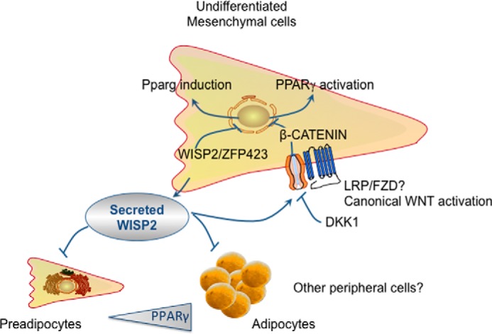
Schematic illustration of the autocrine and paracrine effects of WISP2. Adipogenic differentiation of mesenchymal precursor cells involves both commitments to the adipose lineage with Pparg induction as well as adipose cell differentiation following PPARγ activation. WISP2 is both an intracellular and a secreted protein by mesenchymal precursor cells. Intracellular WISP2 retains ZFP423, the transcriptional activator of Pparg, from entering the nucleus and initiate adipogenic commitment of the precursor cells. Secreted WISP2, in an autocrine manner, activates the canonical WNT pathway through an unknown cellular signaling pathway involving LRP5/6. This prevents PPARγ activation and maintains the precursor cells in an undifferentiated state. This effect of WISP2 is antagonized by the canonical WNT inhibitor DICKKOPF-1 (DKK1). Thus, WISP2 exerts dual effects in the regulation of adipogenesis. As a secreted protein, WISP2 can also target differentiated 3T3-L1 adipocytes, inhibit PPARγ activation, and promote a myofibroblast phenotype. WISP2 may also target other peripheral cells, but this remains to be demonstrated.
This work was supported by grants from the Swedish Medical Research Council (K2013-54X-03506-42-5), the Swedish ALF funds, the Torsten Söderbergs Foundation, the O.E and Edla Johansson Foundation, the Fredrik and Ingrid Thuring Foundation, the Wilhelm and Martina Lundgren Foundation, the Edgar Sjölund Foundation, and the Novo Nordisk Foundation.
- PPARγ
- peroxisome proliferator-activated receptor γ
- WISP2
- WNT1-inducible-signaling pathway protein2
- LRP
- lipoprotein receptor-related proteins
- ERK
- extracellular signal-regulated kinase(s)
- α-SMA
- α-smooth muscle actin.
REFERENCES
- 1. Després J. P., Lemieux I., Bergeron J., Pibarot P., Mathieu P., Larose E., Rodés-Cabau J., Bertrand O. F., Poirier P. (2008) Abdominal obesity and the metabolic syndrome: contribution to global cardiometabolic risk. Arterioscler. Thromb. Vasc. Biol. 28, 1039–1049 [DOI] [PubMed] [Google Scholar]
- 2. Virtue S., Vidal-Puig A. (2010) Adipose tissue expandability, lipotoxicity and the metabolic syndrome–an allostatic perspective. Biochim. Biophys. Acta 1801, 338–349 [DOI] [PubMed] [Google Scholar]
- 3. Gustafson B., Hammarstedt A., Hedjazifar S., Smith U. (2013) Restricted adipogenesis in hypertrophic obesity: the role of WISP2, WNT, and BMP4. Diabetes 62, 2997–3004 [DOI] [PMC free article] [PubMed] [Google Scholar]
- 4. Weisberg S. P., McCann D., Desai M., Rosenbaum M., Leibel R. L., Ferrante A. W., Jr. (2003) Obesity is associated with macrophage accumulation in adipose tissue. J. Clin. Invest. 112, 1796–1808 [DOI] [PMC free article] [PubMed] [Google Scholar]
- 5. Sun K., Kusminski C. M., Scherer P. E. (2011) Adipose tissue remodeling and obesity. J. Clin. Invest. 121, 2094–2101 [DOI] [PMC free article] [PubMed] [Google Scholar]
- 6. Gustafson B., Smith U. (2012) The WNT inhibitor Dickkopf 1 and bone morphogenetic protein 4 rescue adipogenesis in hypertrophic obesity in humans. Diabetes 61, 1217–1224 [DOI] [PMC free article] [PubMed] [Google Scholar]
- 7. Tontonoz P., Spiegelman B. M. (2008) Fat and beyond: the diverse biology of PPARγ. Ann. Rev. Biochem. 77, 289–312 [DOI] [PubMed] [Google Scholar]
- 8. Gustafson B., Gogg S., Hedjazifar S., Jenndahl L., Hammarstedt A., Smith U. (2009) Inflammation and impaired adipogenesis in hypertrophic obesity in man. Am. J. Physiol. Endocrinol. Metab. 297, E999–E1003 [DOI] [PubMed] [Google Scholar]
- 9. Laudes M. (2011) Role of WNT signalling in the determination of human mesenchymal stem cells into preadipocytes. J. Mol. Endocrinol. 46, R65–R72 [DOI] [PubMed] [Google Scholar]
- 10. Rosen E. D., MacDougald O. A. (2006) Adipocyte differentiation from the inside out. Nat. Rev. Mol. Cell Biol. 7, 885–896 [DOI] [PubMed] [Google Scholar]
- 11. Nusse R. (2008) Wnt signaling and stem cell control. Cell Res. 18, 523–527 [DOI] [PubMed] [Google Scholar]
- 12. Waki H., Park K. W., Mitro N., Pei L., Damoiseaux R., Wilpitz D. C., Reue K., Saez E., Tontonoz P. (2007) The small molecule harmine is an antidiabetic cell-type-specific regulator of PPARγ expression. Cell Metab. 5, 357–370 [DOI] [PubMed] [Google Scholar]
- 13. Hammarstedt A., Hedjazifar S., Jenndahl L., Gogg S., Grünberg J., Gustafson B., Klimcakova E., Stich V., Langin D., Laakso M., Smith U. (2013) WISP2 regulates preadipocyte commitment and PPARγ activation by BMP4. Proc. Natl. Acad. Sci. U.S.A. 110, 2563–2568 [DOI] [PMC free article] [PubMed] [Google Scholar]
- 14. Dahlman I., Elsen M., Tennagels N., Korn M., Brockmann B., Sell H., Eckel J., Arner P. (2012) Functional annotation of the human fat cell secretome. Arch. Physiol. Biochem. 118, 84–91 [DOI] [PubMed] [Google Scholar]
- 15. Perbal B. (2013) CCN proteins: A centralized communication network. J. Cell Commun. Signal. 7, 169–177 [DOI] [PMC free article] [PubMed] [Google Scholar]
- 16. Gupta R. K., Arany Z., Seale P., Mepani R. J., Ye L., Conroe H. M., Roby Y. A., Kulaga H., Reed R. R., Spiegelman B. M. (2010) Transcriptional control of preadipocyte determination by Zfp423. Nature 464, 619–623 [DOI] [PMC free article] [PubMed] [Google Scholar]
- 17. Huang H., Song T. J., Li X., Hu L., He Q., Liu M., Lane M. D., Tang Q. Q. (2009) BMP signaling pathway is required for commitment of C3H10T1/2 pluripotent stem cells to the adipocyte lineage. Proc. Natl. Acad. Sci. U.S.A. 106, 12670–12675 [DOI] [PMC free article] [PubMed] [Google Scholar]
- 18. Tang Q. Q., Otto T. C., Lane M. D. (2004) Commitment of C3H10T1/2 pluripotent stem cells to the adipocyte lineage. Proc. Natl. Acad. Sci. U.S.A. 101, 9607–9611 [DOI] [PMC free article] [PubMed] [Google Scholar]
- 19. Gustafson B., Smith U. (2010) Activation of canonical wingless-type MMTV integration site family (Wnt) signaling in mature adipocytes increases β-catenin levels and leads to cell dedifferentiation and insulin resistance. J. Biol. Chem. 285, 14031–14041 [DOI] [PMC free article] [PubMed] [Google Scholar]
- 20. Willert K., Nusse R. (2012) Wnt proteins. Cold Spring Harb. Perspect. Biol. 4, a007864. [DOI] [PMC free article] [PubMed] [Google Scholar]
- 21. Janda C. Y., Waghray D., Levin A. M., Thomas C., Garcia K. C. (2012) Structural basis of Wnt recognition by Frizzled. Science 337, 59–64 [DOI] [PMC free article] [PubMed] [Google Scholar]
- 22. Niehrs C., Shen J. (2010) Regulation of Lrp6 phosphorylation. Cell. Mol. Life Sci. 67, 2551–2562 [DOI] [PMC free article] [PubMed] [Google Scholar]
- 23. Jho E. H., Zhang T., Domon C., Joo C. K., Freund J. N., Costantini F. (2002) Wnt/β-catenin/Tcf signaling induces the transcription of Axin2, a negative regulator of the signaling pathway. Mol. Cell. Biol. 22, 1172–1183 [DOI] [PMC free article] [PubMed] [Google Scholar]
- 24. Fritah A., Saucier C., De Wever O., Bracke M., Bièche I., Lidereau R., Gespach C., Drouot S., Redeuilh G., Sabbah M. (2008) Role of WISP-2/CCN5 in the maintenance of a differentiated and noninvasive phenotype in human breast cancer cells. Mol. Cell. Biol. 28, 1114–1123 [DOI] [PMC free article] [PubMed] [Google Scholar]
- 25. Červenka I., Wolf J., Mašek J., Krejci P., Wilcox W. R., Kozubík A., Schulte G., Gutkind J. S., Bryja V. (2011) Mitogen-activated protein kinases promote WNT/β-catenin signaling via phosphorylation of LRP6. Mol. Cell. Biol. 31, 179–189 [DOI] [PMC free article] [PubMed] [Google Scholar]
- 26. Isakson P., Hammarstedt A., Gustafson B., Smith U. (2009) Impaired preadipocyte differentiation in human abdominal obesity: role of Wnt, tumor necrosis factor-α, and inflammation. Diabetes 58, 1550–1557 [DOI] [PMC free article] [PubMed] [Google Scholar]
- 27. Wei J., Melichian D., Komura K., Hinchcliff M., Lam A. P., Lafyatis R., Gottardi C. J., MacDougald O. A., Varga J. (2011) Canonical Wnt signaling induces skin fibrosis and subcutaneous lipoatrophy: a novel mouse model for scleroderma? Arthritis Rheum. 63, 1707–1717 [DOI] [PMC free article] [PubMed] [Google Scholar]
- 28. Ohkawara B., Glinka A., Niehrs C. (2011) Rspo3 binds syndecan 4 and induces Wnt/PCP signaling via clathrin-mediated endocytosis to promote morphogenesis. Dev. Cell 20, 303–314 [DOI] [PubMed] [Google Scholar]
- 29. Luo Q., Kang Q., Si W., Jiang W., Park J. K., Peng Y., Li X., Luu H. H., Luo J., Montag A. G., Haydon R. C., He T. C. (2004) Connective tissue growth factor (CTGF) is regulated by Wnt and bone morphogenetic proteins signaling in osteoblast differentiation of mesenchymal stem cells. J. Biol. Chem. 279, 55958–55968 [DOI] [PubMed] [Google Scholar]
- 30. He X. (2006) Unwinding a path to nuclear β-catenin. Cell 127, 40–42 [DOI] [PubMed] [Google Scholar]
- 31. Cruciat C. M., Niehrs C. (2013) Secreted and transmembrane wnt inhibitors and activators. Cold Spring Harb. Perspect. Biol. 5, a015081. [DOI] [PMC free article] [PubMed] [Google Scholar]
- 32. Gustafson B., Smith U. (2006) Cytokines promote Wnt signaling and inflammation and impair the normal differentiation and lipid accumulation in 3T3-L1 preadipocytes. J. Biol. Chem. 281, 9507–9516 [DOI] [PubMed] [Google Scholar]
- 33. Talchai C., Xuan S., Lin H. V., Sussel L., Accili D. (2012) Pancreatic β cell dedifferentiation as a mechanism of diabetic β cell failure. Cell 150, 1223–1234 [DOI] [PMC free article] [PubMed] [Google Scholar]
- 34. Ren S., Johnson B. G., Kida Y., Ip C., Davidson K. C., Lin S. L., Kobayashi A., Lang R. A., Hadjantonakis A. K., Moon R. T., Duffield J. S. (2013) LRP-6 is a coreceptor for multiple fibrogenic signaling pathways in pericytes and myofibroblasts that are inhibited by DKK-1. Proc. Natl. Acad. Sci. U.S.A. 110, 1440–1445 [DOI] [PMC free article] [PubMed] [Google Scholar]