
Figure 4.
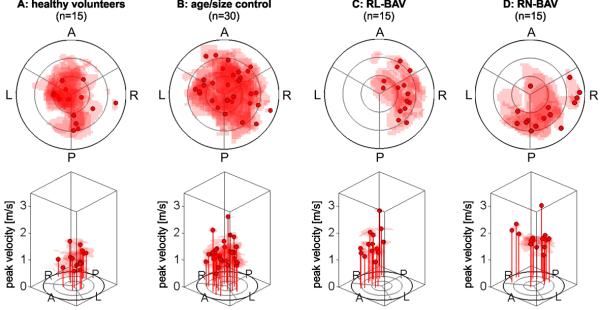
Flow profile asymmetry maps schematically illustrating flow eccentricity using the locations of the upper 15% of systolic velocities for all participants within each cohort at the the sinotubular junction (analysis plane S1). RN-BAV patients showed outflow asymmetry towards the right posterior wall compared to RL-BAV, whose flow profile was directed towards the right-wall. (“A” anterior, “L” left, “P” posterior, “R” right). Note that each subject’s profile map was normalized to their sinotubular junction diameter.