

Abstract
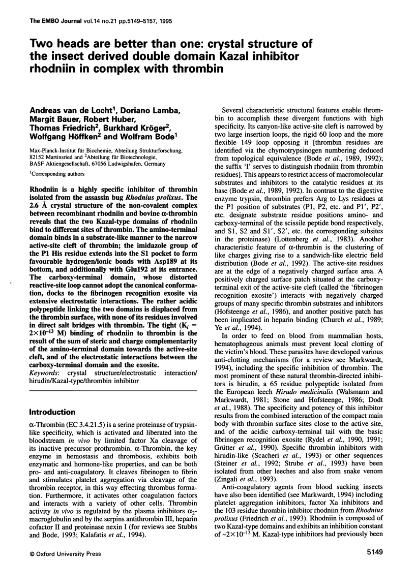
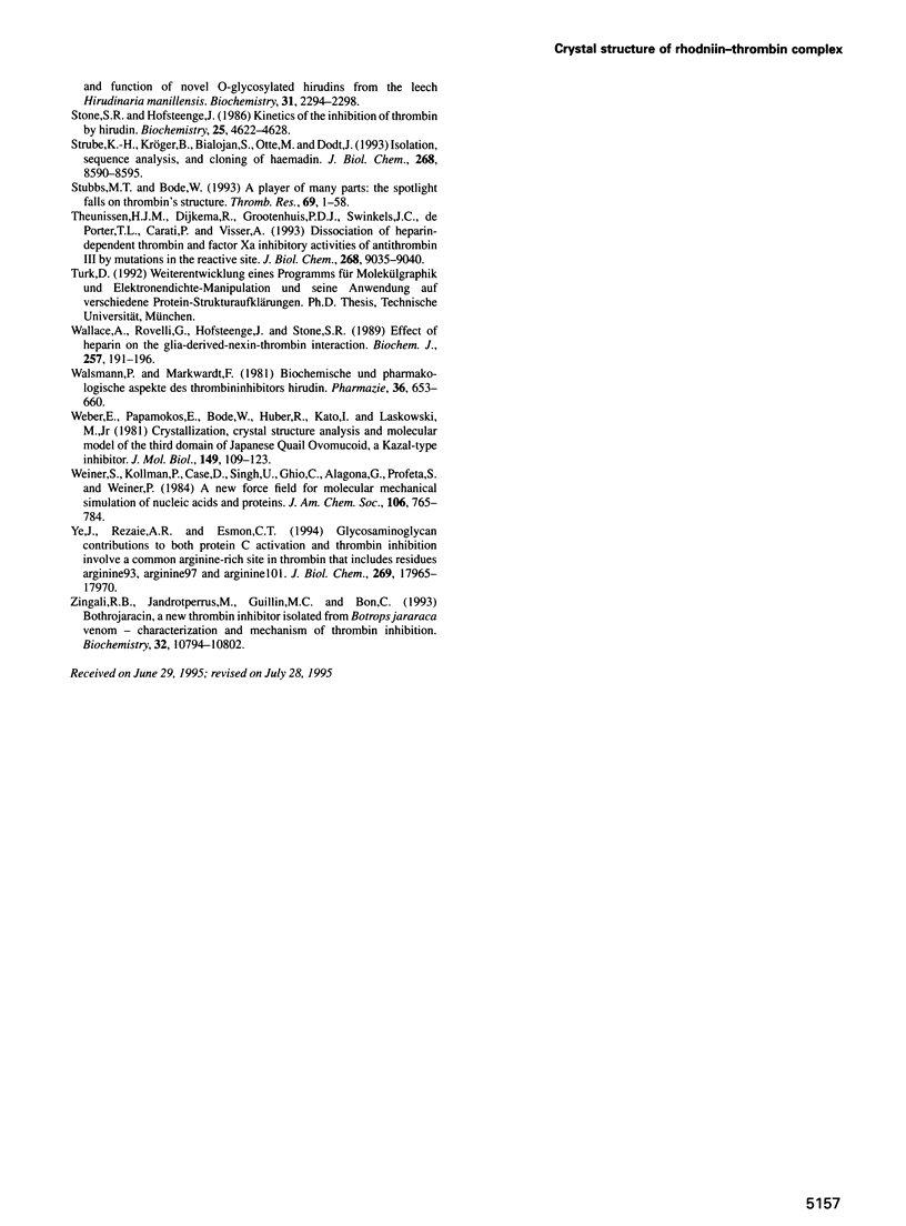
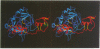
Rhodniin is a highly specific inhibitor of thrombin isolated from the assassin bug Rhodnius prolixus. The 2.6 Angstrum crystal structure of the non-covalent complex between recombinant rhodniin and bovine alpha-thrombin reveals that the two Kazal-type domains of rhodniin bind to different sites of thrombin. The amino-terminal domain binds in a substrate-like manner to the narrow active-site cleft of thrombin; the imidazole group of the P1 His residue extends into the S1 pocket to form favourable hydrogen/ionic bonds with Asp189 at its bottom, and additionally with Glu192 at its entrance. The carboxy-terminal domain, whose distorted reactive-site loop cannot adopt the canonical conformation, docks to the fibrinogen recognition exosite via extensive electrostatic interactions. The rather acidic polypeptide linking the two domains is displaced from the thrombin surface, with none of its residues involved in direct salt bridges with thrombin. The tight (Ki = 2 x 10(-13) M) binding of rhodniin to thrombin is the result of the sum of steric and charge complementarity of the amino-terminal domain towards the active-site cleft, and of the electrostatic interactions between the carboxy-terminal domain and the exosite.
Full text
PDF
Images in this article
Selected References
These references are in PubMed. This may not be the complete list of references from this article.
- Barton G. J. ALSCRIPT: a tool to format multiple sequence alignments. Protein Eng. 1993 Jan;6(1):37–40. doi: 10.1093/protein/6.1.37. [DOI] [PubMed] [Google Scholar]
- Bode W., Huber R. Natural protein proteinase inhibitors and their interaction with proteinases. Eur J Biochem. 1992 Mar 1;204(2):433–451. doi: 10.1111/j.1432-1033.1992.tb16654.x. [DOI] [PubMed] [Google Scholar]
- Bode W., Mayr I., Baumann U., Huber R., Stone S. R., Hofsteenge J. The refined 1.9 A crystal structure of human alpha-thrombin: interaction with D-Phe-Pro-Arg chloromethylketone and significance of the Tyr-Pro-Pro-Trp insertion segment. EMBO J. 1989 Nov;8(11):3467–3475. doi: 10.1002/j.1460-2075.1989.tb08511.x. [DOI] [PMC free article] [PubMed] [Google Scholar]
- Bode W., Turk D., Karshikov A. The refined 1.9-A X-ray crystal structure of D-Phe-Pro-Arg chloromethylketone-inhibited human alpha-thrombin: structure analysis, overall structure, electrostatic properties, detailed active-site geometry, and structure-function relationships. Protein Sci. 1992 Apr;1(4):426–471. doi: 10.1002/pro.5560010402. [DOI] [PMC free article] [PubMed] [Google Scholar]
- Bode W., Walter J., Huber R., Wenzel H. R., Tschesche H. The refined 2.2-A (0.22-nm) X-ray crystal structure of the ternary complex formed by bovine trypsinogen, valine-valine and the Arg15 analogue of bovine pancreatic trypsin inhibitor. Eur J Biochem. 1984 Oct 1;144(1):185–190. doi: 10.1111/j.1432-1033.1984.tb08447.x. [DOI] [PubMed] [Google Scholar]
- Bolognesi M., Gatti G., Menagatti E., Guarneri M., Marquart M., Papamokos E., Huber R. Three-dimensional structure of the complex between pancreatic secretory trypsin inhibitor (Kazal type) and trypsinogen at 1.8 A resolution. Structure solution, crystallographic refinement and preliminary structural interpretation. J Mol Biol. 1982 Dec 25;162(4):839–868. doi: 10.1016/0022-2836(82)90550-2. [DOI] [PubMed] [Google Scholar]
- Brandstetter H., Turk D., Hoeffken H. W., Grosse D., Stürzebecher J., Martin P. D., Edwards B. F., Bode W. Refined 2.3 A X-ray crystal structure of bovine thrombin complexes formed with the benzamidine and arginine-based thrombin inhibitors NAPAP, 4-TAPAP and MQPA. A starting point for improving antithrombotics. J Mol Biol. 1992 Aug 20;226(4):1085–1099. doi: 10.1016/0022-2836(92)91054-s. [DOI] [PubMed] [Google Scholar]
- Church F. C., Pratt C. W., Noyes C. M., Kalayanamit T., Sherrill G. B., Tobin R. B., Meade J. B. Structural and functional properties of human alpha-thrombin, phosphopyridoxylated alpha-thrombin, and gamma T-thrombin. Identification of lysyl residues in alpha-thrombin that are critical for heparin and fibrin(ogen) interactions. J Biol Chem. 1989 Nov 5;264(31):18419–18425. [PubMed] [Google Scholar]
- Dodt J., Köhler S., Baici A. Interaction of site specific hirudin variants with alpha-thrombin. FEBS Lett. 1988 Feb 29;229(1):87–90. doi: 10.1016/0014-5793(88)80803-2. [DOI] [PubMed] [Google Scholar]
- Evans S. V. SETOR: hardware-lighted three-dimensional solid model representations of macromolecules. J Mol Graph. 1993 Jun;11(2):134-8, 127-8. doi: 10.1016/0263-7855(93)87009-t. [DOI] [PubMed] [Google Scholar]
- Friedrich T., Kröger B., Bialojan S., Lemaire H. G., Höffken H. W., Reuschenbach P., Otte M., Dodt J. A Kazal-type inhibitor with thrombin specificity from Rhodnius prolixus. J Biol Chem. 1993 Aug 5;268(22):16216–16222. [PubMed] [Google Scholar]
- Grütter M. G., Priestle J. P., Rahuel J., Grossenbacher H., Bode W., Hofsteenge J., Stone S. R. Crystal structure of the thrombin-hirudin complex: a novel mode of serine protease inhibition. EMBO J. 1990 Aug;9(8):2361–2365. doi: 10.1002/j.1460-2075.1990.tb07410.x. [DOI] [PMC free article] [PubMed] [Google Scholar]
- Guinto E. R., Ye J., Le Bonniec B. F., Esmon C. T. Glu192-->Gln substitution in thrombin yields an enzyme that is effectively inhibited by bovine pancreatic trypsin inhibitor and tissue factor pathway inhibitor. J Biol Chem. 1994 Jul 15;269(28):18395–18400. [PubMed] [Google Scholar]
- Hofsteenge J., Taguchi H., Stone S. R. Effect of thrombomodulin on the kinetics of the interaction of thrombin with substrates and inhibitors. Biochem J. 1986 Jul 1;237(1):243–251. doi: 10.1042/bj2370243. [DOI] [PMC free article] [PubMed] [Google Scholar]
- Huber R., Kukla D., Bode W., Schwager P., Bartels K., Deisenhofer J., Steigemann W. Structure of the complex formed by bovine trypsin and bovine pancreatic trypsin inhibitor. II. Crystallographic refinement at 1.9 A resolution. J Mol Biol. 1974 Oct 15;89(1):73–101. doi: 10.1016/0022-2836(74)90163-6. [DOI] [PubMed] [Google Scholar]
- Jones T. A., Zou J. Y., Cowan S. W., Kjeldgaard M. Improved methods for building protein models in electron density maps and the location of errors in these models. Acta Crystallogr A. 1991 Mar 1;47(Pt 2):110–119. doi: 10.1107/s0108767390010224. [DOI] [PubMed] [Google Scholar]
- Kabsch W., Sander C. Dictionary of protein secondary structure: pattern recognition of hydrogen-bonded and geometrical features. Biopolymers. 1983 Dec;22(12):2577–2637. doi: 10.1002/bip.360221211. [DOI] [PubMed] [Google Scholar]
- Kalafatis M., Swords N. A., Rand M. D., Mann K. G. Membrane-dependent reactions in blood coagulation: role of the vitamin K-dependent enzyme complexes. Biochim Biophys Acta. 1994 Nov 29;1227(3):113–129. doi: 10.1016/0925-4439(94)90086-8. [DOI] [PubMed] [Google Scholar]
- Lane D. A., Olds R. J., Boisclair M., Chowdhury V., Thein S. L., Cooper D. N., Blajchman M., Perry D., Emmerich J., Aiach M. Antithrombin III mutation database: first update. For the Thrombin and its Inhibitors Subcommittee of the Scientific and Standardization Committee of the International Society on Thrombosis and Haemostasis. Thromb Haemost. 1993 Aug 2;70(2):361–369. [PubMed] [Google Scholar]
- Laskowski M., Jr, Apostol I., Ardelt W., Cook J., Giletto A., Kelly C. A., Lu W. Y., Park S. J., Qasim M. A., Whatley H. E. Amino acid sequences of ovomucoid third domain from 25 additional species of birds. J Protein Chem. 1990 Dec;9(6):715–725. doi: 10.1007/BF01024766. [DOI] [PubMed] [Google Scholar]
- Le Bonniec B. F., Esmon C. T. Glu-192----Gln substitution in thrombin mimics the catalytic switch induced by thrombomodulin. Proc Natl Acad Sci U S A. 1991 Aug 15;88(16):7371–7375. doi: 10.1073/pnas.88.16.7371. [DOI] [PMC free article] [PubMed] [Google Scholar]
- Le Bonniec B. F., MacGillivray R. T., Esmon C. T. Thrombin Glu-39 restricts the P'3 specificity to nonacidic residues. J Biol Chem. 1991 Jul 25;266(21):13796–13803. [PubMed] [Google Scholar]
- Lottenberg R., Hall J. A., Blinder M., Binder E. P., Jackson C. M. The action of thrombin on peptide p-nitroanilide substrates. Substrate selectivity and examination of hydrolysis under different reaction conditions. Biochim Biophys Acta. 1983 Feb 15;742(3):539–557. doi: 10.1016/0167-4838(83)90272-8. [DOI] [PubMed] [Google Scholar]
- Markwardt F. Coagulation inhibitors from blood-sucking animals. A new line of developing antithrombotic drugs. Pharmazie. 1994 May;49(5):313–316. [PubMed] [Google Scholar]
- Mathews I. I., Padmanabhan K. P., Ganesh V., Tulinsky A., Ishii M., Chen J., Turck C. W., Coughlin S. R., Fenton J. W., 2nd Crystallographic structures of thrombin complexed with thrombin receptor peptides: existence of expected and novel binding modes. Biochemistry. 1994 Mar 22;33(11):3266–3279. doi: 10.1021/bi00177a018. [DOI] [PubMed] [Google Scholar]
- Qiu X., Padmanabhan K. P., Carperos V. E., Tulinsky A., Kline T., Maraganore J. M., Fenton J. W., 2nd Structure of the hirulog 3-thrombin complex and nature of the S' subsites of substrates and inhibitors. Biochemistry. 1992 Dec 1;31(47):11689–11697. doi: 10.1021/bi00162a004. [DOI] [PubMed] [Google Scholar]
- Rydel T. J., Ravichandran K. G., Tulinsky A., Bode W., Huber R., Roitsch C., Fenton J. W., 2nd The structure of a complex of recombinant hirudin and human alpha-thrombin. Science. 1990 Jul 20;249(4966):277–280. doi: 10.1126/science.2374926. [DOI] [PubMed] [Google Scholar]
- Rydel T. J., Tulinsky A., Bode W., Huber R. Refined structure of the hirudin-thrombin complex. J Mol Biol. 1991 Sep 20;221(2):583–601. doi: 10.1016/0022-2836(91)80074-5. [DOI] [PubMed] [Google Scholar]
- Scacheri E., Nitti G., Valsasina B., Orsini G., Visco C., Ferrera M., Sawyer R. T., Sarmientos P. Novel hirudin variants from the leech Hirudinaria manillensis. Amino acid sequence, cDNA cloning and genomic organization. Eur J Biochem. 1993 May 15;214(1):295–304. doi: 10.1111/j.1432-1033.1993.tb17924.x. [DOI] [PubMed] [Google Scholar]
- Southan C., Lane D. A., Bode W., Henschen A. Thrombin-induced fibrinopeptide release from a fibrinogen variant (fibrinogen Sydney I) with an Aalpha Arg-16----His substitution. Eur J Biochem. 1985 Mar 15;147(3):593–600. doi: 10.1111/j.0014-2956.1985.00593.x. [DOI] [PubMed] [Google Scholar]
- Steiner V., Knecht R., Börnsen K. O., Gassmann E., Stone S. R., Raschdorf F., Schlaeppi J. M., Maschler R. Primary structure and function of novel O-glycosylated hirudins from the leech Hirudinaria manillensis. Biochemistry. 1992 Mar 3;31(8):2294–2298. doi: 10.1021/bi00123a012. [DOI] [PubMed] [Google Scholar]
- Stone S. R., Hofsteenge J. Kinetics of the inhibition of thrombin by hirudin. Biochemistry. 1986 Aug 12;25(16):4622–4628. doi: 10.1021/bi00364a025. [DOI] [PubMed] [Google Scholar]
- Strube K. H., Kröger B., Bialojan S., Otte M., Dodt J. Isolation, sequence analysis, and cloning of haemadin. An anticoagulant peptide from the Indian leech. J Biol Chem. 1993 Apr 25;268(12):8590–8595. [PubMed] [Google Scholar]
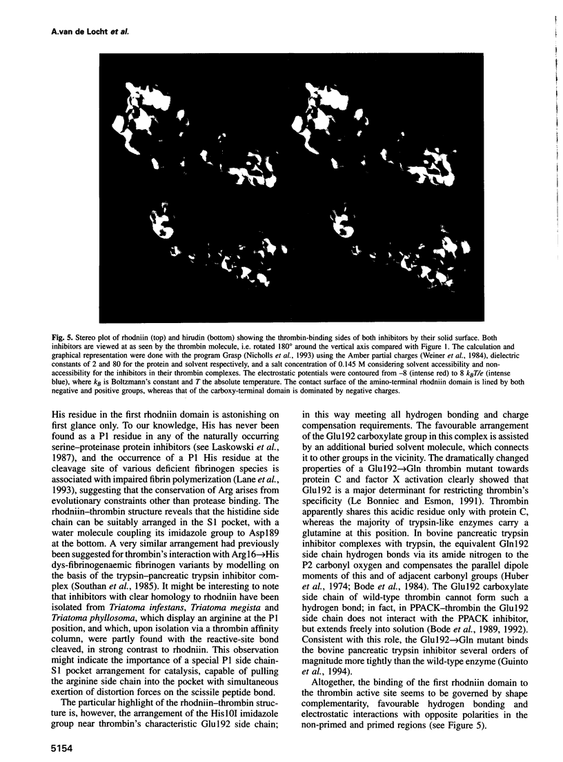
- Stubbs M. T., Bode W. A player of many parts: the spotlight falls on thrombin's structure. Thromb Res. 1993 Jan 1;69(1):1–58. doi: 10.1016/0049-3848(93)90002-6. [DOI] [PubMed] [Google Scholar]
- Theunissen H. J., Dijkema R., Grootenhuis P. D., Swinkels J. C., de Poorter T. L., Carati P., Visser A. Dissociation of heparin-dependent thrombin and factor Xa inhibitory activities of antithrombin-III by mutations in the reactive site. J Biol Chem. 1993 Apr 25;268(12):9035–9040. [PubMed] [Google Scholar]
- Wallace A., Rovelli G., Hofsteenge J., Stone S. R. Effect of heparin on the glia-derived-nexin-thrombin interaction. Biochem J. 1989 Jan 1;257(1):191–196. doi: 10.1042/bj2570191. [DOI] [PMC free article] [PubMed] [Google Scholar]
- Walsmann P., Markwardt F. Biochemische und pharmakologische Aspekte des Thrombininhibitors Hirudin. Pharmazie. 1981 Oct;36(10):653–660. [PubMed] [Google Scholar]
- Weber E., Papamokos E., Bode W., Huber R., Kato I., Laskowski M., Jr Crystallization, crystal structure analysis and molecular model of the third domain of Japanese quail ovomucoid, a Kazal type inhibitor. J Mol Biol. 1981 Jun 15;149(1):109–123. doi: 10.1016/0022-2836(81)90263-1. [DOI] [PubMed] [Google Scholar]
- Ye J., Rezaie A. R., Esmon C. T. Glycosaminoglycan contributions to both protein C activation and thrombin inhibition involve a common arginine-rich site in thrombin that includes residues arginine 93, 97, and 101. J Biol Chem. 1994 Jul 8;269(27):17965–17970. [PubMed] [Google Scholar]
- Zingali R. B., Jandrot-Perrus M., Guillin M. C., Bon C. Bothrojaracin, a new thrombin inhibitor isolated from Bothrops jararaca venom: characterization and mechanism of thrombin inhibition. Biochemistry. 1993 Oct 12;32(40):10794–10802. doi: 10.1021/bi00091a034. [DOI] [PubMed] [Google Scholar]