Abstract
Recent studies have uncovered novel mechanisms underlying the breakdown of periodontal host-microbe homeostasis, which can precipitate dysbiosis and periodontitis in susceptible hosts. Dysbiotic microbial communities of keystone pathogens and pathobionts are thought to exhibit synergistic virulence whereby not only can they endure the host response but can also thrive by exploiting tissue-destructive inflammation, which fuels a self-feeding cycle of escalating dysbiosis and inflammatory bone loss, potentially leading to tooth loss and systemic complications. Here we discuss new paradigms in our understanding of periodontitis, which may shed light into other polymicrobial inflammatory disorders. In addition, we highlight gaps in knowledge required for an integrated picture of the interplay between microbes and innate and adaptive immune elements that initiate and propagate chronic periodontal inflammation.
Periodontitis: an inflammatory dialogue when things get out of balance
Periodontitis is a biofilm-induced chronic inflammatory disease that leads to the destruction of the periodontium, i.e., the tooth-supporting structures such as the gingiva and the underlying alveolar bone [1]. The tooth-associated biofilm or dental plaque is required but not sufficient to induce periodontitis, as it is the host inflammatory response to this microbial challenge that ultimately can cause the destruction of the periodontium [1].
The latest epidemiological data in the U.S. have corroborated the high prevalence of periodontitis (>47% of adults) [2]. In addition to being a common cause of tooth loss, severe periodontitis (8.5% of adults [2]) can adversely affect systemic health, as it increases the patients’ risk for atherosclerosis, diabetes, rheumatoid arthritis, and adverse pregnancy outcomes [3-7]. Fossil evidence attests that periodontitis is an ancient disease that became more prevalent after the domestication of plants and animals in Neolithic societies. This is a time (≈10,000 years ago) when Porphyromonas gingivalis and other periodontitis-associated bacteria became more common than they were in hunter-gatherer societies, according to a recent sequencing project of ancient calcified dental plaque [8].
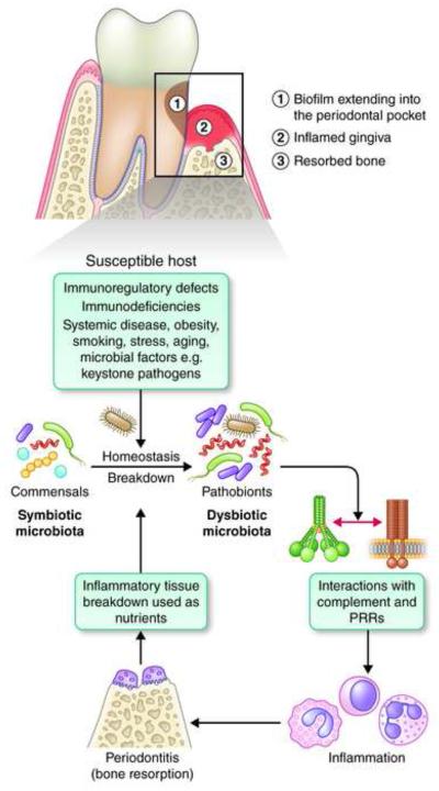
Early cultural analyses and current culture-independent molecular analyses of the periodontal microbiota have revealed profound ecological shifts in community structure associated with the transition from health to disease (reviewed in ref. [9]). Until relatively recently, the prevailing paradigm was that specific organisms were involved in the etiology of periodontitis, the more prominent being the ‘red complex’ bacteria, P. gingivalis, Treponema denticola, and Tannerella forsythia (reviewed in ref. [10]). This notion was in part fueled by the bias of culture-based methods to overestimate the importance of the easily grown species, such as P. gingivalis, which additionally could induce inflammatory bone loss in animal models. However, recent advances based on independent metagenomic and mechanistic approaches [11-18] collectively suggest that the pathogenesis of periodontitis involves polymicrobial synergy and dysbiosis (the ‘PSD model’) [10]. The dysbiosis of the periodontal microbiota signifies a change in the relative abundance of individual components of the bacterial community compared to their abundance in health, leading to alterations in the host-microbial crosstalk sufficient to mediate destructive inflammation and bone loss [18,19](Fig. 1).
Fig. 1. Polymicrobial synergy and dysbiosis in susceptible hosts causes periodontitis.
Periodontal health requires a controlled inflammatory state that can maintain host-microbe homeostasis in the periodontium. However, defects in the immuno-inflammatory status of the host or predisposing conditions and environmental factors (collectively defining a ‘susceptible host’) can shift the balance towards dysbiosis, a state in which former commensals behave as pro-inflammatory pathobionts. The presence of keystone pathogens can similarly tip the balance toward dysbiosis even in hosts without apparent predisposing genetic or environmental factors (at least in mice). The inflammation caused by the dysbiotic microbiota depends in great part on crosstalk signaling between complement and PRRs and has two major and interrelated effects: it causes inflammatory destruction of periodontal tissue (including bone loss, the hallmark of periodontitis) which in turn provides nutrients (tissue breakdown peptides and other products) that further promote dysbiosis and hence tissue destruction, thereby generating a self-perpetuating pathogenic cycle. It should be noted that host susceptibility might not simply be a determinant of the transition from a symbiotic to a dysbiotic microbiota but it may also underlie the predisposition of the host to develop inflammation sufficient to cause irreversible tissue damage. For instance, at least in principle, there might be individuals who can tolerate the conversion of a symbiotic microbiota into a dysbiotic state (such hosts would be susceptible to dysbiosis but not to periodontal bone loss).
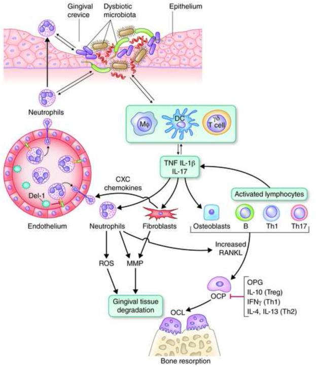
The late downstream events that activate osteoclasts to resorb alveolar bone are well established in both human and animal models and predominantly involve mechanisms dependent on RANKL [20] (Fig. 2), a member of the tumor necrosis factor cytokine family that is also implicated in rheumatoid arthritis [21]. However, much less is known about the associated initiating mechanisms. Indeed, it is only poorly understood how a dysbiotic microbiota induces deregulated or non-resolving periodontal inflammation that could eventually culminate in pathologic bone resorption. Similarly uncertain is how dysbiosis arises in the first place and whether dysbiosis is a cause or a consequence of the disease process. An even more formidable challenge is to understand the precise roles of innate and adaptive immune components in the inflammatory dialogue between the host and the microbiota in periodontitis. This Review does not aspire to give a definite answer to all these questions but rather to critically evaluate available literature and constructively synthesize it into working models that can guide productive future investigations. The basic premise or conceptual framework of this paper is that periodontitis, though undoubtedly an inflammatory disease, can be better understood mechanistically if it is seen as fundamentally a disruption of host-microbe homeostasis [1,10].
Fig. 2. Inflammatory mechanisms leading to bone loss in periodontitis.
Recruited neutrophils to the gingival crevice fail to control a dysbiotic microbiota, which can thus invade the connective tissue and interact with additional immune cell types, such as macrophages (Mφ), dendritic cells (DC), and γδ T cells, a subset of innate-like lymphocytes. These cells produce proinflammatory mediators (such as the bone-resorptive cytokines TNF, IL-1β, and IL-17) and also regulate the development of Th cell types, which also contribute to and exacerbate the inflammatory response. IL-17, a signature cytokine of Th17 (though also produced by innate cell sources), acts on innate immune and connective tissue cell types, such as neutrophils, fibroblasts, and osteoblasts. Through these interactions, IL-17 induces the production of CXC chemokines (which recruit neutrophils in a Del-1–dependent manner), matrix metalloproteinases (MMPs) and other tissue-destructive molecules (e.g., ROS), as well as osteoblast expression of RANKL, which drives the maturation of osteoclast precursors (OCP). Activated lymphocytes (B and T cells, specifically Th1 and Th17) play a major role in pathologic bone resorption through the same RANKL-dependent mechanism, whereas osteoprotegerin (OPG) is a soluble decoy receptor that inhibits the interaction of RANKL with its functional receptor (RANK) on OCP. The RANKL/OPG ratio increases with increasing inflammatory activity. Activated neutrophils express membrane-bound RANKL and could directly stimulate osteoclastogenesis if they are within sufficient proximity to the bone. The anti-inflammatory cytokine IL-10 (produced by Tregs), as well as IFNγ (produced by Th1 cells) and IL-4 plus IL-13 (produced by Th2 cells) can suppress osteoclastogenesis. The innate-adaptive cell interplay is considerably more complex than depicted here but serves to illustrate major destructive mechanisms operating in the context of unresolved periodontal infection and inflammation.
The microbial challenge: keystones and pathobionts
The gram-negative asaccharolytic bacterium P. gingivalis has long been associated with human periodontitis and its capacity to induce the disease in rodent or non-human primate models appeared to confirm its role as a causative organism [22]. However, the virulence credentials of P. gingivalis were more consistent with its being a manipulator of the host response [23] rather than a potent inducer of inflammation, an activity normally associated with a bacterium involved in an inflammatory disease [22]. This paradox was reconciled by a recent study that demonstrated the obligatory participation of the commensal microbiota in P. gingivalis-instigated inflammation and bone loss [14]. Specifically, by subverting innate immune signaling including the crosstalk between complement and Toll-like receptors (TLR) [23,24], P. gingivalis can impair host defenses in ways that alter the growth and development of the entire microbial community, thereby triggering a destructive change in the normally homeostatic relationship with the host [14]. Therefore, P. gingivalis orchestrates rather than directly causes inflammatory bone loss, which is largely mediated by pathobionts, i.e., commensals that under conditions of disrupted homeostasis have the potential to cause deregulated inflammation and disease [25] (Fig. 1).
P. gingivalis comprises <0.01% of the total bacterial count in experimental mouse periodontitis [14], consistent with its being a low-abundance constituent also in human periodontitis-associated biofilms [18]. The ability of the low-abundant P. gingivalis to instigate inflammatory disease through community-wide supportive effects has prompted its designation as a keystone pathogen, in analogy to the role of the literal keystone as the central supporting stone at the apex of an arch [14,22]. It should be noted that the terms ‘keystone pathogen’ and ‘pathobiont’ represent distinct concepts. Pathobionts are not necessarily low-abundance species and require hosts with specific genetic or environmental alterations (e.g., compromised immune system) to cause inflammatory pathology, whereas the virulence of a keystone pathogen is not necessarily reliant upon an already disrupted homeostasis. Since keystone pathogens can cause or contribute to homeostasis breakdown, pathobionts in principle act downstream of keystone species. Certain periodontal bacteria, such as T. denticola, T. forsythia, and Aggregatibacter actinomycetemcomitans are strongly associated with destructive inflammatory responses and additionally subvert the host response in ways that could, at least in principle, enhance the survival of also bystander species [1,26-28]. Therefore, although ‘keystone’ and ‘pathobiont’ are useful terms that can accurately describe the role of many disease-associated species, certain other bacteria may have mixed roles. For instance, T. denticola is a very minor component of the subgingival biofilm in periodontal health but it thrives to high abundance in diseased periodontal pockets, consistent with its being a pathobiont [28]. Nevertheless, its demonstrated capacity to manipulate the host response could contribute to homeostasis breakdown, similar to the role of a keystone pathogen [1,28]. Keystone or keystone-like pathogens appear to be involved also in other polymicrobial inflammatory diseases (e.g., inflammatory bowel disease) [19] and are considered as potentially important players in the gut ecosystem [29]. It is moreover thought that the elucidation of the complex interplay between the host and mucosal commensal bacteria in homeostatic and diseased states “will require a better understanding of the keystone bacterial species that control the immune responses at individual sites” [30].
Recent studies suggest that P. gingivalis could additionally modify the adaptive immune response. Specifically, the interaction of P. gingivalis with dendritic cells induces a cytokine pattern that favors T helper 17 (Th17) polarization at the expense of the Th1 lineage [31] (see Box 1 for T cell subsets). Moreover, P. gingivalis inhibits gingival epithelial cell production of Th1-recruiting chemokines [32] as well as T cell production of IFNγ [33]. It could thus be hypothesized that the keystone effects of P. gingivalis also include the manipulation of T cell development in ways that favor Th17-mediated inflammation (more below) in the absence of effective Th1-dependent cell-mediated immunity, which promotes immune clearance of P. gingivalis [23].
BOX 1. CD4+ T cell subsets and inflammatory disease.
On the basis of distinct cytokine production patterns and functions, CD4+ T cells can be classified into several subsets including the following (cytokines in parenthesis denote signature cytokines produced from the particular subset): 1) T helper type 1 or Th1 (IFN-γ); 2) Th2 (IL-4, IL-5, and IL-13); 3) Th17 (IL-17 and IL-22); and 4) T regulatory cells or Treg (IL-10 and transforming growth factor β [TGF-β]). Th1 cells are primarily responsible for cell-mediated immunity to intracellular pathogens (bacteria, protozoans, viruses), and have been implicated in delayed-type hypersensitivity and inflammatory diseases. Th2 cells mediate humoral immunity including production of IgE and activate mast cells, which mediate immune responses to helminths. This subset is implicated in allergic reactions. The differentiation of Th1 and Th2 populations are driven by IL-12 and IL-4, respectively. The key transcription factors driving their differentiation are T-bet (Th1) and GATA3 (Th2). The recently described Th17 subset mediates responses that reinforce neutrophil and innate immunity against extracellular bacteria and fungi. They are implicated in autoimmune and inflammatory diseases, some of which involve bone pathology. TGFβ, IL-6, IL-1, and IL-21 are important for the differentiation of Th17, whereas IL-23 is required for Th17 cell expansion and survival. The retinoic acid-related orphan receptor γt (RORγt) is the key transcription factor driving the differentiation of Th17 cells. CD4+ Foxp3+ regulatory T cells prevent excessive inflammation by suppressing effector functions of Th1, Th2, and Th17, in part through the production of IL-10 and TGF-β. The Th1/Th2 paradigm, established in the late 1980s, elegantly explained much about T cells and immunity, although in many cases diseases of immunological etiology were pigeonholed into one category or the other, often without adequate supportive evidence. The discovery of the Th17 subset has prompted a re-examination of the role of T cells in inflammatory diseases. For detailed information, the reader is referred to recent dedicated reviews [54,69].
If P. gingivalis is a conductor (rather than a musician) in the orchestration of inflammatory bone loss, it would be instructive to consider the credentials of the ‘musicians’ in this orchestra. Commensals or symbionts share similar microbe-associated molecular patterns (e.g., lipopolysaccharide, peptidoglycan, and lipoproteins) with pathogens. Notwithstanding their symbiotic relationship with the host, symbionts have therefore the potential to induce inflammation through activation of pattern-recognition receptors (PRRs). This notion is consistent with in vitro findings that TLR-dependent inflammatory responses can readily be induced by human dental plaque regardless of whether it is isolated from healthy or diseased sites [34]. In vivo, however, such potential pathobionts should, at a minimum, be able to withstand the harsh inflammatory environment of the periodontal pockets, if not to take advantage of it. A recent study has identified a pathobiont in the mouse oral cavity (designated NI1060), which selectively accumulates at damaged periodontal tissue, ostensibly to procure nutrients from inflammatory tissue breakdown components [15]. Moreover, NI1060 proactively causes destructive periodontal inflammation by activating the intracellular PRR Nod1. In contrast, other commensals, such as NI440 and NI968, are dominant only in healthy sites and do not behave as pathobionts [15].
The notion that at least some commensals can opportunistically mediate destructive inflammation is consistent with the emerging association with periodontitis of uncultivable or previously underappreciated bacteria, including the Gram-positive Filifactor alocis and Peptostreptococcus stomatis and other species from the genera Prevotella, Megasphaera, Selenomonas, and Desulfobulbus [16-18]. Many of these newly recognized organisms show at least as good a correlation with disease as do the classical red complex bacteria [17,18]. Although most of these species are as yet to be cultivated, the current investigation of the more tractable organisms has revealed virulence features consistent with a pathobiont status. For instance, F. alocis has shown remarkable potential to endure oxidative stress [35] and to cause strong proinflammatory responses [36]. Interestingly, as the bacterial biomass increases with increasing clinical periodontal inflammation, the ecological succession from health to disease is manifested as emergence of newly-dominant community members rather than appearance of novel species [18]. This important finding is consistent with the ecologic plaque hypothesis, which predicted that ‘periodontal pathogens’ are members of the normal microbiota but at levels too low to cause disease, whereas changes in ecologic conditions could favor the outgrowth of such organisms beyond a threshold sufficient to lead to periodontitis [37].
As alluded to above, inflammation is an important source of nutrients (especially for asaccharolytic bacteria that obligatorily rely on non-carbohydrate sources for energy) and, therefore, exerts a powerful influence in the composition of the periodontal microbiota favoring those species that can utilize tissue breakdown products (e.g., degraded proteins/peptides and hemin, a source of essential iron). Conversely, those species that cannot benefit from these environmental changes, or for which host inflammation is detrimental, may have a fitness disadvantage and hence be outcompeted [19]. The selective blooming of ‘inflammophilic’ bacteria, acting as pathobionts, has the potential to set off a self-feeding ‘vicious cycle’ for further tissue destruction and bacterial overgrowth (Fig. 1).
Host susceptibility to periodontitis
Despite its explanatory power for periodontal disease pathogenesis, the concept of keystone pathogens and pathobionts leaves a number of questions unanswered and, moreover, requires certain clarifications. For instance, the breakdown of tissue homeostasis leading to the blooming of inflammophilic pathobionts may not necessarily come from within the microbial community, i.e., by the action of keystone pathogens. The host-microbe homeostasis can also be disrupted by congenital or acquired host immunodeficiencies or immunoregulatory defects, systemic diseases such as diabetes, obesity, environmental factors, such as smoking, diet, and stress, and epigenetic modifications in response to environmental changes, which– alone or in combination– can contribute to unfavorable tipping of the homeostatic balance [38-42] (Fig. 1). Aging is another factor associated with a decline in immune regulation and function, which in turn can predispose to increased susceptibility to periodontitis [43,44].
Since P. gingivalis can also be detected (albeit with decreased frequency) in periodontally healthy individuals [18], a reasonable question is why the presence of P. gingivalis does not always lead to dysbiosis and periodontitis. A plausible hypothesis is that there may be non-susceptible individuals who can either resist or tolerate the conversion of the periodontal microbiota from a symbiotic to a dysbiotic state, by virtue of their intrinsic immuno-inflammatory status. The identification of host-related factors that determine one’s susceptibility to microbial immune subversion could provide useful insights, although such differential vulnerability could additionally or alternatively be explained by strain and virulence diversity within the population structure of the implicated bacteria [45].
Although dental plaque accumulation causes gingivitis (a reversible form of periodontal inflammation that does not involve the alveolar bone), gingivitis in turn does not necessarily lead to periodontitis, suggesting that stable gingivitis represents a protective host response [46]. In this regard, there are cases of individuals who remain free of periodontitis despite massive dental plaque accumulation [38,39]. In cases in which gingivitis can transition to periodontitis, it is likely that plaque accumulation causes gingival inflammation that can progressively exert selective pressure for the development of a dysbiotic and inflammophilic microbiota. This emerging community may include members that can subvert or evade the immune response (e.g., keystone pathogens), thereby contributing to the stabilization of a disease-provoking biofilm dominated by pathobionts. The severity of the ensuing periodontitis may depend, in great part, on host-related parameters (e.g., congenital, environmental, epigenetic, or age-related factors that influence the host’s immune, inflammatory, and regenerative responses).
Cellular and molecular mechanisms in periodontal inflammation and bone loss
One of the hallmarks of periodontitis is the massive accumulation of neutrophils, which can be found in the gingival connective tissue, the junctional epithelium, and especially in the periodontal pocket, where they constitute the overwhelming majority of recruited leukocytes [1,47,48]. The periodontal pocket represents a pathologically deepened gingival crevice (normally the space between the free gingiva and the tooth surface with the attached biofilm). The involvement of neutrophils in the pathogenesis of a chronic disease such as periodontitis may appear surprising, given that they are generally associated with the acute host response to infections. However, neutrophils play an increasingly acknowledged role in chronic inflammatory diseases, such as rheumatoid arthritis and psoriasis [49,50]. Moreover, it is uncertain whether the ‘chronic’ nature of periodontitis represents a constant pathologic process or a persistent series of brief acute insults (bursts) separated by periods of remission [46]. Whether either or both of these models are relevant warrants further research, although the ‘burst model’ is consistent with the involvement of neutrophils in periodontitis.
Literally any deviation from normal neutrophil activity (diminished or excessive recruitment; impaired function or hyperactivity) causes disruption of periodontal tissue homeostasis leading to distinct forms of the disease ranging from early-onset periodontitis in children with congenital defects (e.g., leukocyte adhesion deficiency) to chronic periodontitis in adults [48,51]. The concept that neutrophils are key to periodontal health is also evident from mechanistic studies in mice: Mice deficient in the LFA-1 integrin fail to recruit neutrophils to the periodontium [14]. Conversely, mice deficient in Del-1, an endothelial cell-secreted antagonist of LFA-1, show unrestrained neutrophil infiltration in the periodontium [43]. Intriguingly, both LFA-1–deficient mice and Del-1–deficient mice develop naturally occurring dysbiosis and periodontitis, whereas their wild-type littermate siblings remain healthy [14,43]. The development of dysbiosis due to single-gene deficiencies also indicates that host genetics exert a selective pressure on the microbiota.
Neutrophils can cause periodontal tissue destruction through the release of degradative enzymes (e.g., matrix metalloproteinases) or cytotoxic substances (e.g., reactive oxygen species) [47,48] (Fig. 2). It has also been proposed that neutrophils can induce osteoclastic bone resorption through the expression of membrane-bound RANKL [52]. However, since neutrophils release no soluble RANKL [52], they could mediate periodontal bone resorption only if they are in close proximity to the bone. Neutrophils might additionally mediate indirect destructive effects by mediating the chemotactic recruitment of IL-17–producing CD4+ T helper cells (Th17) [53] (Fig. 3), which have been implicated in autoimmune and inflammatory conditions [21,54] (Box 1). The mechanism of neutrophil-mediated recruitment of Th17 cells appears to involve neutrophil production of CCL2 and CCL20 chemokines, which are ligands respectively for CCR2 and CCR6, chemokine receptors that are characteristically expressed by Th17 cells [53].
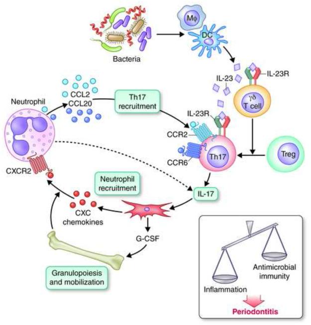
Fig. 3. Cellular cross-talk interactions of Th17 that can shift the balance towards periodontitis.
Bacterially induced IL-23 production by periodontal innate immune cells, such as dendritic cells (DC) and macrophages (Mφ), can not only promote the survival and expansion of Th17 cells but additionally activate γδ T cells in ways that restrain Tregs and shift the balance in favor of Th17 cells. Acting as a link between innate and adaptive immunity, Th17 secrete IL-17 which, by acting mainly through fibroblast upregulation of G-CSF and CXC chemokines, can orchestrate bone marrow production and release of neutrophils and their chemotactic recruitment to the periodontium. Recruited neutrophils, in turn, can produce CCL2 and CCL20 chemokines, which can selectively recruit more Th17 cells by acting on Th17-expressed CCR2 and CCR6. These cellular crosstalk interactions can sustain a positively reinforced feedback for high-level production of IL-17 (possibly also expressed by the neutrophils themselves, according to some studies) sufficient to tip the balance from host protection to inflammatory periodontitis.
Th17 cells can function as a dedicated osteoclastogenic subset that links T-cell activation to bone loss [21]. Therefore, the study of Th17 cells, as well as of regulatory T cells, may provide fresh insights into the role of the adaptive response in periodontal pathogenesis, which could hardly fit into the original Th1/Th2 model (Box 1). Indeed, although there is adequate evidence to implicate CD4+ - but not CD8+ - T cells in periodontal bone destruction, the disease cannot be adequately described in simple Th1 vs. Th2 dichotomous terms, despite over two decades of intensive research [55]. Under the Th1/Th2 paradigm, Seymour and colleagues proposed an elegant model that was, however, consistent with only a subset of the clinical and experimental data. Specifically, it was proposed that Th1 cells predominate in stable lesions (i.e., when there is a balance between the host and the microbiota), whereas Th2 cells are associated with the progression of periodontitis, featuring an inflammatory infiltrate rich in B cells and antibody-secreting plasma cells [56]. The role of the B cell/plasma cell response is not fully understood in periodontitis, although it is thought that the antibody response is not protective [56]. In fact, the increased deposition of immune complexes along with complement fragments in diseased gingiva suggests that plasma cell-secreted antibodies could be involved in inflammatory responses [57]. Moreover, the capacity of B cells to produce inflammatory cytokines and matrix metalloproteinases could further contribute to tissue damage [56,58]. Perhaps more importantly, B cells constitute, along with T cells, a major source of membrane-bound and secreted RANKL in the bone resorptive lesions of periodontitis [57] (Fig. 2). The postulated protective role of Th1 cells is consistent with the negative correlation of Th1-related cytokines (IFNγ and IL-12) with the severity of periodontitis in some studies, and the capacity of the same cytokines to promote cell-mediated immunity [56] and to inhibit osteoclastogenesis [55]. However, other studies have attributed destructive effects to IFNγ and Th1 cells in periodontitis, consistent with the capacity of activated Th1 cells to also express RANKL (reviewed in refs. [55,59]) (Fig. 2). Such discrepancies therefore might, in part, be attributed to opposing roles played by the same T cell subset in periodontitis. In the same vein, Th2 cells, which are thought to support destructive B cell responses [56], can also secrete IL-4 and IL-13 that can inhibit osteoclastogenesis [60] (Fig. 2).
As alluded to above, a more integrated understanding of the role of periodontal T cells could be achieved by studying Th17 and the CD4+ Foxp3+ regulatory T cells (Tregs) (Box 1). In experimental mouse periodontitis, Tregs appear in high numbers after the peak appearance of RANKL-expressing CD4+ T cells [61] and systemic antibody-mediated depletion of Tregs leads to increased inflammation and bone loss [59]. Tregs may thus serve to attenuate inflammatory tissue damage, although their anti-inflammatory potential may be compromised in the inflamed periodontium. Indeed, Tregs seem to convert into IL-17-producing Th17 cells in human periodontal lesions, which also contain IL-17+/Foxp3+ double-positive cells, which are suggestive of an intermediate stage in this process [62]. The mechanistic basis for this observation is uncertain, although a plausible mechanism might involve the capacity of IL-23– activated γδ T cells to restrain Tregs and shift the balance in favor of effector T helper cells [63] (Fig. 3). IL-23 can additionally mediate the clonal expansion of Th17 cells and to stimulate their IL-17 production [54]. In human periodontal lesions, the number of IL-23–expressing macrophages correlates positively with both inflammation and the abundance of IL-17–expressing T cells, which represent the predominant T cell subset in the lesions [64]. It should be noted that other Th17-promoting cytokines, specifically IL-6 and IL-1β, can also regulate the Th17/Treg balance in favor of Th17 [65].
Despite their potent proinflammatory properties, the net effect of Th17 or IL-17 in a microbially-induced inflammatory disease cannot be predicted a priori. This is because IL-17 can stimulate protective innate immunity [66], in part by orchestrating G-CSF–dependent granulopoiesis and the chemotactic recruitment and activation of neutrophils [67,68] (Fig. 3). Furthermore, IL-22, also produced by Th17 cells, can stimulate epithelial cell production of antimicrobial peptides [69]. However, Th17 persistence at sites of inflammation and chronic IL-17 signaling can turn an acute inflammatory response into chronic immunopathology. In the context of bone-related diseases such as rheumatoid arthritis and periodontitis, IL-17 can potentially induce the expression of matrix metalloproteinases in fibroblasts, endothelial cells, and epithelial cells, as well as RANKL expression in osteoblasts [21,55] (and possibly in T cells [70]), thereby mediating destruction of both connective tissue and the underlying bone (Fig. 2). Moreover, Th17 cells are now recognized as effective B-cell helpers for antibody responses linked to inflammatory conditions [71,72]. Therefore the association of Th2 cells with the destructive B cell-dominated periodontal lesion (see above) needs to be re-examined in parallel with a possible new role for Th17.
Recently, IL-17 was causally linked to inflammatory periodontal bone loss in mice [43] and its levels correlate with the severity of periodontitis in humans [59,62,64,73]. Whether IL-17 is crucially involved in the pathogenesis of human periodontitis remains to be established in future clinical trials using local anti-IL-17 or IL-17 receptor blockade treatments. Taking advantage of the high prevalence of periodontitis, this question could alternatively be addressed by monitoring this oral disease in subjects undergoing IL-17–targeted interventions for systemic diseases such as psoriasis.
Although a signature cytokine of Th17 cells, IL-17 is also expressed by a variety of other RORγt-expressing cell types/subsets, including innate lymphoid cells, γδ T cells, and perhaps neutrophils [74-76]. It should be noted that the presence and role of innate lymphoid cells in periodontitis has yet to be addressed, and that the available data for neutrophil expression of IL-17 are stronger for mouse than human cells. IL-17–expressing periodontal neutrophils could thus self-amplify their recruitment and exacerbate inflammation in an IL-17-dependent manner (Fig. 3). The γδ T cell subset is present in abundance at mucosal sites and can produce IL-17 in response to innate signals (IL-1β and IL-23), without a requirement for TCR engagement [77]. At least in mice, γδ T cells are an important source of periodontal IL-17 [43], which is maximally induced in the tissue by co-activation of complement and TLR signaling, probably indirectly through the induction of IL-1β and IL-23 production by phagocytes [78].
The dissection of the periodontal host response, in terms of protective and destructive aspects, seems to be confounded by the very nature of the disease, in which a potentially protective antimicrobial response could be offset by collateral inflammatory tissue damage. This is possibly also true for other inflammatory diseases with complex polymicrobial etiology. Currently, therefore, it is not possible to attribute definite roles to effector T cell subsets in periodontitis. Perhaps the strongest case could be proposed for Th17 cells, which, in cooperation with γδ T cells and neutrophils, have the potential to act as important effectors of periodontal inflammation and tissue damage (Fig. 3).
Therapeutic implications
Although antimicrobial approaches can potentially contribute to the treatment of periodontitis, the fact that the irreversible tissue damage is ultimately inflicted by the host response has prompted many investigators to focus on strategies that target host signaling pathways [79,80]. Importantly, anti-inflammatory modalities can also indirectly exert antimicrobial effects, since periodontal dysbiosis is crucially dependent upon an inflammatory environment [43,79,81] (Fig. 1). Whereas the precise mechanisms initiating and sustaining periodontitis are yet to be fully understood, adequate knowledge is available for rational therapeutic intervention at the experimental level. Successful interventions that inhibited periodontitis in preclinical animal models have targeted diverse but interconnected inflammatory pathways, ranging from upstream events (e.g., inflammatory cell recruitment [43]), intermediate signaling pathways that amplify and propagate inflammation (e.g., complement [78] and proinflammatory cytokines [82]), to downstream events (e.g., RANKL-dependent osteoclastogenesis [83]). Other successful approaches have targeted the resolution of periodontal inflammation through the use of specific pro-resolution agonists, such as the small-lipid molecules lipoxins and resolvins [79]. Future safety and efficacy clinical studies will show which of these candidate strategies can find application for the treatment of human periodontitis.
Concluding Remarks
Periodontal tissue homeostasis could be likened to an ‘armed peace’ between the host and the periodontal microbiota, with occasional microbial attacks that are readily subdued by immune defenses. This controlled inflammatory state is likely represented by stable gingivitis, which would therefore reflect a protective host response. The transition to periodontitis requires both a dysbiotic microbiota and a susceptible host (Fig. 1), which engage into a complex inflammatory dialogue (Fig. 2). Dysbiotic microbial communities exhibit synergistic interactions for enhanced colonization, nutrient procurement, and persistence in an inflammatory environment that promotes their adaptive fitness. Whereas keystone pathogens, such as P. gingivalis, can subvert the host response and contribute to homeostasis breakdown (Fig. 1), other bacteria can act as pathobionts that trigger destructive inflammation involving both innate and adaptive immune elements (Figs. 2 and 3). From a microbial standpoint, the importance of inflammation lies in its providing a source of essential nutrients, although it can cause collateral damage to the periodontal tissues. The inhibition of inflammation, therefore, appears to be central to the treatment of periodontitis, although a fully integrated model of periodontal pathogenesis warrants further research (Box 2).
BOX 2. Further research and outstanding questions.
Mechanistic studies (e.g., using cell lineage–specific conditional knockout models) to better define the roles and crosstalk of T cell subsets in periodontitis.
Genome-wide studies to better define and correlate the transcriptomes and epigenomes of host cells in periodontal health and disease.
Is dysbiosis a cause or a consequence of the periodontal disease process?
What are the molecular mechanisms by which periodontal bacteria inhibit antimicrobial or killing mechanisms without suppressing the overall inflammatory response?
Does the progression of human periodontitis represent a linear process, or consists of periods of exacerbation and remission?
Can the findings from successful preclinical interventions be translated into effective therapeutic modalities for human periodontitis?
Highlights.
Periodontitis requires a dysbiotic microbiota and a susceptible host
Disruption of periodontal tissue homeostasis leads to inflammation
Periodontal inflammation involves a complex innate-adaptive immune interplay
Inflammation causes tissue damage and perpetuates dysbiosis
Acknowledgments
I thank Dana T. Graves (University of Pennsylvania) and Marco A. Cassatella (University of Verona) for comments and Debbie Maizels (Zoobotanica Scientific Illustration) for drawing the figures in this paper. Supported by grants from the U.S. National Institutes of Health (DE015254, DE017138, DE021580, and DE021685). I regret that a number of important studies could only indirectly be acknowledged through comprehensive reviews, due to space and reference number limitations.
GLOSSARY BOX
- Asaccharolytic
A microorganism unable to metabolize carbohydrates and therefore must use other carbon sources (e.g., peptides) for energy.
- Commensal
A microorganism that lives in close contact with a host and benefits from this association, whereas the host is not adversely affected.
- Dysbiosis
An imbalance in the relative abundance of microbial species within an ecosystem that is associated with a disease (e.g., inflammatory bowel disease). Dysbiosis can be either the cause or the consequence of disease.
- Homeostasis
A condition of equilibrium or stability in a system, which is maintained by adjusting physiological processes to counteract external changes; a balanced relationship between a host tissue and the resident microbiota that prevents destructive inflammation or disease.
- Keystone species
A species that has a disproportionately large effect on its environment relative to its abundance, analogous to the role of a keystone in an arch.
- Keystone pathogen
A keystone microbial species that remodels a microbial community in ways that promote disease pathogenesis.
- Microbiota
A complex and diverse community of microorganisms living within a given anatomical niche, e.g., an environmentally exposed surface of a multicellular eukaryotic organism.
- Pathobiont
A normally harmless symbiont that can become pathogenic under certain environmental conditions, e.g., perturbation of tissue homeostasis or in immunocompromised hosts.
- Periodontitis
A biofilm-induced chronic inflammatory disease, which affects the integrity of the tissues that surround and support the teeth (periodontal ligament, gingiva, and alveolar bone, collectively known as the periodontium) and may exert an adverse impact on systemic health.
- Symbiosis (and variations thereof)
A close association of two different species (e.g., a microbe and a mammalian host) that live together without necessarily implying that either partner benefits. Parasitism represents symbiosis in which one species benefits (increases its fitness) at the expense of the other species, whereas in Mutualism both species benefit. Commensalism represents symbiosis in which one species benefits without adversely affecting the other species.
Footnotes
Publisher's Disclaimer: This is a PDF file of an unedited manuscript that has been accepted for publication. As a service to our customers we are providing this early version of the manuscript. The manuscript will undergo copyediting, typesetting, and review of the resulting proof before it is published in its final citable form. Please note that during the production process errors may be discovered which could affect the content, and all legal disclaimers that apply to the journal pertain.
References
- 1.Darveau RP. Periodontitis: a polymicrobial disruption of host homeostasis. Nat Rev Microbiol. 2010; 8:481–490. doi: 10.1038/nrmicro2337. [DOI] [PubMed] [Google Scholar]
- 2.Eke PI, et al. Prevalence of periodontitis in adults in the United States: 2009 and 2010. J Dent Res. 2012; 91:914–920. doi: 10.1177/0022034512457373. [DOI] [PubMed] [Google Scholar]
- 3.Genco RJ, Van Dyke TE. Prevention: Reducing the risk of CVD in patients with periodontitis. Nat Rev Cardiol. 2010; 7:479–480. doi: 10.1038/nrcardio.2010.120. [DOI] [PubMed] [Google Scholar]
- 4.Lundberg K, et al. Periodontitis in RA-the citrullinated enolase connection. Nat Rev Rheumatol. 2010; 6:727–730. doi: 10.1038/nrrheum.2010.139. [DOI] [PubMed] [Google Scholar]
- 5.Lalla E, Papapanou PN. Diabetes mellitus and periodontitis: a tale of two common interrelated diseases. Nat Rev Endocrinol. 2011; 7:738–748. doi: 10.1038/nrendo.2011.106. [DOI] [PubMed] [Google Scholar]
- 6.Kebschull M, et al. “Gum bug leave my heart alone”–Epidemiologic and mechanistic evidence linking periodontal infections and atherosclerosis. J Dent Res. 2010; 89:879–902. doi: 10.1177/0022034510375281. [DOI] [PMC free article] [PubMed] [Google Scholar]
- 7.Madianos PN, et al. Adverse pregnancy outcomes (APOs) and periodontal disease: pathogenic mechanisms. J Periodontol. 2013; 84:S170–180. doi: 10.1902/jop.2013.1340015. [DOI] [PubMed] [Google Scholar]
- 8.Adler CJ, et al. Sequencing ancient calcified dental plaque shows changes in oral microbiota with dietary shifts of the Neolithic and Industrial revolutions. Nat Genet. 2013; 45:450–455. doi: 10.1038/ng.2536. [DOI] [PMC free article] [PubMed] [Google Scholar]
- 9.Wade WG. Has the use of molecular methods for the characterization of the human oral microbiome changed our understanding of the role of bacteria in the pathogenesis of periodontal disease? J Clin Periodontol. 2011; 38(Suppl 11):7–16. doi: 10.1111/j.1600-051X.2010.01679.x. [DOI] [PubMed] [Google Scholar]
- 10.Hajishengallis G, Lamont RJ. Beyond the red complex and into more complexity: The Polymicrobial Synergy and Dysbiosis (PSD) model of periodontal disease etiology. Mol Oral Microbiol. 2012; 27:409–419. doi: 10.1111/j.2041-1014.2012.00663.x. [DOI] [PMC free article] [PubMed] [Google Scholar]
- 11.Orth RK, et al. Synergistic virulence of Porphyromonas gingivalis and Treponema denticola in a murine periodontitis model. Mol Oral Microbiol. 2011; 26:229–240. doi: 10.1111/j.2041-1014.2011.00612.x. [DOI] [PubMed] [Google Scholar]
- 12.Ramsey MM, et al. Metabolite cross-feeding enhances virulence in a model polymicrobial infection. PLoS Pathog. 2011; 7:e1002012. doi: 10.1371/journal.ppat.1002012. [DOI] [PMC free article] [PubMed] [Google Scholar]
- 13.Settem RP, et al. Fusobacterium nucleatum and Tannerella forsythia induce synergistic alveolar bone loss in a mouse periodontitis model. Infect Immun. 2012; 80:2436–2443. doi: 10.1128/IAI.06276-11. [DOI] [PMC free article] [PubMed] [Google Scholar]
- 14.Hajishengallis G, et al. Low-abundance biofilm species orchestrates inflammatory periodontal disease through the commensal microbiota and complement. Cell Host Microbe. 2011; 10:497–506. doi: 10.1016/j.chom.2011.10.006. [DOI] [PMC free article] [PubMed] [Google Scholar]
- 15.Jiao Y, et al. Induction of bone loss by pathobiont-mediated nod1 signaling in the oral cavity. Cell Host Microbe. 2013; 13:595–601. doi: 10.1016/j.chom.2013.04.005. [DOI] [PMC free article] [PubMed] [Google Scholar]
- 16.Dewhirst FE, et al. The human oral microbiome. J Bacteriol. 2010; 192:5002–5017. doi: 10.1128/JB.00542-10. [DOI] [PMC free article] [PubMed] [Google Scholar]
- 17.Griffen AL, et al. Distinct and complex bacterial profiles in human periodontitis and health revealed by 16S pyrosequencing. ISME J. 2012; 6:1176–1185. doi: 10.1038/ismej.2011.191. [DOI] [PMC free article] [PubMed] [Google Scholar]
- 18.Abusleme L, et al. The subgingival microbiome in health and periodontitis and its relationship with community biomass and inflammation. ISME J. 2013; 7:1016–1025. doi: 10.1038/ismej.2012.174. [DOI] [PMC free article] [PubMed] [Google Scholar]
- 19.Hajishengallis G, et al. The keystone-pathogen hypothesis. Nat Rev Microbiol. 2012; 10:717–725. doi: 10.1038/nrmicro2873. [DOI] [PMC free article] [PubMed] [Google Scholar]
- 20.Belibasakis GN, Bostanci N. The RANKL-OPG system in clinical periodontology. J Clin Periodontol. 2012; 39:239–248. doi: 10.1111/j.1600-051X.2011.01810.x. [DOI] [PubMed] [Google Scholar]
- 21.Miossec P, Kolls JK. Targeting IL-17 and TH17 cells in chronic inflammation. Nat Rev Drug Discov. 2012; 11:763–776. doi: 10.1038/nrd3794. [DOI] [PubMed] [Google Scholar]
- 22.Darveau RP, et al. Porphyromonas gingivalis as a potential community activist for disease. J Dent Res. 2012; 91:816–820. doi: 10.1177/0022034512453589. [DOI] [PMC free article] [PubMed] [Google Scholar]
- 23.Hajishengallis G, Lambris JD. Microbial manipulation of receptor crosstalk in innate immunity. Nat Rev Immunol. 2011; 11:187–200. doi: 10.1038/nri2918. [DOI] [PMC free article] [PubMed] [Google Scholar]
- 24.Barth K, et al. Disruption of immune regulation by microbial pathogens and resulting chronic inflammation. J Cell Physiol. 2013; 228:1413–1422. doi: 10.1002/jcp.24299. [DOI] [PMC free article] [PubMed] [Google Scholar]
- 25.Round JL, Mazmanian SK. The gut microbiota shapes intestinal immune responses during health and disease. Nat Rev Immunol. 2009; 9:313–323. doi: 10.1038/nri2515. [DOI] [PMC free article] [PubMed] [Google Scholar]
- 26.Schreiner H, et al. A Comparison of Aggregatibacter actinomycetemcomitans (Aa) Virulence Traits in a Rat Model for Periodontal Disease. PLoS One. 2013; 8:e69382. doi: 10.1371/journal.pone.0069382. [DOI] [PMC free article] [PubMed] [Google Scholar]
- 27.Sharma A. Virulence mechanisms of Tannerella forsythia. Periodontol 2000. 2010; 54:106–116. doi: 10.1111/j.1600-0757.2009.00332.x. [DOI] [PMC free article] [PubMed] [Google Scholar]
- 28.Frederick JR, et al. Molecular signaling mechanisms of the periopathogen, Treponema denticola. J Dent Res. 2011; 90:1155–1163. doi: 10.1177/0022034511402994. [DOI] [PMC free article] [PubMed] [Google Scholar]
- 29.Stecher B, et al. ‘Blooming’ in the gut: how dysbiosis might contribute to pathogen evolution. Nat Rev Microbiol. 2013; 11:277–284. doi: 10.1038/nrmicro2989. [DOI] [PubMed] [Google Scholar]
- 30.Belkaid Y, Naik S. Compartmentalized and systemic control of tissue immunity by commensals. Nat Immunol. 2013; 14:646–653. doi: 10.1038/ni.2604. [DOI] [PMC free article] [PubMed] [Google Scholar]
- 31.Moutsopoulos NM, et al. Porphyromonas gingivalis promotes Th17 inducing pathways in chronic periodontitis. J Autoimmun. 2012; 39:294–303. doi: 10.1016/j.jaut.2012.03.003. [DOI] [PMC free article] [PubMed] [Google Scholar]
- 32.Jauregui CE, et al. Suppression of T-cell chemokines by Porphyromonas gingivalis. Infect Immun. 2013; 81:2288–2295. doi: 10.1128/IAI.00264-13. [DOI] [PMC free article] [PubMed] [Google Scholar]
- 33.Gaddis DE, et al. Role of TLR2 dependent-IL-10 production in the inhibition of the initial IFN-γ T cell response to Porphyromonas gingivalis. J Leukoc Biol. 2012; 93:21–31. doi: 10.1189/jlb.0512220. [DOI] [PMC free article] [PubMed] [Google Scholar]
- 34.Yoshioka H, et al. Analysis of the activity to induce toll-like receptor (TLR)2- and TLR4-mediated stimulation of supragingival plaque. J Periodontol. 2008; 79:920–928. doi: 10.1902/jop.2008.070516. [DOI] [PubMed] [Google Scholar]
- 35.Aruni AW, et al. Filifactor alocis has virulence attributes that can enhance its persistence under oxidative stress conditions and mediate invasion of epithelial cells by Porphyromonas gingivalis. Infect Immun. 2011; 79:3872–3886. doi: 10.1128/IAI.05631-11. [DOI] [PMC free article] [PubMed] [Google Scholar]
- 36.Moffatt CE, et al. Filifactor alocis interactions with gingival epithelial cells. Mol Oral Microbiol. 2011; 26:365–373. doi: 10.1111/j.2041-1014.2011.00624.x. [DOI] [PMC free article] [PubMed] [Google Scholar]
- 37.Marsh PD. Are dental diseases examples of ecological catastrophes? Microbiology. 2003; 149:279–294. doi: 10.1099/mic.0.26082-0. [DOI] [PubMed] [Google Scholar]
- 38.Stabholz A, et al. Genetic and environmental risk factors for chronic periodontitis and aggressive periodontitis. Periodontol 2000. 2010; 53:138–153. doi: 10.1111/j.1600-0757.2010.00340.x. [DOI] [PubMed] [Google Scholar]
- 39.Laine ML, et al. Genetic susceptibility to periodontitis. Periodontol 2000. 2012; 58:37–68. doi: 10.1111/j.1600-0757.2011.00415.x. [DOI] [PubMed] [Google Scholar]
- 40.Lindroth AM, Park YJ. Epigenetic biomarkers: a step forward for understanding periodontitis. J Periodontal Implant Sci. 2013; 43:111–120. doi: 10.5051/jpis.2013.43.3.111. [DOI] [PMC free article] [PubMed] [Google Scholar]
- 41.Zhou Q, et al. Signaling mechanisms in the restoration of impaired immune function due to diet-induced obesity. Proc Natl Acad Sci U S A. 2011; 108:2867–2872. doi: 10.1073/pnas.1019270108. [DOI] [PMC free article] [PubMed] [Google Scholar]
- 42.Divaris K, et al. Exploring the genetic basis of chronic periodontitis: a genomewide association study. Hum Mol Genet. 2013; 22:2312–2324. doi: 10.1093/hmg/ddt065. [DOI] [PMC free article] [PubMed] [Google Scholar]
- 43.Eskan MA, et al. The leukocyte integrin antagonist Del-1 inhibits IL-17-mediated inflammatory bone loss. Nat Immunol. 2012; 13:465–473. doi: 10.1038/ni.2260. [DOI] [PMC free article] [PubMed] [Google Scholar]
- 44.Hajishengallis G. Too old to fight? Aging and its toll on innate immunity. Mol Oral Microbiol. 2010; 25:25–37. doi: 10.1111/j.2041-1014.2009.00562.x. [DOI] [PMC free article] [PubMed] [Google Scholar]
- 45.Han YW, Wang X. Mobile microbiome: oral bacteria in extra-oral infections and inflammation. J Dent Res. 2013; 92:485–491. doi: 10.1177/0022034513487559. [DOI] [PMC free article] [PubMed] [Google Scholar]
- 46.Graves DT, et al. Review of osteoimmunology and the host response in endodontic and periodontal lesions. J Oral Microbiol. 2011; 3 doi: 10.3402/jom.v3i0.5304. 10.3402/jom.v3403i3400.5304. [DOI] [PMC free article] [PubMed] [Google Scholar]
- 47.Nussbaum G, Shapira L. How has neutrophil research improved our understanding of periodontal pathogenesis? J Clin Periodontol. 2011; 38:49–59. doi: 10.1111/j.1600-051X.2010.01678.x. [DOI] [PubMed] [Google Scholar]
- 48.Ryder MI. Comparison of neutrophil functions in aggressive and chronic periodontitis. Periodontol 2000. 2010; 53:124–137. doi: 10.1111/j.1600-0757.2009.00327.x. [DOI] [PubMed] [Google Scholar]
- 49.Kolaczkowska E, Kubes P. Neutrophil recruitment and function in health and inflammation. Nat Rev Immunol. 2013; 13:159–175. doi: 10.1038/nri3399. [DOI] [PubMed] [Google Scholar]
- 50.Caielli S, et al. Neutrophils come of age in chronic inflammation. Curr Opin Immunol. 2012; 24:671–677. doi: 10.1016/j.coi.2012.09.008. [DOI] [PMC free article] [PubMed] [Google Scholar]
- 51.Hajishengallis E, Hajishengallis G. Neutrophil homeostasis and periodontal health in children and adults. J Dent Res. 2013; 92 doi: 10.1177/0022034513507956. in press. [DOI] [PMC free article] [PubMed] [Google Scholar]
- 52.Chakravarti A, et al. Surface RANKL of Toll-like receptor 4-stimulated human neutrophils activates osteoclastic bone resorption. Blood. 2009; 114:1633–1644. doi: 10.1182/blood-2008-09-178301. [DOI] [PubMed] [Google Scholar]
- 53.Pelletier M, et al. Evidence for a cross-talk between human neutrophils and Th17 cells. Blood. 2009; 115:335–343. doi: 10.1182/blood-2009-04-216085. [DOI] [PubMed] [Google Scholar]
- 54.Weaver CT, et al. The Th17 pathway and inflammatory diseases of the intestines, lungs, and skin. Annu Rev Pathol. 2013; 8:477–512. doi: 10.1146/annurev-pathol-011110-130318. [DOI] [PMC free article] [PubMed] [Google Scholar]
- 55.Gaffen SL, Hajishengallis G. A new inflammatory cytokine on the block: rethinking periodontal disease and the Th1/Th2 paradigm in the context of Th17 cells and IL-17. J Dent Res. 2008; 87:817–828. doi: 10.1177/154405910808700908. [DOI] [PMC free article] [PubMed] [Google Scholar]
- 56.Gemmell E, et al. The role of T cells in periodontal disease: homeostasis and autoimmunity. Periodontology 2000. 2007; 43:14–40. doi: 10.1111/j.1600-0757.2006.00173.x. [DOI] [PubMed] [Google Scholar]
- 57.Kajiya M, et al. Role of periodontal pathogenic bacteria in RANKL-mediated bone destruction in periodontal disease. J Oral Microbiol. 2010; 2 doi: 10.3402/jom.v2i0.5532. 10.3402/jom.v3402i3400.5532. [DOI] [PMC free article] [PubMed] [Google Scholar]
- 58.Berglundh T, et al. B cells in periodontitis: friends or enemies? Periodontol 2000. 2007; 45:51–66. doi: 10.1111/j.1600-0757.2007.00223.x. [DOI] [PubMed] [Google Scholar]
- 59.Garlet GP. Destructive and protective roles of cytokines in periodontitis: A reappraisal from host defense and tissue destruction viewpoints. J Dent Res. 2010; 89:1349–1363. doi: 10.1177/0022034510376402. [DOI] [PubMed] [Google Scholar]
- 60.McInnes IB, Schett G. Cytokines in the pathogenesis of rheumatoid arthritis. Nat Rev Immunol. 2007; 7:429–442. doi: 10.1038/nri2094. [DOI] [PubMed] [Google Scholar]
- 61.Kobayashi R, et al. Induction of IL-10-producing CD4+ T-cells in chronic periodontitis. J Dent Res. 2011; 90:653–658. doi: 10.1177/0022034510397838. [DOI] [PMC free article] [PubMed] [Google Scholar]
- 62.Okui T, et al. The presence of IL-17+/FOXP3+ double-positive cells in periodontitis. J Dent Res. 2012; 91:574–579. doi: 10.1177/0022034512446341. [DOI] [PubMed] [Google Scholar]
- 63.Petermann F, et al. γδ T cells enhance autoimmunity by restraining regulatory T cell responses via an interleukin-23-dependent mechanism. Immunity. 2010; 33:351–363. doi: 10.1016/j.immuni.2010.08.013. [DOI] [PMC free article] [PubMed] [Google Scholar]
- 64.Allam JP, et al. IL-23-producing CD68(+) macrophage-like cells predominate within an IL-17-polarized infiltrate in chronic periodontitis lesions. J Clin Periodontol. 2011; 38:879–886. doi: 10.1111/j.1600-051X.2011.01752.x. [DOI] [PubMed] [Google Scholar]
- 65.Afzali B, et al. The role of T helper 17 (Th17) and regulatory T cells (Treg) in human organ transplantation and autoimmune disease. Clin Exp Immunol. 2007; 148:32–46. doi: 10.1111/j.1365-2249.2007.03356.x. [DOI] [PMC free article] [PubMed] [Google Scholar]
- 66.Conti HR, et al. Th17 cells and IL-17 receptor signaling are essential for mucosal host defense against oral candidiasis. J Exp Med. 2009; 206:299–311. doi: 10.1084/jem.20081463. [DOI] [PMC free article] [PubMed] [Google Scholar]
- 67.Iwakura Y, et al. Functional specialization of interleukin-17 family members. Immunity. 2011; 34:149–162. doi: 10.1016/j.immuni.2011.02.012. [DOI] [PubMed] [Google Scholar]
- 68.von Vietinghoff S, Ley K. Homeostatic regulation of blood neutrophil counts. J Immunol. 2008; 181:5183–5188. doi: 10.4049/jimmunol.181.8.5183. [DOI] [PMC free article] [PubMed] [Google Scholar]
- 69.Korn T, et al. IL-17 and Th17 Cells. Annu Rev Immunol. 2009; 27:485–517. doi: 10.1146/annurev.immunol.021908.132710. [DOI] [PubMed] [Google Scholar]
- 70.Ju JH, et al. Oral administration of type-II collagen suppresses IL-17-associated RANKL expression of CD4+ T cells in collagen-induced arthritis. Immunol Lett. 2008; 117:16–25. doi: 10.1016/j.imlet.2007.09.011. [DOI] [PubMed] [Google Scholar]
- 71.Mitsdoerffer M, et al. Proinflammatory T helper type 17 cells are effective B-cell helpers. Proc Natl Acad Sci U S A. 2010; 107:14292–14297. doi: 10.1073/pnas.1009234107. [DOI] [PMC free article] [PubMed] [Google Scholar]
- 72.Hickman-Brecks CL, et al. Th17 cells can provide B cell help in autoantibody induced arthritis. J Autoimmun. 2011; 36:65–75. doi: 10.1016/j.jaut.2010.10.007. [DOI] [PMC free article] [PubMed] [Google Scholar]
- 73.Ohyama H, et al. The involvement of IL-23 and the Th17 pathway in periodontitis. J Dent Res. 2009; 88:633–638. doi: 10.1177/0022034509339889. [DOI] [PubMed] [Google Scholar]
- 74.Cua DJ, Tato CM. Innate IL-17-producing cells: the sentinels of the immune system. Nat Rev Immunol. 2010; 10:479–489. doi: 10.1038/nri2800. [DOI] [PubMed] [Google Scholar]
- 75.Tan Z, et al. RORγt+IL-17+ neutrophils play a critical role in hepatic ischemiareperfusion injury. J Mol Cell Biol. 2013; 5:143–146. doi: 10.1093/jmcb/mjs065. [DOI] [PMC free article] [PubMed] [Google Scholar]
- 76.Mantovani A, et al. Neutrophils in the activation and regulation of innate and adaptive immunity. Nat Rev Immunol. 2011; 11:519–531. doi: 10.1038/nri3024. [DOI] [PubMed] [Google Scholar]
- 77.Sutton CE, et al. IL-17-producing gammadelta T cells and innate lymphoid cells. Eur J Immunol. 2012; 42:2221–2231. doi: 10.1002/eji.201242569. [DOI] [PubMed] [Google Scholar]
- 78.Abe T, et al. Local complement-targeted intervention in periodontitis: proof-ofconcept using a C5a receptor (CD88) antagonist. J Immunol. 2012; 189:5442–5448. doi: 10.4049/jimmunol.1202339. [DOI] [PMC free article] [PubMed] [Google Scholar]
- 79.Hasturk H, et al. Paradigm shift in the pharmacological management of periodontal diseases. Front Oral Biol. 2012; 15:160–176. doi: 10.1159/000329678. [DOI] [PMC free article] [PubMed] [Google Scholar]
- 80.Tonetti MS, Chapple IL. Biological approaches to the development of novel periodontal therapies-consensus of the Seventh European Workshop on Periodontology. J Clin Periodontol. 2011; 38(Suppl 11):114–118. doi: 10.1111/j.1600-051X.2010.01675.x. [DOI] [PubMed] [Google Scholar]
- 81.Hajishengallis G, et al. Role of complement in host-microbe homeostasis of the periodontium. Semin Immunol. 2013; 25:65–72. doi: 10.1016/j.smim.2013.04.004. [DOI] [PMC free article] [PubMed] [Google Scholar]
- 82.Graves D. Cytokines that promote periodontal tissue destruction. J Periodontol. 2008; 79:1585–1591. doi: 10.1902/jop.2008.080183. [DOI] [PubMed] [Google Scholar]
- 83.Han X, et al. Porphyromonas gingivalis infection-associated periodontal bone resorption is dependent on receptor activator of NF-κB ligand. Infect Immun. 81:1502–1509. doi: 10.1128/IAI.00043-13. [DOI] [PMC free article] [PubMed] [Google Scholar]


