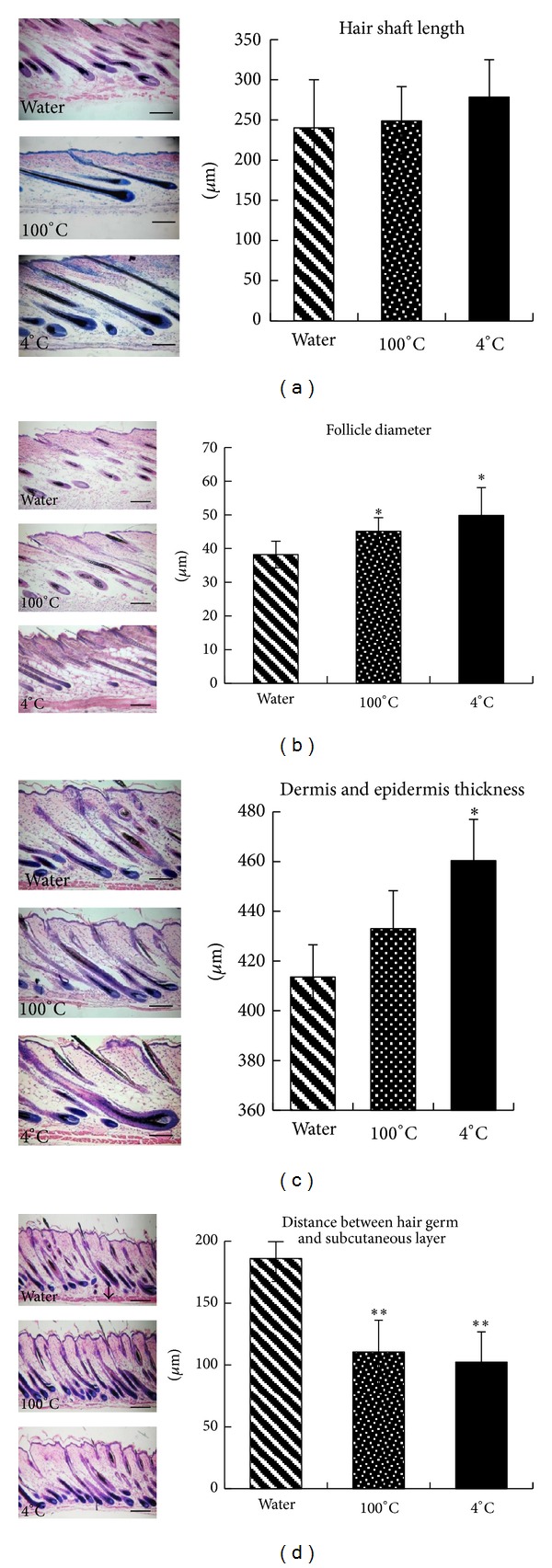
Figure 1.

HE staining and quantitative morphologic analysis of hair follicles. HE-stained paraffin sections of dorsal skin of control (water) and 100°C or 4°C deer antler extract-treated mice were examined at 100x magnification. (a) Quantitative analysis of the hair shaft length. (b) Hair follicle diameter of the different groups. (c) Thickness of the dermis and epidermis in the different groups. (d) Distance between the hair germ and the subcutaneous layer in the different groups. ((a)–(d) left part) Sections of the back skins were stained, and representative photomicrographs of skin sections are shown. Bars are 100 μm. Values are mean ± standard deviation (SD) (n = 10/mouse; *P < 0.05 and **P < 0.01 compared to control). Nine sections were reduplicated in each group.
