Abstract
Muscle volume is known to correlate with maximal joint torque in humans, but the role of muscle moment arm in determining maximal torque is less clear. Moderate correlations have been reported between maximal isometric knee extensor torque and knee extensor moment arm, but no such observations have been made for the ankle joint. It has been suggested that smaller muscle moment arms may enhance force generation at high rates of joint rotation, but this has not yet been observed for ankle muscles in vivo. The purpose of the present study was to correlate plantar flexor moment arm and plantar flexor muscle volume with maximal plantar flexor torque measured at different rates of plantar flexion. Magnetic resonance imaging was used to quantify the plantar flexor moment arm and muscle volume of the posterior compartment in 20 healthy young men. Maximal plantar flexor torque was measured isometrically and at three plantar flexion speeds using an isokinetic dynamometer. Plantar flexor torque was significantly correlated with muscle volume (0.222 < R2 < 0.322) and with muscle moment arm at each speed (0.323 < R2 < 0.494). While muscle volume was strongly correlated with body mass and stature, moment arm was not. The slope of the torque-moment arm regression line decreased as the rate of joint rotation increased, indicating that subjects with small moment arms experienced smaller reductions in torque at high speeds. The findings of this study suggest that plantar flexor moment arm is a determinant of joint strength that is at least as important as muscle size.
Keywords: plantar flexors, joint torque, moment arm, force-velocity
muscle-tendon units are linear actuators that generate movement only through their ability to produce or resist joint rotation. The joint moment produced by a muscle is the product of the force carried by the muscle's tendon and the moment arm of the muscle-tendon unit about the center of joint rotation. The capacity for joint rotation in an individual human subject thus depends on both muscle force generation and muscle leverage, but the influence of only one of these factors, muscle force, has been investigated thoroughly. There are many published studies documenting correlations between joint strength (i.e., maximum joint torque) and muscle size, which we understand to be a direct determinant of muscle force. Examples of such studies relating strength to size may be found for muscles crossing nearly every major upper and lower extremity joint. At the ankle, for example, interindividual variation in triceps surae muscle volume has been shown to explain 69–80% of the variance in maximal plantar flexor torque (11, 21). Very strong correlations have been found between both elbow flexor and extensor muscle volume and their associated maximal joint torques (R2 = 0.87 and R2 = 0.85, respectively) (9).
In contrast to effects of muscle size, the influence of muscle moment arm on variation in strength across individuals has received little attention. We know of only one such study (6), in which a moderate positive correlation (R2 = 0.25) between maximal isometric knee extensor torque and knee extensor moment arm was reported for young healthy subjects. A stronger correlation was found between maximal isometric extensor torque and extensor muscle volume (R2 = 0.60) in the same study. While direct correlations between strength and moment arm have not been reported in other studies, several recent investigations have identified possible links between athletic performance and ankle muscle moment arm. Running has been found to be less energetically costly for runners with short heels (24, 26), perhaps because the higher muscle forces necessary for running with a short plantar flexor moment arm result in greater elastic energy storage in the Achilles tendon. Other studies have reported comparisons between trained sprinters and height-matched nonsprinters, revealing that sprinters have significantly smaller plantar flexor moment arms (5, 16).
A longer Achilles tendon moment arm will, by definition, allow a greater plantar flexor moment to be generated for a given muscle force, but the role of this moment arm in determining ankle joint strength may require consideration of the effects of joint angular velocity as well as mechanical advantage. Examination of human wrist extensors (18) has shown that some muscles are well suited for generating force during rapid joint rotations because they have longer muscle fibers and shorter moment arms, both of which would tend to favor muscle force by reducing sarcomere shortening velocities. Shorter plantar flexor moment arms among human sprinters (5, 16) and observations that the propulsive muscles of animals specialized for sprinting have smaller moment arms relative to the lengths of their associated muscle fascicles (3, 32) also suggest that muscular joint moment and power may be maximized by trading leverage for force generation. Computer simulations using musculoskeletal models have also been used to demonstrate that short plantar flexor moment arms may augment plantar flexor force during rapid plantar flexion by reducing plantar flexor shortening velocity (5, 16, 23), but these simulations did not model adaptations in muscle architecture that may accompany variation in moment arm (14).
The purpose of this study was to use a dynamometer to measure maximal isometric and isokinetic plantar flexor moments in healthy young male subjects to determine whether muscular joint strength was correlated with Achilles tendon moment arm at varying rates of plantar flexion. Achilles tendon moment arm and plantar flexor muscle volume were estimated from magnetic resonance (MR) image data. We hypothesized that maximum plantar flexor moment would be positively correlated with both muscle moment arm and muscle volume during isometric and slow isokinetic contractions. We further hypothesized that the dependence of strength on moment arm would be reduced during faster isokinetic plantar flexions because subjects with larger plantar flexor moment arms would see greater torque reductions at high speeds due to force-velocity effects.
MATERIALS AND METHODS
Twenty healthy young adult men participated in this study. The average age of the participants was 26.0 ± 3.5 yr, and their mean stature and body mass were 177.7 ± 7.7 cm and 76.3 ± 15.6 kg, respectively. All human subjects testing procedures were approved by the Institutional Review Board of The Pennsylvania State University. Written, informed consent was obtained from all participants.
Plantar flexor strength was measured with subjects seated in a System 3 isokinetic dynamometer (Biodex Medical Systems, Shirley, NY) with the right foot unshod and secured to the dynamometer foot plate with stiff nylon straps to prevent foot movement. The lateral malleolus was aligned with the dynamometer motor spindle. The right knee was fully extended, and the right thigh was strapped to the seat to prevent knee flexion. Neutral ankle position (0°) was defined to occur when a right angle was formed by the foot plate and the long axis of the shank. Dorsiflexion was defined as negative, and plantar flexion was defined as positive. To minimize heel motion during testing, the dynamometer seat was moved toward the foot plate until the investigator could not easily advance the seat further. During testing, no visible motion of the heel with respect to the footplate was apparent. To set the maximum dorsiflexion angle, subjects' ankles were slowly rotated into dorsiflexion until subjects reported slight plantar flexor tightness; this occurred between 10 and 13° dorsiflexion for all subjects. The maximum plantar flexion angle was set to 30° for all subjects.
Maximal plantar flexor torque was measured under both isometric and isokinetic conditions. In isometric tests, subjects were instructed to push against the footplate maximally for 2 s as isometric plantar flexor torque was measured with the ankle in the neutral position. These maximal isometric contractions were performed with rest periods of at least 5 s between each effort. Three maximal isometric contractions were collected for each subject, and the best trial was used to define peak isometric torque. During isokinetic tests, maximal plantar flexor contractions were performed as the foot plate was rotated in the plantar flexion direction at 30, 120, 210, and 300°/s. Subjects used a handheld switch to initiate foot plate rotation manually and were instructed to “simultaneously press the hand switch and press as hard and fast as possible against the foot plate.” To permit acclimation to the isokinetic trials, subjects performed at least three practice contractions at each velocity. Subjects then completed a set of five maximal contractions at each of the four plantar flexion speeds in randomized orders. Five seconds of rest were provided within each speed contraction, and 1 min of rest was provided between conditions. Peak torques did not consistently occur during the first trial of each condition, suggesting that fatigue effects were minimal. During each contraction, subjects received verbal encouragement from the same investigator to plantar flex maximally. The peak torque measured as the ankle passed through the neutral position was analyzed for each condition. Because five subjects were unable to consistently accelerate the footplate to 300°/s at neutral position, this speed was excluded from subsequent analysis.
To quantify plantar flexor moment arm and muscle volume, MR images of each subject were acquired with a 3.0-T Siemens Trio scanner (Siemens, Erlangen, Germany) while subjects remained passive. Subjects were positioned supine on the scanner bed with both knees fully extended. The right ankle was placed on an MR-compatible ankle-positioning device (5) that was constructed from plastic and fastened to the scanner bed before imaging. This device supported the foot while allowing for manual positioning of the ankle. Stiff straps secured the right foot to the ankle-positioning device to minimize movement during scans. Images were acquired with the ankle positioned at 10° dorsiflexion, neutral ankle position, and 10° plantar flexion (three-dimensional isotropic T1 weighted sequence; echo time: 1.31 ms, repetition time: 3.96 ms, 300-mm field of view, 0.6-mm voxel size). The complete volume of the lower leg was acquired with the ankle in neutral position (three-dimensional isotropic T1 weighted; echo time: 1.09 ms, repetition time: 3.96 mm, 500-mm field of view, 0.9-mm voxel size). In addition to MR scanning, B-mode ultrasonography (Aloka 1100; transducer: SSD-625, 7.5 MHz and 39-mm scan width; Wallingford, CT) was used to measure the right medial gastrocnemius thickness and pennation angle, from which fascicle length was calculated, using methods previously employed by Lee and Piazza (16). During the ultrasound scans, subjects were standing with the ankle in neutral position.
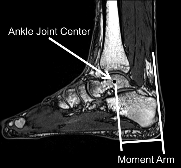
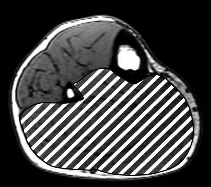
MR image data were processed using Osirix software (Pixmeo, Geneva, Switzerland). Quasi-sagittal plane images of the ankle were reconstructed and printed onto transparent sheets. The center of rotation between the tibia and talus at the neutral position (Fig. 1) was determined using a modified Reuleaux method that has been described in detail elsewhere (5, 8, 19, 25). Plantar flexor moment arm was defined as the shortest (perpendicular) distance between the midline of the Achilles tendon and the center of ankle rotation. To measure plantar flexor muscle volume, 1-mm-thick axial slices of the shank were reconstructed in Osirix viewer, and every 10th image was analyzed to obtain 1-cm-thick axial slices (10). The margins of the triceps surae were difficult to identify on some images, so the entire posterior compartment of the leg was outlined on each slice using ImageJ software (version 1.45s; National Institutes of Health, Bethesda, MD). Total plantar flexor muscle volume was found as the sum of the products of the slice areas and the slice thickness (Fig. 2). To assess reliability, estimation of moment arm and muscle volume were repeated for three subjects using image data collected on a second day. The day-to-day differences in moment arm and muscle volume averaged across the three subjects were 2.7 and 0.8%, respectively.
Fig. 1.
Magnetic resonance image of a sagittal cross section of the foot and ankle showing the center of ankle rotation and plantar flexor moment arm.
Fig. 2.
Magnetic resonance image of a cross section of the lower leg showing the area of the posterior compartment filled in with a striped pattern.
Simple linear regressions were performed to test for correlations between torques and either plantar flexor moment arm or muscle volume, and torques were also regressed simultaneously upon plantar flexor moment arm and muscle volume in multiple linear regressions. Moment arm and muscle volume were also regressed against one another and against body mass and stature. A repeated-measures one-way ANOVA with post hoc Bonferroni-corrected pairwise mean comparisons was performed to determine whether there were differences in plantar flexor torque across the four velocity conditions (isometric and 30, 120, and 210°/s). To test the hypothesis that moment arm influences ankle torque, a linear mixed model was developed with maximal plantar flexor torque as the outcome variable, moment arm as the predictor variable, and muscle volume as a covariate. This analysis controlled for muscle volume while testing whether the slope of the moment arm-torque regression line at each nonzero speed (30, 120, and 210°/s) differed significantly from the slope found for the isometric regression model. The linear mixed model was performed in the R statistical computing environment (version 2.15.1, R Development Core Team; www.r-project.org), while ANOVA was performed using IBM SPSS (version 20, Armonk, NY). The level of significance was set at α = 0.05.
RESULTS
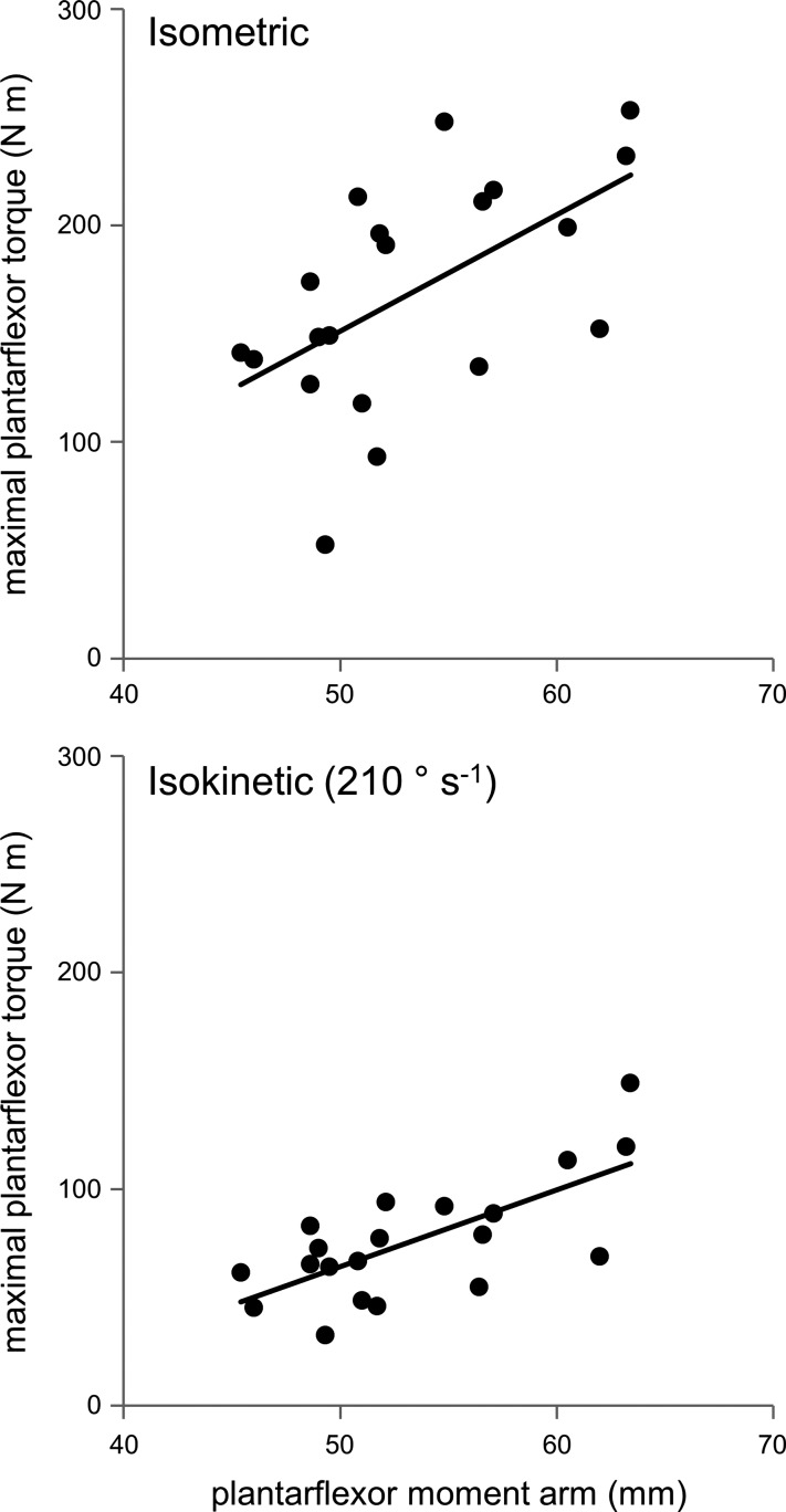
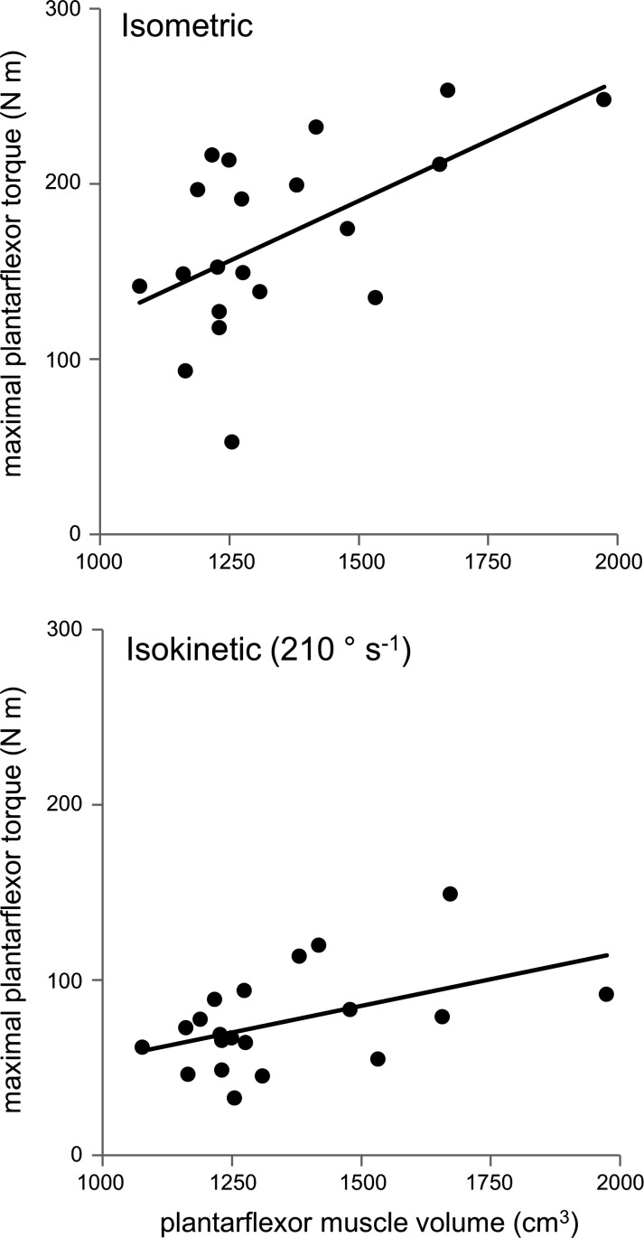
Maximal isometric plantar flexor torque was moderately and significantly correlated with both plantar flexor moment arm and plantar flexor muscle volume (P ≤ 0.01 for both; Table 1; Figs. 3 and 4). Maximal plantar flexor torque measured during isokinetic tests was moderately and significantly correlated (all P ≤ 0.036) with plantar flexor volume at each of the three speeds tested (Table 1; Fig. 4). Stronger correlations between maximal isokinetic torque and plantar flexor moment arm were found at each of the speeds tested (all P ≤ 0.002; Table 1; Fig. 3). Multiple regression results showed strong correlations between plantar flexor torque and both moment arm and muscle volume during isometric and isokinetic contractions (all P ≤ 0.007; Table 1).
Table 1.
Coefficients of determination (R2) for correlations between maximal torques and plantar flexor MA and volume
| Independent Variable(s) |
|||
|---|---|---|---|
| Plantar Flexion Speed | VOL | MA | VOL and MA |
| Isometric | 0.322 (0.009) | 0.315 (0.010) | 0.445 (0.007) |
| 30°/s | 0.226 (0.034) | 0.465 (0.001) | 0.506 (0.003) |
| 120°/s | 0.243 (0.027) | 0.438 (0.001) | 0.491 (0.003) |
| 210°/s | 0.222 (0.036) | 0.484 (0.001) | 0.520 (0.002) |
Nos. in parentheses are the P values for each correlation. VOL, plantar flexor (posterior compartment) muscle volume; MA, plantar flexor moment arm.
Fig. 3.
Maximal plantar flexor torque measured at neutral position plotted against plantar flexor moment arm for isometric trials (top) and the 210°/s isokinetic trials (bottom). Significant correlations were found between torque and moment arm for both conditions (R2 = 0.315, P = 0.01; and R2 = 0.484, P = 0.001, respectively).
Fig. 4.
Maximal plantar flexor torque measured at neutral position plotted against posterior compartment muscle volume for isometric trials (top) and the 210°/s isokinetic trials (bottom). Significant correlations were found between torque and muscle volume for both conditions (R2 = 0.322, P = 0.009; and R2 = 0.222, P = 0.036, respectively).
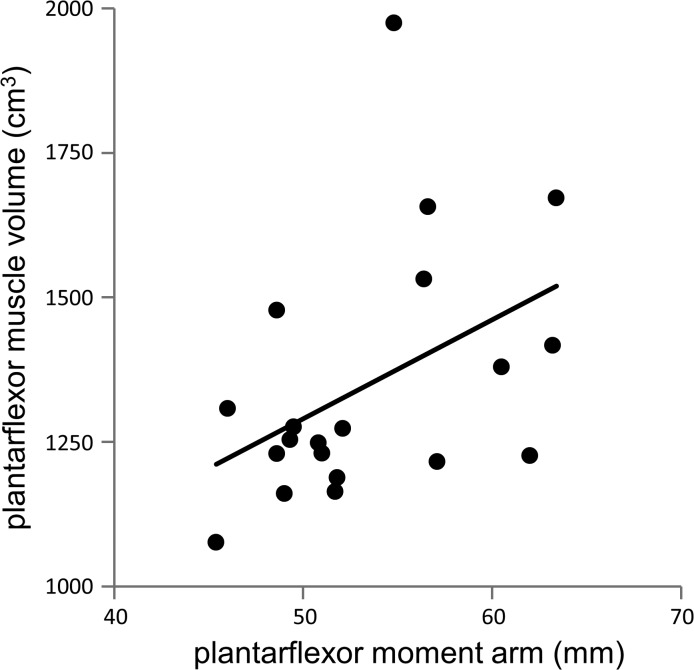
Strong, positive correlations were found between muscle volume and body mass (R2 = 0.757, P < 0.001) and between muscle volume and stature (R2 = 0.562, P < 0.001), but the correlations between moment arm and body mass (R2 = 0.140, P = 0.105) and between moment arm and stature (R2 = 0.156, P = 0.085) were weaker and insignificant. A weak correlation that approached significance was found between muscle volume and moment arm (Fig. 5; R2 = 0.191, P = 0.054).
Fig. 5.
Posterior compartment muscle volume plotted against plantar flexor moment arm for all subjects. A correlation was found between muscle volume and moment arm that nearly reached the level of significance (R2 = 0.191, P = 0.054).
ANOVA revealed that plantar flexion velocity had a significant effect (P < 0.001) on maximal plantar flexor torque, with torque averaged across subjects decreasing from 169.4 ± 52.9 N·m during isometric contractions to 76.1 ± 28.1 N·m during isokinetic contractions at 210°/s (Table 2). Post hoc mean comparisons of torque between speed conditions showed that each torque was different from every other (all P < 0.001).
Table 2.
Mean MA, muscle volume, maximal torques, and MG characteristics
| Measure | Value |
|---|---|
| Plantar flexor MA, mm | 53.4 ± 5.6 |
| Plantar flexor muscle volume, cm3 | 1348 ± 219 |
| Isometric torque, N·m | 169.4 ± 52.9 |
| Isokinetic torque at 30°/s, N·m | 129.6 ± 47.5 |
| Isokinetic torque at 120°/s, N·m | 92.0 ± 35.4 |
| Isokinetic torque at 210°/s, N·m | 76.1 ± 28.1 |
| MG pennation angle, ° | 19.5 ± 6.6 |
| MG fascicle length, mm | 51.9 ± 5.7 |
Values are means ± SD. MG, medial gastrocnemius.
The linear mixed model (Table 3) revealed that plantar flexor moment arm had a significant effect on plantar flexor torque when controlling for muscle volume (P = 0.002). At fast isokinetic contractions of 210°/s, a smaller moment arm had a positive effect on torque compared with isometric contractions (P = 0.048; Table 3). However, during slower isokinetic contractions of 30°/s and of 120°/s, the moment arm's effect on torque did not differ from isometric (P = 0.272 and 0.078, respectively).
Table 3.
Results of multiple regression model using muscle volume and MA as predictors of torque (R2) and the linear mixed model demonstrating the effect of MA on isokinetic ankle torque
| Plantar Flexion Speed | R2 | MA Effects on TOR |
|---|---|---|
| Isometric | 0.445 (0.007) | |
| 30°/s | 0.506 (0.003) | ↑ (0.272) |
| 120°/s | 0.491 (0.003) | ↓ (0.078) |
| 210°/s | 0.520 (0.002) | ↓ (0.048) |
Nos. in parentheses are P values for the multiple regression model and the effects of MA on isokinetic ankle torque (TOR), respectively. Directional arrows in column showing the effects of MA on TOR indicate whether slope of regression line increased or decreased with respect to isometric torque regression.
DISCUSSION
The results of the present study supported our hypotheses that maximal plantar flexor torque would be positively correlated with both plantar flexor moment arm and plantar flexor muscle volume during both isometric and isokinetic contractions (Figs. 3 and 4). While muscle volume was strongly correlated with both body mass and stature, no significant correlation was noted between moment arm and either body mass or stature, suggesting that the influence of moment arm on plantar flexor torque is more complex than a simple expression of body size. Furthermore, moment arm and muscle volume were not strongly or significantly correlated with one another, and correlation between torque and moment arm was evident even when controlling for muscle volume. The results tended to support our hypothesis that the association between plantar flexor moment arm and plantar flexor torque would weaken as the rate of ankle rotation increased, although the magnitude of plantar flexor torque did not increase in subjects with smaller moment arms. In addition, we found stronger correlations between torque and moment arm during isokinetic trials (Tables 1 and 3). To our knowledge, the present study is the first to establish a correlation between maximal plantar flexor torque and plantar flexor muscle moment arm in humans.
The magnitudes of our strength measurements are similar to values reported previously. The average peak isometric plantar flexor torque of 169 ± 53 N·m measured in the present study compares favorably to means of 145 ± 9 N·m (20), 120 ± 8 N·m (29), and 171 ± 32 N·m (22) measured for young male subjects of similar size. As would be expected due to force-velocity effects, the mean isokinetic torques we measured decreased with increasing plantar flexion speed, with mean peak torque of 76 ± 28 N·m measured at 210°/s (Table 2); comparable reductions in peak isokinetic torques with speed have been reported (29) for subjects of similar size, with mean peak torque of 57 ± 9 N·m occurring at 250°/s.
The magnitudes of the plantar flexor moment arms we measured (5.3 ± 0.6 cm) (Table 2) were comparable to those reported by two previous investigators (8, 25) who used similar methods to measure moment arms in healthy young men. These studies reported moment arms at neutral position of 5.2 ± 0.4 cm (8) and 5.4 ± 0.3 cm (25). During image processing, it was difficult to clearly identify the outline of the triceps surae, so the posterior compartment of the leg was reported instead, and this compartment includes popliteus, which is not a plantar flexor. Furthermore, this choice excluded the volumes of the plantar flexors fibularis longus and fibularis brevis. The effects of these choices were mitigated somewhat by the fact that these muscles are small relative to the other plantar flexors that comprise the posterior compartment (30, 31). We were able to compare lateral and medial gastrocnemius muscle volumes from our subjects to those reported in previous studies. The mean lateral and medial gastrocnemius volumes of 167.2 ± 29.8 and 304.0 ± 56.7 cm3, respectively, that we measured are comparable to values of 140.8 ± 27.7 and 243.7 ± 33.0 cm3 measured previously using similar methods (10). The correlation we found between muscle volume for the posterior compartment and maximal isometric torque (R2 = 0.322), however, was weaker than the very strong correlation (R2 = 0.80) previously reported between triceps surae volume and torque in for young male subjects (21).
Our results show a weak and insignificant correlation between muscle volume and moment arm (R2 = 0.191, P = 0.054). A much stronger correlation has been reported between elbow extensor muscle cross-sectional area and moment arm (R2 = 0.645) (28). In that study, the authors also found a similarly strong correlation between upper arm circumference and elbow extensor moment arm (R2 = 0.493). We found a similar but weaker correlation between calf circumference and moment arm (R2 = 0.234).
The results of the present study raise interesting questions regarding the factors that influence plantar flexor strength. The correlations we found between torque and moment arm were stronger than those found between torque and muscle volume, suggesting that muscle leverage is at least as important a determinant of strength as the volume of the plantar flexor muscles. Large calf muscles are indicators of plantar flexor strength that are plainly evident to the eye, but a large Achilles tendon moment arm is just as effective, if much less obvious, predictor of strength. Muscle moment arms are likely to be determined by joint structure and tendon paths, and for this reason it may be that moment arms are less readily altered than muscle size once skeletal maturity is reached. Plantar flexor moment arm may determine the potential for plantar flexor strength, or the potential gains that may be achieved through participation in a strength training program.
Plantar flexor moment arm may also be a predictor of age-related decline in mobility. Previous work in our laboratory (16) has shown that walking velocity in slower older adults is strongly and positively correlated with plantar flexor moment arm. As elderly subjects lose muscle mass due to age-related sarcopenia, they use an increasing fraction of their available strength when performing activities such as brisk walking that are not commonly regarded as requiring maximal effort (13). As was the case with young subjects performing maximal plantar flexions in the present study, ankle muscle leverage (moment arm) may become a more important determinant of walking velocity in elderly subjects when muscle force is limited (17).
Subjects with smaller plantar flexor moment arms exhibited smaller reductions in torque at high rates of joint rotation than did subjects with larger moment arms (Table 3). However, these differences were not as clear as those predicted by computer simulations (5, 16, 23). Previous findings of differences in moment arm between sprinters and nonsprinters (5, 16) and the predictions of musculoskeletal computer simulations (5, 16, 23) led us to hypothesize that a small moment arm might enhance torque production at high rates of plantar flexion. It may be that the findings for our subjects, who were not regular participants in activities like sprinting, do not generalize well to sprinters. Adaptations to training among sprinters may place the muscle at different operating points on the force-length and force-velocity curves (1, 2, 15, 16). A high ratio of muscle fiber length to moment arm, for example, may reduce muscle shortening velocity and enhance force production at faster rates of rotation (18, 23).
The design of our experiment did not allow full consideration of the effects of muscular adaptation or force-length effects. Previous investigators have surgically altered the moment arms of animals to elicit acute muscle adaptations (7, 14), but the subjects in this study received no treatment to alter their moment arms. While it is possible that the optimal fiber lengths were adapted to compensate for interindividual differences in moment arm, we found no relationships between medial gastrocnemius fascicle length and either moment arm (R2 = 0.003) or peak torque measured at any of the speeds tested (all R2 ≤ 0.027). We attempted to control for muscle length effects by assessing torque at neutral ankle position, but we cannot know what point this position represents on the force-length curve. Additional analyses revealed that the correlations between the peak torques that occurred at any point in the ankle motion and moment arm (0.452 ≤ R2 ≤ 0.472) were very similar in strength to correlations between peak torque at neutral position and moment arm (0.438 ≤ R2 ≤ 0.484). Furthermore, we failed to find correlations between the ankle angles at which peak torques occurred and moment arm (all R2 ≤ 0.179).
Certain additional limitations affected our study. Plantar flexor moment arm was determined using a two-dimensional analysis conducted in a quasi-sagittal plane. While this method has been employed by several previous investigators (5, 8, 19, 25), rotations out of the plane of the image could not be quantified, and it is possible that more sophisticated three-dimensional determination of moment arm (12, 27) would have yielded different results. MR images were acquired with the ankles unloaded in this study, and moment arms measured with the joint under load are likely to be larger (19) and may have greater physiological relevance. Because of limited scan time, we were only able to quantify plantar flexor moment arm at neutral ankle position. Our experimental design involved correlation of joint torques generated by several muscles with the plantar flexor moment arm of only a subset of those muscles (i.e., the triceps surae, which insert on the Achilles tendon). While we attempted to minimize heel lift during dynamometer tests, we did not attempt to account for this movement using methods such as those proposed by Arampatzis et al. (4), and we cannot state for certain that our torque measurements were not affected by this artifact.
Only about one-half of the variance in torque could be accounted for in the present study by variation in moment arm and muscle volume. Maximal joint torque should be affected by several factors not considered in this study, including tendon stiffness, where the plantar flexors were operating on their muscle force-length curves, physiological cross-sectional area, motor unit activation, and muscle composition. We did not ensure that subjects gave maximum effort during the dynamometer trials using electromyography, nor did we attempt to assess true muscle strength using twitch interpolation, and these choices may have led to variation in effort that affected our correlations.
Further study is needed to understand better the determinants of joint kinetic performance in populations such as elderly adults and persons with movement disorders for whom mobility is impaired by compromised joint function. These studies should take into account other factors, such as tendon dynamics and muscle fiber length patterns, to obtain a clearer understanding of the influence of variation in joint structure on joint function.
DISCLOSURES
No conflicts of interest, financial or otherwise, are declared by the author(s).
AUTHOR CONTRIBUTIONS
Author contributions: J.R.B. and S.J.P. conception and design of research; J.R.B. performed experiments; J.R.B. analyzed data; J.R.B. and S.J.P. interpreted results of experiments; J.R.B. and S.J.P. prepared figures; J.R.B. drafted manuscript; J.R.B. and S.J.P. edited and revised manuscript; J.R.B. and S.J.P. approved final version of manuscript.
ACKNOWLEDGMENTS
The authors acknowledge Dr. Amanda Hyde for assistance with the statistical analyses and the Social, Life, and Engineering Sciences Imaging Center at The Pennsylvania State University for imaging time.
REFERENCES
- 1.Abe T, Fukashiro S, Harada Y, Kawamoto K. Relationship between sprint performance and muscle fascicle length in female sprinters. J Physiol Anthropol Appl Human Sci 20: 141–147, 2001 [DOI] [PubMed] [Google Scholar]
- 2.Abe T, Kumagai K, Brechue WF. Fascicle length of leg muscles is greater in sprinters than distance runners. Med Sci Sports Exerc 32: 1125–1129, 2000 [DOI] [PubMed] [Google Scholar]
- 3.Alexander RM, Jayes AS, Maloiy GMO, Wathuta EM. Allometry of the leg muscles of mammals. J Zool 194: 539–552, 1981 [Google Scholar]
- 4.Arampatzis A, Stafilidis S, DeMonte G, Karamanidis K, Morey-Klapsing G, Brüggemann GP. Strain and elongation of the human gastrocnemius tendon and aponeurosis during maximal plantarflexion effort. J Biomech 38: 833–841, 2005 [DOI] [PubMed] [Google Scholar]
- 5.Baxter JR, Novack TA, Van Werkhoven H, Pennell DR, Piazza SJ. Ankle joint mechanics and foot proportions differ between human sprinters and non-sprinters. Proc Biol Sci 279: 2018–2024, 2012 [DOI] [PMC free article] [PubMed] [Google Scholar]
- 6.Blazevich AJ, Coleman DR, Horne S, Cannavan D. Anatomical predictors of maximum isometric and concentric knee extensor moment. Eur J Appl Physiol 105: 869–878, 2009 [DOI] [PubMed] [Google Scholar]
- 7.Burkholder TJ, Lieber RL. Sarcomere number adaptation after retinaculum transection in adult mice. J Exp Biol 201: 309–316, 1998 [PubMed] [Google Scholar]
- 8.Fath F, Blazevich AJ, Waugh CM, Miller SC, Korff T. Direct comparison of in vivo Achilles tendon moment arms obtained from ultrasound and MR scans. J Appl Physiol 109: 1644–1652, 2010 [DOI] [PubMed] [Google Scholar]
- 9.Fukunaga T, Miyatani M, Tachi M, Kouzaki M, Kawakami Y, Kanehisa H. Muscle volume is a major determinant of joint torque in humans. Acta Physiol Scand 172: 249–255, 2001 [DOI] [PubMed] [Google Scholar]
- 10.Fukunaga T, Roy RR, Shellock FG, Hodgson JA, Day MK, Lee PL, Kwong-Fu H, Edgerton VR. Physiological cross-sectional area of human leg muscles based on magnetic resonance imaging. J Orthop Res 10: 928–934, 1992 [DOI] [PubMed] [Google Scholar]
- 11.Gadeberg P, Andersen H, Jakobsen J. Volume of ankle dorsiflexors and plantar flexors determined with stereological techniques. J Appl Physiol 86: 1670–1675, 1999 [DOI] [PubMed] [Google Scholar]
- 12.Hashizume S, Iwanuma S, Akagi R, Kanehisa H, Kawakami Y, Yanai T. In vivo determination of the Achilles tendon moment arm in three-dimensions. J Biomech 45: 409–413, 2012 [DOI] [PubMed] [Google Scholar]
- 13.Judge JO, Davis RB, 3rd, Ounpuu S. Step length reductions in advanced age: the role of ankle and hip kinetics. J Gerontol A Biol Sci Med Sci 51: M303–M312, 1996 [DOI] [PubMed] [Google Scholar]
- 14.Koh TJ, Herzog W. Increasing the moment arm of the tibialis anterior induces structural and functional adaptation: implications for tendon transfer. J Biomech 31: 593–599, 1998 [DOI] [PubMed] [Google Scholar]
- 15.Kumagai K, Abe T, Brechue WF, Ryushi T, Takano S, Mizuno M. Sprint performance is related to muscle fascicle length in male 100-m sprinters. J Appl Physiol 88: 811–816, 2000 [DOI] [PubMed] [Google Scholar]
- 16.Lee SSM, Piazza SJ. Built for speed: musculoskeletal structure and sprinting ability. J Exp Biol 212: 3700–3707, 2009 [DOI] [PubMed] [Google Scholar]
- 17.Lee SSM, Piazza SJ. Correlation between plantarflexor moment arm and preferred gait velocity in slower elderly men. J Biomech 45: 1601–1606, 2012 [DOI] [PubMed] [Google Scholar]
- 18.Lieber RL, Fridén J. Functional and clinical significance of skeletal muscle architecture. Muscle Nerve 23: 1647–1666, 2000 [DOI] [PubMed] [Google Scholar]
- 19.Maganaris CN, Baltzopoulos V, Sargeant AJ. Changes in Achilles tendon moment arm from rest to maximum isometric plantarflexion: in vivo observations in man. J Physiol 510: 977–985, 1998 [DOI] [PMC free article] [PubMed] [Google Scholar]
- 20.Miaki H, Someya F, Tachino K. A comparison of electrical activity in the triceps surae at maximum isometric contraction with the knee and ankle at various angles. Eur J Appl Physiol Occup Physiol 80: 185–191, 1999 [DOI] [PubMed] [Google Scholar]
- 21.Morse CI, Thom JM, Davis MG, Fox KR, Birch KM, Narici MV. Reduced plantarflexor specific torque in the elderly is associated with a lower activation capacity. Eur J Appl Physiol 92: 219–226, 2004 [DOI] [PubMed] [Google Scholar]
- 22.Morse CI, Thom JM, Reeves ND, Birch KM, Narici MV. In vivo physiological cross-sectional area and specific force are reduced in the gastrocnemius of elderly men. J Appl Physiol 99: 1050–1055, 2005 [DOI] [PubMed] [Google Scholar]
- 23.Nagano A, Komura T. Longer moment arm results in smaller joint moment development, power and work outputs in fast motions. J Biomech 36: 1675–1681, 2003 [DOI] [PubMed] [Google Scholar]
- 24.Raichlen DA, Armstrong H, Lieberman DE. Calcaneus length determines running economy: implications for endurance running performance in modern humans and Neandertals. J Hum Evol 60: 299–308, 2011 [DOI] [PubMed] [Google Scholar]
- 25.Rugg SG, Gregor RJ, Mandelbaum BR, Chiu L. In vivo moment arm calculations at the ankle using magnetic resonance imaging (MRI). J Biomech 23: 495–501, 1990 [DOI] [PubMed] [Google Scholar]
- 26.Scholz MN, Bobbert MF, van Soest AJ, Clark JR, van Heerden J. Running biomechanics: shorter heels, better economy. J Exp Biol 211: 3266–3271, 2008 [DOI] [PubMed] [Google Scholar]
- 27.Sheehan FT. The 3D in vivo Achilles' tendon moment arm, quantified during active muscle control and compared across sexes. J Biomech 45: 225–230, 2012 [DOI] [PMC free article] [PubMed] [Google Scholar]
- 28.Sugisaki N, Wakahara T, Miyamoto N, Murata K, Kanehisa H, Kawakami Y, Fukunaga T. Influence of muscle anatomical cross-sectional area on the moment arm length of the triceps brachii muscle at the elbow joint. J Biomech 43: 2844–2847, 2010 [DOI] [PubMed] [Google Scholar]
- 29.Thom JM, Morse CI, Birch KM, Narici MV. Triceps surae muscle power, volume, and quality in older versus younger healthy men. J Gerontol A Biol Sci Med Sci 60: 1111–1117, 2005 [DOI] [PubMed] [Google Scholar]
- 30.Ward SR, Eng CM, Smallwood LH, Lieber RL. Are current measurements of lower extremity muscle architecture accurate? Clin Orthop Relat Res 467: 1074–1082, 2009 [DOI] [PMC free article] [PubMed] [Google Scholar]
- 31.Wickiewicz TL, Roy RR, Powell PL, Edgerton VR. Muscle architecture of the human lower limb. Clin Orthop Relat Res 179: 275–283, 1983 [PubMed] [Google Scholar]
- 32.Williams SB, Wilson AM, Daynes J, Peckham K, Payne RC. Functional anatomy and muscle moment arms of the thoracic limb of an elite sprinting athlete: the racing greyhound (Canis familiaris). J Anat 213: 373–382, 2008 [DOI] [PMC free article] [PubMed] [Google Scholar]


