a remarkable example of physiological regulation is the coordination between metabolic rate and local blood flow in microscopic volumes of an organ. In skeletal muscle an increase in oxygen consumption (V̇o2) over a wide range evokes a proportional (∼5 to 6 × V̇o2) increase in blood flow (29). In the brain, neurovascular coupling is the basis for noninvasive blood flow methods (e.g., fMRI) for imaging functionally active brain regions with high spatial and temporal resolution. However, the phenomenon of local functional hyperemia in skeletal muscle and in brain still has no reliable mechanistic rationale.
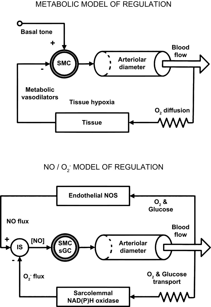
The historically established metabolic theory of local blood flow regulation assumes the existence of basal tone in arterioles (Fig. 1, top). An increase in cell respiration leads to a drop in tissue/cellular Po2 and then to the production of vasodilator metabolites by parenchymal cells, which increase local blood flow via a negative feedback control (30, 31, 33). However, this mechanism implies that the restoration of an adequate oxygen supply stops the production of the metabolic signal, which should then reduce local blood flow. The drawback of this model is the inevitable maintenance of tissue hypoxia. A key issue for the traditional metabolic theory is a failure to confirm the necessary components for this mechanism of regulation: an active closure of capillaries and significant change in capillary density (14, 18, 28), a functional role for precapillary sphincters (10), and certain metabolic vasodilator(s) produced by parenchymal cells during hypoxia (31, 32). Because of its reliance on tissue-produced vasodilators, the old metabolic theory cannot incorporate new knowledge about the action of the signaling radical superoxide (O2−) and nitric oxide (NO), a strong vasodilator produced by the vascular endothelium and not by tissue cells.
Fig. 1.
Top: metabolic model of regulation. When the O2 supply to a tissue whose activity increases is insufficient to meet the increased O2 demand, tissue hypoxia ensues and metabolic vasodilators are released from the active tissue into the interstitium, where they diffuse to the nearby arterioles, producing a negative feedback signal, causing them to dilate. The increased blood flow brings an elevated oxygen supply that relieves the tissue hypoxia (and the stimulus for producing the vasodilators). Bottom: NO/O2− model of regulation. When O2 and glucose content in the blood is sufficient, eNOS produces a constant flux of NO into the perivascular space. This provides a positive feedback signal for the smooth muscle cells (SMC) of the arteriolar wall. This NO flux serves as a reference signal (“set point”) and is constant if Po2 in the blood is high enough. Transport of O2 and glucose to parenchymal cells is restricted by diffusion and transmembrane glucose transporters. This is presented as a resistor in the diagram. The parenchymal cells and vascular wall are consumers of glucose and O2 and are sensors of their adequate supply. With sufficient or excessive delivery of glucose, the membrane NAD(P)H oxidase is supplied with its substrates NADH and NADPH from the cytosol to generate extracellular O2−. Thus the negative feedback signal reporting the achievement of a high level of supply is encoded as the rate of O2− emission into the interstitial space (IS). The periarteriolar NO concentration ([NO]) in the IS is determined by the interaction of NO and O2− fluxes. The superoxide flux depends on a sufficient supply of O2 and glucose to parenchymal cells, and the NO flux is determined by the high content of oxygen and glucose in blood. The interaction of these fluxes in the interstitial space is similar to the subtraction of part of the NO flux due to its neutralization by superoxide. The result of this subtraction is the error signal, represented as [NO] at the SMCs of the arteriolar wall. These cells serve as a controller of vascular wall tone via the soluble guanylyl cyclase (sGC) pathway, changing the inner diameter and hydraulic resistance of arterioles to adjust the local blood flow. At rest, the perivascular [NO] is low and arterioles are constricted. Activation of oxidative metabolism reduces the production of O2−, thus opening the arterioles with high [NO]. The presence of ecSOD in the interstitium accelerates removal of the residual O2− and accelerates the development of hyperemia.
An alternative approach to the regulation of local blood flow began developing a quarter of a century ago, when the mechanism of vasodilation by NO produced by the endothelium was established (11, 15, 27). At the same time, the direct inhibitory effect of O2− on NO was demonstrated (13, 15, 23, 27, 35, 36). It was found that O2− is a specific antagonist of NO, which reacts with NO at a rate limited by diffusion: from 6.7 × 10⁹ M/s (4) to 1.9 × 10¹⁰ M/s (17). This knowledge formed the basis for understanding the NO/O2− system and the contributions of the constitutive enzymes eNOS and NAD(P)H oxidase and extracellular superoxide dismutase (ecSOD) to the control of microvascular tone by a radical signaling mechanism (1–3, 5, 7, 8, 19, 20, 22, 24, 25, 34, 37). A detailed review describing the numerous studies leading to the formulation of the NO/O2− signaling mechanism of local blood flow regulation was recently published (12).
For a new perspective on the regulation of local blood flow, the key assumptions of the metabolic hypothesis need to be critically re-examined: the maintenance of basal tone in the arteriolar wall and the action of metabolic vasodilators produced by parenchymal cells in response to hypoxia. Quite the contrary, the proposed mechanism involving NO/O2− coupling is based on the concept that functional activity is the normal physiological state for a tissue; the corresponding normal state for its vasculature is dilation (12). This eliminates the problem of basal tone, because the basal dilation state is supported by continuous production of NO by the constitutive enzyme eNOS in microvascular endothelial cells. Active regulation of local blood flow comes about when the rate of oxygen and glucose supply reaches or exceeds the tissue demand, thus leading to an abundance of cytosolic reducing agents (NADH and NADPH) and extracellular oxygen that are the substrates for membrane NAD(P)H oxidase in parenchymal cells and the vascular wall (Fig. 1, bottom). That circumstance causes the production of O2− into the interstitial space and subsequent neutralization of some of the interstitial NO, which leads to constriction of the arterioles and decreased local blood flow.
An increase of the functional activity of an organ activates the mitochondria, which causes the import of reducing equivalents from the cytosol into mitochondria and at the same time reduces the oxygen tension on the surface of parenchymal cells. This reduces the production of O2− into the interstitial space by NAD(P)H oxidase. Extracellular SOD, having a lower rate of O2− removal compared with NO (2–4 × 10⁹ M/s), degrades the remaining interstitial O2−. A low level of O2− opens the interstitial space for the diffusive flux of NO to the smooth muscle cells in arterioles, causing them to dilate and increase local blood flow. Thus the interstitial concentration of NO acts as an error signal whose magnitude is related to the mismatch between oxygen and glucose delivery and their consumption (Fig. 1, bottom). To start active hyperemia, the tissue cells need not produce a metabolic vasodilator(s), but just stop the emission of the NO inhibitor (O2−) into the interstitial space. Reactive hyperemia in the NO/O2−-based model is mechanistically different from active hyperemia, because the blood flow interruption also affects the positive feedback loop, stopping the basal production of NO (Fig. 1, bottom). After reperfusion, the microvascular endothelium immediately receives nutrients and oxygen to produce NO, whereas diffusion and metabolic limitations lead to a delay in the release of O2− from the tissue cells. Therefore, the response of the system during reperfusion is determined by the duration of the ischemia (6, 16, 21) and the presence of oxygen in the blood (9).
It should be emphasized that the NO/O2−-based model of regulation is aimed to control the upper level of local blood flow so that the constriction of arterioles during low metabolic activity prevents the cells in a tissue from being exposed to an excessive supply of oxygen and substrates. Thus the goal of regulation is maintaining a sufficient, but not excessive, supply of oxygen and substrates to the cells. Increasing blood flow simply requires a termination of the inhibitory signal (O2−) instead of the production of metabolic vasodilators. NO signaling, currently considered a redundant and stand-alone vasodilation pathway, becomes the key mechanism for regulation of local blood flow. This hypothesis explains why the vasodilation signal is generated by microvessels and how the parenchymal cells block this signal when local blood flow is sufficient. The proposed mechanism consists of well studied components: the enzymes eNOS, NAD(P)H oxidase, ecSOD and the two rapidly diffusing and interacting signaling radicals. Because O2− is compartmentalized in a small extracellular space, its interstitial concentration can change quickly. Production of O2− by membrane NAD(P)H oxidase depends on the balance of glycolysis and oxidative phosphorylation via the availability of NADH and NAD(P)H in the cytosol and the interstitial Po2. Thus the coupling of tissue metabolic activity with local blood flow is achieved.
The NO/O2− regulation hypothesis provides conceptual support for the successful practical application of NO donors and other pharmacologic agents used to manipulate the NO signaling pathway. This model underpins the application of pharmacologic modulators of angiotensin II signaling for the correction of hypertension. It is known that most of the actions of angiotensin II are mediated by the AT1 receptor, which can activate membrane NAD(P)H oxidase and increase the production of superoxide into the interstitium (26). The use of blockers of angiotensin converting enzyme and AT1 receptors thus inhibits O2− production and increases NO bioavailability for the SMCs in the arteriolar wall. The proposed mechanism of regulation defines physiologic roles for the signaling radicals NO and O2− as key elements to control an adequate supply of oxygen and glucose in critical organs. By the explanation of local blood flow regulation in healthy organisms, this model provides a solid conceptual tool to support medical research into tissue hypoxia, development of vascular pathologies, tolerance and cross-tolerance to nitrovasodilators, and neurovascular coupling. A key role of glycolysis in maintaining the turnover of cytosolic NADH and O2− production by the sarcolemmal NAD(P)H oxidase also allows glucose metabolism to be considered as an important factor in local blood flow regulation during metabolic syndrome.
GRANTS
The work of the authors included in this article was supported in part by Grant HL18292 from the National Heart, Lung, and Blood Institute.
DISCLOSURES
No conflicts of interest, financial or otherwise, are declared by the author(s).
AUTHOR CONTRIBUTIONS
Author contributions: A.S.G. and R.N.P. conception and design of research; A.S.G. prepared figures; A.S.G. drafted manuscript; A.S.G. and R.N.P. edited and revised manuscript; A.S.G. and R.N.P. approved final version of manuscript.
ACKNOWLEDGMENTS
The authors are grateful to the reviewers of this manuscript for making insightful and thoughtful comments and suggestions which improved the overall presentation of this material.
REFERENCES
- 1.Afanas'ev I. Interplay between superoxide and nitric oxide in aging and diseases. Biogerontology 5: 267–270, 2004 [DOI] [PubMed] [Google Scholar]
- 2.Ahmeda AF, Johns EJ. The regulation of blood perfusion in the renal cortex and medulla by reactive oxygen species and nitric oxide in the anaesthetised rat. Acta Physiol (Oxf) 204: 443–450, 2012 [DOI] [PubMed] [Google Scholar]
- 3.Bagi Z, Koller A, Kaley G. Superoxide-NO interaction decreases flow- and agonist-induced dilations of coronary arterioles in Type 2 diabetes mellitus. Am J Physiol Heart Circ Physiol 285: H1404–H1410, 2003 [DOI] [PubMed] [Google Scholar]
- 4.Beckman JS, Koppenol WH. Nitric oxide, superoxide, peroxynitrite: the good, the bad, ugly. Am J Physiol Cell Physiol 271: C1424–C1437, 1996 [DOI] [PubMed] [Google Scholar]
- 5.Bharadwaj LA, Prasad K. Mechanism of superoxide-anion induced modulation of vascular tone. Int J Angiol 11: 23–29, 2002 [Google Scholar]
- 6.Burton KS, Johnson PC. Reactive hyperemia in individual capillaries of skeletal muscle. Am J Physiol 223: 517–524, 1972 [DOI] [PubMed] [Google Scholar]
- 7.Cosentino F, Sill JC, Katusic ZS. Role of superoxide anions in the mediation of endothelium-dependent contractions. Hypertension 23: 229–235, 1994 [DOI] [PubMed] [Google Scholar]
- 8.Demchenko IT, Boso AE, Bennett PB, Whorton AR, Piantadosi CA. Hyperbaric oxygen reduces cerebral blood flow by inactivating nitric oxide. Nitric Oxide 4: 597–608, 2000 [DOI] [PubMed] [Google Scholar]
- 9.Fairchild HM, Ross J, Guyton AC. Failure of recovery from reactive hyperemia in the absence of oxygen. Am J Physiol 210: 490–492, 1966 [DOI] [PubMed] [Google Scholar]
- 10.Fulton GP, Lutz BR. The neuromotor mechanism of the small blood vessels of the frog. Science 92: 223–224, 1940 [DOI] [PubMed] [Google Scholar]
- 11.Furchgott RF, Zawadzki JV. The obligatory role of endothelial cells in the relaxation of arterial smooth muscle by acetylcholine. Nature 288: 373–376, 1980 [DOI] [PubMed] [Google Scholar]
- 12.Golub AS, Pittman RN. Bang-bang model for regulation of local blood flow. Microcirculation 20: 455–483, 2013 [DOI] [PMC free article] [PubMed] [Google Scholar]
- 13.Gryglewski RJ, Palmer RM, Moncada S. Superoxide anion is involved in the breakdown of endothelium-derived vascular relaxing factor. Nature 320: 454–456, 1986 [DOI] [PubMed] [Google Scholar]
- 14.Hirai DM, Copp SW, Schwagerl PJ, Musch TI, Poole DC. Acute effects of hydrogen peroxide on skeletal muscle microvascular oxygenation from rest to contractions. J Appl Physiol 110: 1290–1298, 2011 [DOI] [PubMed] [Google Scholar]
- 15.Ignarro LJ. Endothelium-derived nitric oxide: actions and properties. FASEB J 3: 31–36, 1989 [DOI] [PubMed] [Google Scholar]
- 16.Johnson PC, Burton KS, Henrich H, Henrich U. Effect of occlusion duration on reactive hyperemia in sartorius muscle capillaries. Am J Physiol 230: 715–719, 1976 [DOI] [PubMed] [Google Scholar]
- 17.Kissner R, Nauser T, Bugnon P, Lye PG, Koppenol WH. Formation and properties of peroxynitrite as studied by laser flash photolysis, high-pressure stopped-flow technique, and pulse radiolysis. Chem Res Toxicol 10: 1285–1292, 1997 [DOI] [PubMed] [Google Scholar]
- 18.Krogh A. The supply of oxygen to the tissues and the regulation of the capillary circulation. J Physiol 52: 457–474, 1919 [DOI] [PMC free article] [PubMed] [Google Scholar]
- 19.Lai EY, Wellstein A, Welch WJ, Wilcox CS. Superoxide modulates myogenic contractions of mouse afferent arterioles. Hypertension 58: 650–656, 2011 [DOI] [PMC free article] [PubMed] [Google Scholar]
- 20.Liochev SI, Fridovich I. Superoxide and nitric oxide: consequences of varying rates of production and consumption: a theoretical treatment. Free Radic Biol Med 33: 137–141, 2002 [DOI] [PubMed] [Google Scholar]
- 21.Lombard JH, Duling BR. Multiple mechanisms of reactive hyperemia in arterioles of the hamster cheek pouch. Am J Physiol Heart Circ Physiol 241: H748–H755, 1981 [DOI] [PubMed] [Google Scholar]
- 22.Mian KB, Martin W. Differential sensitivity of basal and acetylcholine-stimulated activity of nitric oxide to destruction by superoxide anion in rat aorta. Br J Pharmacol 115: 993–1000, 1995 [DOI] [PMC free article] [PubMed] [Google Scholar]
- 23.Moncada S, Palmer RM, Gryglewski RJ. Mechanism of action of some inhibitors of endothelium-derived relaxing factor. Proc Natl Acad Sci USA 83: 9164–9168, 1986 [DOI] [PMC free article] [PubMed] [Google Scholar]
- 24.Munzel T, Heitzer T, Harrison DG. The physiology and pathophysiology of the nitric oxide/superoxide system. Herz 22: 158–172, 1997 [DOI] [PubMed] [Google Scholar]
- 25.Munzel T, Hink U, Heitzer T, Meinertz T. Role for NADPH/NADH oxidase in the modulation of vascular tone. Ann NY Acad Sci 874: 386–400, 1999 [DOI] [PubMed] [Google Scholar]
- 26.Nguyen Dinh Cat A., Montezano A. C, Burger D, Touyz RM. Angiotensin II, NADPH oxidase, and redox signaling in the vasculature. Antioxid Redox Signal 19: 1110–1120, 2013 [DOI] [PMC free article] [PubMed] [Google Scholar]
- 27.Palmer RM, Ferrige AG, Moncada S. Nitric oxide release accounts for the biological activity of endothelium-derived relaxing factor. Nature 327: 524–526, 1987 [DOI] [PubMed] [Google Scholar]
- 28.Poole DC, Brown MD, Hudlicka O. Counterpoint: There is no capillary recruitment in active skeletal muscle during exercise. J Appl Physiol 104: 891–894, 2008 [DOI] [PubMed] [Google Scholar]
- 29.Poole DC, Copp SW, Hirai DM, Musch TI. Dynamics of muscle microcirculatory and blood-myocyte O(2) flux during contractions. Acta Physiol (Oxf) 202: 293–310, 2011 [DOI] [PMC free article] [PubMed] [Google Scholar]
- 30.Rowell LB. Ideas about control of skeletal and cardiac muscle blood flow (1876–2003): cycles of revision and new vision. J Appl Physiol 97: 384–392, 2004 [DOI] [PubMed] [Google Scholar]
- 31.Roy CS, Brown JG. The blood-pressure and its variations in the arterioles, capillaries and smaller veins. J Physiol 2: 323–446, 1880 [DOI] [PMC free article] [PubMed] [Google Scholar]
- 32.Roy CS, Sherrington CS. On the regulation of the blood-supply of the brain. J Physiol 11: 85–158, 1890 [DOI] [PMC free article] [PubMed] [Google Scholar]
- 33.Sarelius I, Pohl U. Control of muscle blood flow during exercise: local factors and integrative mechanisms. Acta Physiol (Oxf) 199: 349–365, 2010 [DOI] [PMC free article] [PubMed] [Google Scholar]
- 34.Thomas DD, Ridnour LA, Espey MG, Donzelli S, Ambs S, Hussain SP, Harris CC, DeGraff W, Roberts DD, Mitchell JB, Wink DA. Superoxide fluxes limit nitric oxide-induced signaling. J Biol Chem 281: 25984–25993, 2006 [DOI] [PubMed] [Google Scholar]
- 35.Wei EP, Christman CW, Kontos HA, Povlishock JT. Effects of oxygen radicals on cerebral arterioles. Am J Physiol Heart Circ Physiol 248: H157–H162, 1985 [DOI] [PubMed] [Google Scholar]
- 36.Wei EP, Kontos HA, Christman CW, DeWitt DS, Povlishock JT. Superoxide generation and reversal of acetylcholine-induced cerebral arteriolar dilation after acute hypertension. Circ Res 57: 781–787, 1985 [DOI] [PubMed] [Google Scholar]
- 37.Zhilyaev SY, Moskvin AN, Platonova TF, Gutsaeva DR, Churilina IV, Demchenko IT. Hyperoxic vasoconstriction in the brain is mediated by inactivation of nitric oxide by superoxide anions. Neurosci Behav Physiol 33: 783–787, 2003 [DOI] [PubMed] [Google Scholar]


