Postmenopausal uterine bleeding can be spontaneous or related to ovarian hormone replacement therapy or to the use of selective estrogen receptor modulators. The guideline development group determined that, for initial management of spontaneous postmenopausal bleeding, primary assessment may be with either endometrial sampling or transvaginal ultrasonography. Guidelines are also provided for patients receiving selective estrogen receptor modulators or hormone replacement therapy.
Abstract
Postmenopausal uterine bleeding is defined as uterine bleeding after permanent cessation of menstruation resulting from loss of ovarian follicular activity. Bleeding can be spontaneous or related to ovarian hormone replacement therapy or to use of selective estrogen receptor modulators (eg, tamoxifen adjuvant therapy for breast carcinoma). Because anovulatory “cycles” with episodes of multimonth amenorrhea frequently precede menopause, no consensus exists regarding the appropriate interval of amenorrhea before an episode of bleeding that allows for the definition of postmenopausal bleeding. The clinician faces the possibility that an underlying malignancy exists, knowing that most often the bleeding comes from a benign source. Formerly, the gold-standard clinical investigation of postmenopausal uterine bleeding was institution-based dilation and curettage, but there now exist office-based methods for the evaluation of women with this complaint. Strategies designed to implement these diagnostic methods must be applied in a balanced way considering the resource utilization issues of overinvestigation and the risk of missing a malignancy with underinvestigation. Consequently, guidelines and recommendations were developed to consider these issues and the diverse spectrum of practitioners who evaluate women with postmenopausal bleeding. The guideline development group determined that, for initial management of spontaneous postmenopausal bleeding, primary assessment may be with either endometrial sampling or transvaginal ultrasonography, allowing patients with an endometrial echo complex thickness of 4 mm or less to be managed expectantly. Guidelines are also provided for patients receiving selective estrogen receptor modulators or hormone replacement therapy, and for an endometrial echo complex with findings consistent with fluid in the endometrial cavity.
Guideline History and Scope
The Abnormal Uterine Bleeding Working Group (AUBWG) was originally created in 2004 to develop evidence-based, consensus guidelines for the management of abnormal uterine bleeding for women in Kaiser Permanente’s Southern California (KPSC) Region (see Sidebar: Members of the Abnormal Uterine Bleeding Working Group). The Southern California Permanente Medical Group (SCPMG) is the only Medical Group contracted to provide care for the more than three million members of the Kaiser Foundation Health Plan in the Southern California Region. The AUBWG determined that the scope of these guidelines included the investigation and initial management of women with postmenopausal bleeding thought or known to emanate from the vagina.a1 The first version of these guidelines, based on evidence published to December 2004, was completed in 2005 and published in an abbreviated format on the Guideline Clearinghouse Web site of the Agency for Healthcare Research and Quality, with the full text made available on request. This version of the guidelines includes the result of a major systematic review of the evidence published since January 2005.
Members of the Abnormal Uterine Bleeding Working Group.
Malcolm G Munro, MD (Chair)
Michael S Amann, MD
Hector E Anguiano, MD
Rosoalie A Bauman, MD
Mon-Lai Cheung, MD
Seth Kivnick, MD
Murali H Kamath, MD
Beatriz R Lauria, MD
Nakia T Mainor, MD
Paula D Richter, MD
Hazim K Shams, MD
Saad Z Solh, MD
Janet J Zhang, MD
Postmenopausal uterine bleeding, either spontaneous or that related to ovarian hormone replacement therapy (HRT) or selective estrogen receptor modulator (SERM) use (eg, tamoxifen adjuvant therapy for breast carcinoma) collectively results in a substantial number of patient encounters. These encounters directly involve physicians and other clinicians in the Departments of Family Medicine, Internal Medicine, Obstetrics and Gynecology, and Oncology and include physicians, nurse practitioners, and other midlevel clinicians. The clinical problem also indirectly affects medical clinicians and resources in the Departments of Radiology and Pathology, particularly because of the use of endometrial sampling and imaging of the uterus. Consequently, recommendations have been developed considering this diverse spectrum of practitioners and departments.
Methods
The Chair of the AUBWG was selected by the Regional Chief of Obstetrics and Gynecology for SCPMG. The remaining members of the AUBWG were selected by the chairs of each of the 12 KPSC Medical Centers on the basis of their interest in abnormal uterine bleeding and ability to contribute to guideline development.
After an introductory discussion on the general and SCPMG-specific issues involved in the investigation of postmenopausal bleeding, the committee met as a whole to review methods of guideline development, agree on terms for evidence classification, define basic terms (Table 1) and to come to consensus on the scope of the guidelines to be developed. (See prior section, Guideline History and Scope; Tables 2 and 3; the Sidebars: Problem Formation and Suggested Contents for Report of Endometrial Echo Complex Evaluation.) The Working Group Chair prepared a template to aid the guideline development process. For the revision process, the existing document underwent a draft revision following a systematic review of the literature.
Table 1.
Definitions of terms
| Term | Definition |
|---|---|
| Postmenopausal bleeding | Spontaneous uterine bleeding that occurs more than one year after the date of the last menstrual period |
| Breakthrough bleeding | Unscheduled uterine bleeding encountered in any postmenopausal woman using hormone replacement therapy |
| Satisfactory endometrial biopsy | Comprises perceptible passage of the sampling device through the cervical canal into the endometrial cavity and appropriate functioning of the aspiration mechanism. Adequacy of the specimen for histologic interpretation is determined by the pathologist. |
Table 2.
Hierarchy of evidencea
| Class | Type of evidence |
|---|---|
| 1ab | Meta-analysis of randomized clinical trials |
| 1b | Randomized clinical trial |
| 2 | Meta-analysis of studies that are not randomized |
| 3 | Nonrandomized, but internally controlled trials. Controls are considered to be “internal” if they are included in the original design of the study. Post hoc or historical comparisons are not considered internal controls. Comparisons of otherwise uncontrolled clinical series are not considered internal controls. |
| 4 | Case-control studies |
| 5 | Cohort studies |
| 6 | Clinical series, without internal comparison |
| 7b | Expert opinion without available clinical studies |
Source: Kaiser Foundation Guideline Development Group, Edition 3, September 1, 2004, using a modification of the system adopted by the US Preventive Services Task Force of the Agency for Health Research and Quality.
Level of evidence added by the Abnormal Uterine Bleeding Working Group.
Table 3.
Support for recommendationsa
| Strength of Evidence | Language | Evidence |
|---|---|---|
| A | Guideline Development Team (GDT) strongly recommends that clinicians routinely provide the intervention to eligible patients | Intervention improves important health outcomes, based on good evidence, and the GDT concludes that benefits substantially outweigh harms and costs |
| B | GDT recommends that clinicians routinely provide the intervention to eligible patients | Intervention improves important health outcomes, based on 1) good evidence that benefits outweigh harms and costs or 2) fair evidence that benefits substantially outweigh harms and costs |
| C | GDT makes no recommendation for or against routine provision of the intervention. At the discretion of the GDT, the recommendation may use the language “option,” but must list all the equivalent options. | Evidence is sufficient to determine the benefits, harms, and costs of an intervention, and there is at least fair evidence that the intervention improves important health outcomes. However, the GDT concludes that the balance of the benefits, harms, and costs is too close to justify a general recommendation. |
| D | GDT recommends against routinely providing the intervention to eligible patients | GDT found at least fair evidence that the intervention is ineffective, or that harms or costs outweigh benefits |
| I | GDT concludes that the evidence is insufficient to recommend for or against routinely providing the intervention. At the discretion of the GDT, the recommendation may use the language “option” but must list all the equivalent options. | Evidence that the intervention is effective is lacking, of poor quality, or conflicting, and the balance of benefits, harms, and costs cannot be determined |
Source: Kaiser Foundation Guideline Development Group, Edition 3, September 1, 2004.
Problem Formulation.
Intended use of the guideline
To assist physicians and other health care professionals in the evaluation and management of uterine bleeding in postmenopausal women (postmenopausal uterine bleeding)
Health problem
Postmenopausal uterine bleeding
Health interventions
Strategies and techniques for investigation of women with postmenopausal uterine bleeding
Population
Postmenopausal women
Practitioners
Physicians, physician assistants, nurse practitioners, and other health care professionals in the Departments of Emergency Medicine, Family Medicine, Internal Medicine, and Obstetrics and Gynecology
Medical setting
Offices, clinics, Emergency Departments, and hospital inpatients
Most important health outcomes
Outcomes of condition:
Potential sign of endometrial or other uterine malignancy or premalignant condition such as atypical endometrial hyperplasia
May reflect benign causes (including idiopathic, endometrial polyps) and iatrogenic sources
Outcomes of intervention:
Identification of women with atypical endometrial hyperplasia and cancer
Identification of women with usually benign lesions such as endometrial polyps
Suggested Contents for Report of Endometrial Echo Complex (EEC) Evaluation.
Normally Configured Uterus, EEC well seen
The uterus measures __ __ .__ cm in length; the corpus__ __ . __cm in width, and __ __ . __ cm in anterior-posterior thickness. The EEC is seen in its entirety in both the sagittal and transverse planes, is consistent in thickness and echogenicity, and measures __ __ . __ mm throughout.
Comment: ________________________________________________
Uterus with EEC well seen, endometrial cavity containing sonolucent fluid
The uterus measures __ __ .__ cm in length; the corpus__ __ . __cm in width, and __ __ . __ cm in anterior-posterior thickness. The EEC is seen in its entirety in both the sagittal and transverse planes, and contains sonolucent fluid. The endometrium on both sides of this fluid is consistent in thickness. The two-layer endometrial thickness calculated by subtracting the thickness of the fluid from the total endometrial echo complex thickness is __ __ . __ mm.
Comment: ________________________________________________
Generic
Uterine length: __ __ . __ cm
Corpus width: __ __ . __ cm
Corpus thickness: __ __ . __ cm
Endometrial fluid: □Yes □No
If Yes: Sonolucent? □Yes □No
Thickness of sonolucent fluid (sagittal): _ _ . __ mm
Thickness of sonolucent fluid __ . __ mm
Entire EEC seen: □Yes □No
EEC consistent throughout: □Yes □No
Double-layer endometrial thickness: Sagittal plane: __ __ . __ mm
Transverse plane: __ __ . __ mm
Net endometrial thickness if sonolucent fluid subtracted: Sagittal plane: __ __ . __ mm
Transverse plane: __ __ . __ mm
Comment: ________________________________________________
For the original review, a subgroup of three individuals was charged with leading the investigation and developing draft documents for meetings of the whole group. Drafts were distributed electronically to the whole group, and monthly face-to-face meetings were used to obtain feedback from each of the members of the committee. For this major revision, all members of the AUBWG were involved in the review and revision process with two face-to-face meetings and teleconferences as appropriate.
Literature searches were performed in MEDLINE and The Cochrane Database of Systematic Reviews between January 1, 2005 and November 19, 2012, using the following search terms: postmenopausal uterine bleeding, transvaginal ultrasound, sonohysterography, saline infusion sonography, hysteroscopy, endometrial biopsy, tamoxifen, and hormone replacement therapy (see Sidebar: Evidence Tables [available from: www.thepermanentejour-nal.org/files/Winter2014/EvidenceSearch.pdf). Also searched were the Web sites of the American College of Obstetricians and Gynecologists (www.acog.org/Resources_And_Publications), the Society of Obstetricians and Gynecologists of Canada (http://sogc.org/clinical-practice-guidelines/), the New Zealand Guidelines Group (www.health.govt.nz/about-ministry/ministry-health-websites/new-zealand-guidelines-group), the National Institute for Health and Clinical Excellence (www.nice.org.uk), and the Geneva Foundation for Medical Education and Research (www.gfmer.ch/000_Homepage_En.htm). Each Web site was a repository of evidence-based and consensus-based guidelines.
The original postmenopausal bleeding guidelines and subsequent revisions were developed using the identified evidence and, where deemed necessary by the participants, a consensus process with which to classify, interpret, and develop components of the guideline not evaluated adequately by the literature. Evidence was classified using a modification of the system adopted by the US Preventive Services Task Force of the Agency for Healthcare Research and Quality (Table 2), adding a subgroup for Class 1 evidence to allow for meta-analysis of randomized controlled trials, such as a Cochrane review. An additional modification was to include the creation of a Class 7 grouping that isolated expert opinion, including that from guidelines or other consensus documents from national or international organizations or from the collective opinion of the AUBWG members. The recommendations were created and classified according to the strength of the evidence and classified according to the system used by KPSC Guideline Development teams (Table 3). The term good evidence used in the text or tables indicates that either Grade A or Grade B evidence was available to support the recommendations (Table 4). The process was designed to be a continuous one, allowing for ongoing modifications and revisions as new, higher quality or otherwise clarifying evidence becomes available.
Table 4.
Summary of recommendationsa
| No. | Recommendation | Method | Strength of evidenceb |
|---|---|---|---|
| 1 | Women with spontaneous postmenopausal bleeding should be primarily evaluated with either endometrial biopsy (EB), or transvaginal ultrasound (TVUS) to measure the thickness of the endometrial echo complex (EEC) | E | A |
| 2 | When TVUS or contrast sonography such as saline infusion sonography (SIS) are used as techniques for assessing the endometrium of women with postmenopausal bleeding, a detailed description of the evaluation should be placed in the patient record with or without representative photographs of the sagittal and transverse planes | C | N/A |
| 3 | Practitioners without adequate training in either office-based EB orTVUS should refer patients with postmenopausal bleeding or breakthrough bleeding to an individual, usually a gynecologist, appropriately trained in these techniques | C | N/A |
| 4 | Women with spontaneous postmenopausal bleeding and an EEC > 4 mm should be further evaluated with endometrial sampling | E | A |
| 5 | If clear endometrial fluid is identified on E the fluid from the EEC measurement | B | |
| 6 | In the presence of sonolucent fluid, women with a measurement of≤3 mm may be managed expectantly. In the presence of postmenopausal bleeding, cervical carcinoma should be considered and evaluated appropriately | E | B |
| 7 | Women with persistent spontaneous postmenopausal bleeding require further evaluation of the endometrial cavity for focal lesions with one or a combination of office-based contrast sonography (eg, SIS) and hysteroscopy. Such an approach is necessary even if there is a satisfactory or adequate EB without evidence of hyperplasia, and regardless of the EEC thickness | E | B |
| 8 | Operating room-based dilation and curettage (D&C) of women with postmenopausal bleeding should be performed only when office-based EB is indicated, cannot be performed for patient comfort or technical reasons, or when it is inconclusive and results of sonographic techniques (TVUS, SIS) are not reassuring | E | B |
| 9 | Women taken to the operating room for D&C should have concomitant hysteroscopy with ancillary instruments that allow for the removal of focal lesions such as endometrial polyps | C | N/A |
| 10 | In the presence of endometrial carcinoma, hysteroscopy does not appear to affect short- or long-term prognosis. Consequently, hysteroscopy is not contraindicated in the evaluation of women with postmenopausal bleeding, including cases suggestive of cancer | E | A |
| 11 | Only selected women with bleeding associated with estrogen and progestin-containing hormone replacement therapy (HRT) require assessment of the endometrium. Uterine bleeding or spotting may be expected depending in part on the dose of HRT administered, in part on the schedule of progestin administration, and in part on the duration of therapy | E | B |
| 12 | It is not necessary to routinely evaluate the endometrium of women with uterine spotting or light uterine bleeding in the first six months of continuous estrogen and progestin HRT. Endometrial assessment of such women is recommended if spotting or bleeding persists beyond six months, although there is a very low incidence of endometrial hyperplasia or neoplasia | E | B |
| 13 | Women receiving doses of unopposed estrogen have a much higher incidence of endometrial hyperplasia and carcinoma, and require appropriate investigation of the endometrium | E | A |
| 14 | Women receiving estrogen and cyclical progestins can be expected to have indefinite progestin withdrawal bleeding and require no further investigation provided that the dose and duration of cyclic progestins is adequate | E | A |
| 15 | For women using cyclic progestins, bleeding outside the time of progestin withdrawal is considered abnormal and requires appropriate investigation | E | B |
| 16 | It is apparent that EEC thresholds used for spontaneous bleeding can be applied to patients with HRT-related bleeding, but with a higher incidence of false-positive findings | E | B |
| 17 | Women experiencing uterine bleeding while receiving tamoxifen (usually used as an adjuvant therapy for breast cancer) should be assessed primarily with endometrial sampling because, in such patients, TVUS is neither sensitive nor specific for neoplasia | E | A |
| 18 | Women with persistent bleeding during tamoxifen therapy, and who have already undergone endometrial sampling, should be assessed with one or a combination of contrast sonography (such as SIS) and hysteroscopy with appropriate sampling or excision of polyps if found | C | N/A |
| 19 | Women with repeated bleeding during tamoxifen therapy, and who have been demonstrated to have normal histologic findings and a structurally normal endometrial cavity, should have EB repeated annually | E | B |
| 20 | Postmenopausal bleeding can be a presenting symptom of cancer in the cervical canal. Consequently, if there is no endometrial explanation for postmenopausal bleeding, appropriate steps to evaluate patients for cervical cancer should be undertaken considering the results of a Papanicolaou test, colposcopy, and curettage of the endocervical canal | C | N/A |
Recommendations are evidence based (E) unless sufficient evidence is not available and consensus-based (C) recommendations are provided.
See Table 2; strength of evidence is not applicable (N/A) to consensus-based recommendations.
The original consensus document was created, approved by the members of the AUBWG in October 2004, and then submitted to the SCPMG Regional Chiefs for review, comment, and approval. Specifically, the Chiefs selected the 5-mm endometrial echo complex (EEC) thickness as the evidence-based threshold above which endometrial sampling is recommended. Following the 2012 review of the available evidence, the recommended threshold was changed to 4 mm. Minor modifications of the guideline were made following presentation to the SCPMG Regional Chiefs of Obstetrics and Gynecology. The revised guidelines were unanimously approved by the Regional Chiefs of Obstetrics and Gynecology on March 20, 2013.
Rationale
Introduction
Postmenopausal bleeding is a common patient complaint that is encountered by all physicians and other clinicians of gynecologic care. The clinician faces the possibility that there exists an underlying malignancy, while knowing that, in most instances, the bleeding comes from a benign source. In years past, the gold standard of clinical investigation was the institution-based dilation and curettage (D&C), but there now exists a number of office-based methods for the evaluation of women with this complaint. The post-menopausal use of gonadal steroids for the treatment of menopausal symptoms (HRT) is often associated with endometrial bleeding. Although the number of women using these agents decreased in the first decade of the 21st century, reevaluation of the literature and the release of new studies have collectively suggested that such therapy may provide benefit in at least selected instances.1–3
As a result, clinicians will continue to be challenged by the issue of HRT-associated postmenopausal bleeding in the near future. It is known that tamoxifen and other SERMs may increase the chance of endometrial neoplasia developing. However, the proportion of postmenopausal women using such agents as adjuvant treatment of breast cancer is decreasing with the introduction of aromatase inhibitors3 (Grade C). Nevertheless, there will continue to exist a number of women in their late reproductive years with unknown or uncertain ovarian endocrine status who experience abnormal bleeding in association with the use of these agents. Such women may have to be considered to have postmenopausal bleeding associated with the use of a SERM.
The clinician should appreciate that although the focus of investigation of postmenopausal bleeding is on the endometrium, bleeding in the postmenopausal woman may arise from a number of extraendometrial gynecologic and nongynecologic sites, such as the cervix, vagina, and urologic and gastrointestinal tracts. As a result, it is incumbent on the clinician to consider all these possibilities when evaluating a patient with postmenopausal bleeding or apparent unscheduled (“breakthrough”) bleeding who is receiving HRT. Consequently, should the results of appropriately indicated and performed evaluation of the endometrium be normal, reevaluation for these potential causes is a prudent approach.
Definition of Postmenopausal Bleeding
The World Health Organization defines menopause as the permanent cessation of menstruation resulting from the loss of ovarian follicular activity.4 Because anovulatory “cycles” with episodes of multimonth amenorrhea frequently precede menopause, there is no consensus regarding the appropriate interval of amenorrhea preceding an episode of bleeding that would allow for the definition of postmenopausal uterine bleeding. For the purposes of this guideline, an episode of bleeding 12 months after the last menstrual period will be deemed to constitute postmenopausal bleeding. However, and especially in the perimenopausal years, the definition of “last period” may be difficult to ascertain, so liberal use of assays for estradiol and follicle stimulating hormone will help to determine when bleeding is occurring absent measurable ovarian function.
For women receiving HRT, bleeding is common, and its frequency and timing depends in part on the scheduling of the gonadal steroids used, particularly the progestational agent5 (Class 5). Breakthrough bleeding is unscheduled uterine bleeding that occurs in women receiving either estrogen alone or both estrogen and progestin therapy, which does not include the withdrawal bleeding that occurs following the cyclic withdrawal of progestin treatment. About 50% of women using continuous estrogen-progestin combined replacement regimens experience breakthrough bleeding, with most cases resolving 6 months after initiation of therapy6 (Class 4).
The following definitions of abnormal bleeding during HRT are suggestedb:
For women using cyclically administered progestins in combination with a cyclical or continuous estrogen: 1) occurs at an unscheduled time or 2) occurs at the anticipated time after progestin withdrawal but is heavy or prolonged.
For women using continuous combined estrogen and progestin-containing regimens: 1) commences six months or more after initiation or 2) commences after amenorrhea has been established.
Risk of Endometrial Cancer
In the US, endometrial cancer is the most commonly diagnosed reproductive tract malignancy and is the fourth most common cancer among women, trailing only cancers of the breast, lung, and colorectal origin7 (Class 5). Although endometrial cancer accounts for 6% of all female cancers, a number of intrinsic clinical features, including a propensity for early diagnosis (because of investigation of abnormal or postmenopausal bleeding) and the prompt use of effective therapy, it causes only about 3% of all cancer-related deaths.
There are 2 distinct types of endometrial cancer; most are Type 1, which develops secondary to unopposed estrogen-induced endometrial hyperplasia with an endometrioid appearance on histopathologic evaluation. Type 2 lesions comprise the minority, are of serous or clear cell origin, are not related to estrogen exposure, and are associated with a relatively poor prognosis. Older literature would suggest that Type 2 endometrial cancers comprise between 16% and 35% of endometrial cancer cases8,9 (Class 5), but more recent data from the US reveal that approximately 90% are Type 1 and about 10% are Type 2 lesions10 (Class 5).
For women with postmenopausal bleeding who do not use HRT, the incidence of endometrial cancer ranges from 4.9% to 11.5%11–14 (Class 5). There appears to be a greater risk of endometrial cancer in women who present with post-menopausal bleeding 10 or more years following menopause. One relatively large study found that postmenopausal bleeding over the age of 60 years was associated with a 13% risk of endometrial cancer11 (Class 5). Another large cohort study of slightly more than 3000 women with postmenopausal bleeding identified a peak in the age group from 60 through 64 years, of whom 7% were found to have endometrial cancer; the risk was lower for those in the younger and older age groups14 (Class 5).
There are no reliable data regarding the incidence of endometrial carcinoma in women using estrogen and progestin-containing HRT regimens. However, the estimated proportions range from 0.02% to 0.05% per annum15–17 (Class 5). For women receiving tamoxifen, the risk of endometrial cancer is likely above 10% depending in part on the dose and in part on the duration of therapy18–21 (Class 4).
Relationship to Age
The national registries of nations with government-supported health care programs provide information regarding demographic variables associated with the risk of endometrial carcinoma. The incidence of endometrial cancer is very low below the age of 50 years, and especially before 40 years. The Scottish national data suggest that the incidence of endometrial carcinoma is about 6 to 8/100,000 women each year after the age of 50 years22 (Class 4). However, these data do not describe the risk of endometrial cancer in women presenting with postmenopausal bleeding. Swedish data demonstrate that the incidence of postmenopausal bleeding declines with succeeding years after menopause and that the incidence of endometrial carcinoma increases as the age of the patient with postmenopausal bleeding increases12 (Class 4).
Hormone Replacement Therapy
The relative risk of development of endometrial cancer for a woman receiving unopposed estrogen preparations in historically typical doses is about 5 times that for nonusers17 (Class 4). This increased risk is almost, if not totally, eliminated if appropriate cyclic or continuous progestins are added to the regimen17 (Class 4),23 (Class 1a),24 (Class 1b). However, a number of features may affect these risks, including the dose of the estrogen, and the dose, route, and regimen used to administer the progestin.
Most of the high-quality evidence relating to estrogen and progestin type and dosing and the risks of endometrial neoplasia has been obtained from randomized trials using endometrial hyperplasia as a surrogate outcome, studies that are well summarized in The Cochrane Database of Systematic Reviews25 (Class 1a). Unopposed estrogen, compared with placebo, is associated with an odds ratio (OR) for the short-term (18- to 24-months) development of endometrial hyperplasia that is 2.42 (95% confidence interval [CI], 1.19–4.92), 11.86 (CI, 7.76–18.14), and 13.06 (CI, 5.88–29.02) for low-, moderate-, and high-dose formulations, respectively. When estrogen is administered in conjunction with a progestin, these risks are reduced substantially if an adequate dose is administered for an appropriate time.
When the progestin of a combined HRT regimen is administered cyclically, optimal results are obtained with 10 to 14 days/month because there is evidence that for women who use cyclic progestin therapy for less than 10 days per cycle, the risk of endometrial cancer is raised (OR, 2.9; CI, 1.8–4.6) 17 (Class 4). The minimum cycle duration has generally been considered to be 1 month. However, there is fair evidence that quarterly or even semiannual cycling for 14 days of medroxyprogesterone acetate (10 mg/day) is adequate to prevent most cases of endometrial hyperplasia or neoplasia, at least with conjugated equine estrogen (or equivalent) doses up to 0.625 mg/day26 (Class 1b),27,28 (Class 3). Nevertheless, in the best available case-control study evaluating the role of cyclical therapy, long-cycle progestin replacement was associated with a 1.63 OR (CI, 1.12–2.38) at 5 to 10 years and a 2.95 OR (CI, 2.42–3.62) at 10 or more years compared with women not receiving HRT29 (Class 4). These increases were not seen with continuous progestin regimens, including the use of the intra-uterine progestin releasing system. There is relatively recent evidence that ultralow doses of unopposed estrogen may not increase the incidence of endometrial carcinoma, but these preparations are not yet in widespread use30,31 (Class 3). Furthermore, there is good evidence that even doses as low as 0.3 mg/day of unopposed equine estrogens are associated with an increased risk of endometrial hyperplasia25 (Class 1a).
The addition of a progestin to an estrogen-based HRT regimen is frequently associated with bleeding—generally predictable with cyclic progestin therapy and unpredictable for women receiving continuous treatment with progestin preparations32,33 (Class 4). As a result, the proportion of women having underlying endometrial carcinoma can be expected to be much lower in women using combined HRT regimens than it is for those using estrogen alone or, depending on the formulation and schedule, for those receiving no HRT at all34 (Class 4).
Endometrial Cancer and Tamoxifen
Tamoxifen is a nonsteroidal compound in the family of SERM35 that possesses weak estrogen activity similar to clomiphene citrate. Each of these agents is used to flood estrogen receptors with relatively inactive hormones, an approach designed to diminish estrogen-related cellular function. For those women using tamoxifen as adjuvant therapy for breast cancer, there is a substantial reduction in the rate of recurrence of breast cancer, but there is also a 3 to 6 times increase in the incidence of endometrial carcinoma18–21 (Class 4). This risk is proportional both to the dose of the drug and the duration of therapy, with those women treated for 5 or more years experiencing a fourfold risk36,37 (Class 4). Some evidence exists that, for women in whom endometrial carcinoma develops following prolonged tamoxifen use, both the grade and stage of the tumor may be higher, thereby potentially compromising survival38 (Class 4). Formerly there was controversy regarding the most appropriate approach to monitoring the endometrium in women using tamoxifen, with some investigators suggesting that routine endometrial sampling be performed on an annual basis. However, current evidence suggests that such an approach consumes resources without improving survival rates39,40 (Class 3). Consequently, and at least for the present, only women receiving tamoxifen who experience uterine bleeding should be investigated.
Although not strictly within the scope of this guideline, the management of nonbleeding women with a thickened endometrium (thick EEC) who are receiving tamoxifen is a potential issue. Evidence from studies evaluating this issue fails to support the notion of routine sonographic screening41 (Class 5), at least in part because there is no evidence for a clinically useful EEC threshold in tamoxifen-treated patients40 (Class 3). These findings, often cystic in nature, represent a unique, reversible, tamoxifen-induced change42 (Class 5).
Relationship to Hereditary Nonpolyposis Colonic Cancer
A number of other factors are associated with an increased risk of endometrial cancer. Hereditary nonpolyposis colorectal cancer is a relatively common, autosomal dominant syndrome originally described by Henry Lynch, and it is still known in some quarters by the name Lynch syndrome, particularly if there is a known DNA mismatch pair. Endometrial cancer is the most common extracolonic cancer found in women with this syndrome. The estimated lifetime risk of endometrial cancer in women with Lynch syndrome is 42% to 60%43,44 (Class 5) and, unlike spontaneous endometrial cancer, these malignancies often present in the premenopausal years45 (Class 4).
Other Risk Factors for Endometrial Cancer
The evidence linking other risk factors to the development of endometrial cancer is relatively weak. These include obesity, hypertension, and a history of either endogenous or exogenous hyperestrogenism46 (Class 5).
Clinical Investigation of Women with Postmenopausal Vaginal Bleeding
Physical Examination
Pelvic examination should be performed searching for visual evidence of lesions or bleeding from gynecologic (eg, vulva, vagina, exocervix) and non-gynecologic (eg, perineum, periurethral, perianal) sources.
Evaluation of the Endometrium
Histologic Assessment
Sampling of the endometrium can be accomplished by devices designed for office use, by D&C, or under hysteroscopic direction. Office-based sampling is usually performed with disposable catheters that allow for the application of suction to obtain a specimen. Any method of sampling the endometrial cavity misses a proportion of endometrial cancers24 (Class 1b),47,48 (Class 3),49 (Class 5).
Dilation and Curettage
D&C should no longer be the first method of sampling the endometrium in most cases. Comparison of office-based endometrial sampling with the Pipelle (CooperSurgical Inc; Trumbull, CT; USA) device with combined curettage and hysteroscopy reveals that each blind procedure is an acceptable screening tool but does miss usually benign lesions such as endometrial polyps50 (Class 2). However, it is not completely clear that D&C is equivalent to endometrial sampling with a suction catheter. One group of investigators from Sweden demonstrated that D&C was slightly superior to the Endorette endometrial sampler (Cooper-Surgical Inc; Trumbull, CT; USA), a suction catheter device similar to the Pipelle, for diagnosing endometrial cancer when the EEC measured 7 mm or more51 (Class 1b).
Procedure and Tissue Yield Failure Rates of Office-Based Endometrial Sampling
When inadequate tissue is obtained to allow histologic assessment, the specimen is typically called “nondiagnostic.” Evidence suggests that such patients may have underlying intrauterine lesions, including malignancy, especially if results of transvaginal ultrasonography (TVUS) are nondiagnostic or in excess of an acceptable threshold value such as 5 mm52 (Class 5). Office-based sampling of the endometrium is associated with a procedure failure rate of approximately 10% and a tissue yield failure rate that is historically approximately also 10%34,49,53 (Class 3).
Collectively, these data would suggest that women with “nondiagnostic” endometrial biopsy specimens should have additional uterine evaluations with TVUS, contrast sonography, hysteroscopy, or a combination of any of these procedures.
Comparison of Office-Based Sampling Systems
There are a number of issues to consider when comparing office-based endometrial sampling catheters, including ease of use, procedure-associated pain, frequency of obtaining adequate samples, number of passes required to obtain those samples, and cost. The Pipelle device has been shown, in the context of a randomized trial, to be superior to the Vabra aspirator (Berkeley Medevices Inc; Richmond, CA; USA), with adequate tissue being found in 73.3% vs 52.4% of cases54 (Class 1b). The Pipelle device has also been compared with the Explora catheter (CooperSurgical Inc; Trumbull, CT; USA) in a randomized trial; results showed the Explora to be slightly superior regarding proportion of adequate samples (97% vs 91%)55 (Class 1b). However, in randomized trials from Scotland, the Tao Brush (Cook Medical; Bloomington, IN; USA) was found to be superior to the Pipelle catheter in obtaining a satisfactory endometrial sample (72% to 43%)56,57 (Class 1b). Endometrial cytology with liquid-based techniques has also been described to have a high sensitivity for carcinoma, but it has not been compared with blind sampling58 (Class 3). Clearly, more comparative trials are necessary to determine the optimum combination of effectiveness, cost, and procedure-related pain.
Blind Endometrial Sampling
Blind methods of endometrial sampling frequently fail to identify focal pathology of the endometrium. Although blind sampling, either by endometrial biopsy or D&C, is a satisfactory first-line technique for the detection of endometrial neoplasia that affects the entire endometrial surface, it is inadequate at detecting localized lesions such as endometrial polyps, which may be malignant59–61 (Class 3). Indeed, a well-designed prospective study demonstrated that even D&C performed in the operating room missed endometrial polyps about half the time60 (Class 3). This information would suggest that in the presence of symptoms or evidence of a focal lesion, or, following blind sampling, if symptoms of postmenopausal bleeding persist, imaging of the endometrial cavity should be performed with one or a combination of the following: TVUS, contrast sonography, or hysteroscopy.
Transvaginal Ultrasonography
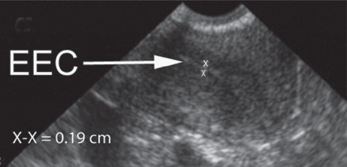
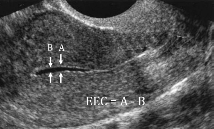
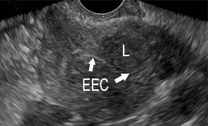
Compared with premenopausal women, the measured thickness of the endometrium (by convention a double layer in the midsagittal view; Figure 1) should be thinner in postmenopausal women not receiving HRT (Figure 2). This double-thickness layer is called the EEC. If there is uniformly sonolucent fluid—generally a nonconcerning finding—the thickness of the fluid echo is subtracted from the measurement from baseline to baseline to obtain the EEC (Figure 3). The morphologic features should be uniform and, if there are irregularities, or if the EEC cannot be adequately evaluated (Figure 4), other investigations are warranted, such as contrast sonography, hysteroscopy, and/or endometrial sampling. At this time, available evidence suggests that there are no advantages offered by the use of 3-dimensional ultrasonography, as compared with standard 2-dimensional ultrasonography62 (Class 3).
Figure 1.
Endometrial echo complex (EEC) measurement in the sagittal plane.
Study should include coronal or transverse plane as well. For demonstration purposes, the image is from a premenopausal woman.
Figure 2.
Typical measurement of normal postmenopausal endometrial echo complex (EEC) (< 4 mm) (arrowhead).
Figure 3.
Measurement of endometrial echo complex (EEC) when there is fluid in the cavity.
Thickness of fluid (B) is subtracted from distance between base of opposing layers of endometrium (A). These should be in the same plane; they are separated slightly here for demonstration purposes.
Figure 4.
Inadequate measurement of endometrial echo complex (EEC) (between symbols).
In general, thicker EECs are associated with a greater likelihood of endometrial or intracavitary pathology, including endometrial polyps, hyperplasia, and cancer63 (Class 3). On the other hand, the reliability of TVUS allows the clinician to identify a group of women with postmenopausal bleeding who have a thin endometrium and thus a very low likelihood of hyperplasia or neoplasia. Unless there is a recurrence of bleeding, this group of women with postmenopausal bleeding and a thin EEC generally require no more investigation64–66 (Class 2). There is some evidence that TVUS may be less sensitive for the detection of Type 2 endometrial carcinomas regardless of the EEC threshold. A cohort study demonstrated that 17% of patients with postmenopausal bleeding and Type 2 endometrial cancer had an EEC of less than 4 mm67 (Class 5).
A number of EEC thickness thresholds have been published to guide clinicians in discriminating between women who require endometrial sampling and those who do not, at least for the first episode of postmenopausal bleeding (Table 5). The thinner the threshold, the fewer cases of hyperplasia and cancer missed, but with higher sensitivity, specificity is reduced and more endometrial sampling must be performed. Furthermore, the use of HRT can have a variable impact on the measurements depending on the use of a progestin. Continuous estrogen-progestin regimens tend to cause a hypotrophic and sonographically thin endometrium, whereas the EEC in women receiving regimens of continuous estrogen and cyclically administered progestins will vary according to the date of the TVUS and the cycle date in the HRT regimen. Available evidence suggests that women with postmenopausal bleeding and a thickened endometrium are less likely to have endometrial pathology when they are receiving HRT than when they are not65,68 (Class 2). Presumably, the endometrial thickness in women receiving estrogen and cyclic progestin therapy would be lowest shortly after the cessation of the progestins, a time that might be best suited for measurement of EEC thickness in women who experience unscheduled bleeding.
Table 5.
Meta-analyses of sensitivity and specificity of endometrial echo complex for detection of endometrial cancer in women with postmenopausal bleeding
| Author, year | Method | Number of included studies | Number of subjects | Number with EEC | Sensitivity by EEC thickness (95% CI) | Specificity by EEC thickness (95% CI) | ||||
|---|---|---|---|---|---|---|---|---|---|---|
| 3 mm | 4 mm | 5 mm | 3 mm | 4 mm | 5 mm | |||||
| Timmermans et al,72 2010 | Original dataset | 13 | 2896 | 259 | 97.9 (90.1–99.6) | 94.8 (86.1–98.2) | 90.3 (80.0–95.5) | 35.4 (29.3–41.9) | 46.7 (38.3–55.2) | 54.0 (46.7–61.2) |
| Gupta et al,66 2002 | Summary data | 57 | 8890 | 1243 | 99.3 (96.8–99.8) | 98.9 (97.1–99.6) | 97.7 (95.2–98.8) | 25.3 (22.8–27.9) | 24.2 (19.7–29.2) | 26.1 (21.1–31.6) |
| Smith-Bindman et al,65 1998 | Summary data | 35 | 5892 | 759 | 100 (89–100) | 96 (93–98) | 96 (94–98) | 38 (32–45) | 53 (51–55) | 61 (59–63) |
CI = confidence interval; EEC = endometrial echo complex.
Normal Thickness of Endometrial Echo Complex
Meta-analytic research using “summary data” has been performed using high-quality studies evaluating the utility of TVUS-obtained EEC thickness for the assessment of women with postmenopausal bleeding. The authors of the largest meta-analysis to date, with just more than 9000 patients, demonstrated that an EEC of 3 mm or less would provide a posttest probability of 0.4% for endometrial cancer; a 4-mm threshold, 1.2%; and a 5-mm threshold, 2.3%66 (Class 2). In this study, the best quality evidence was that for the 5-mm threshold.66 The authors of a second large meta-analysis of nearly 6000 women reported that an EEC of 5 mm or less was associated with a 4% chance of endometrial cancer. This sensitivity did not vary in women using HRT65 (Class 2).
Meta-analysis using the original data-sets is thought to provide more accurate conclusions than using the summary data provided in the published manuscript because of a number of factors, including publication bias, method of analysis, and length of follow-up69,70 (technical descriptions). One such analysis employed this technique but, rather than using fixed cutoffs for EEC, used multiples of the mean for each different study and determined that if the median EEC thickness was used, the sensitivity would be approximately 96% and specificity would be 50%. However, such an approach is difficult to evaluate and implement because it does not provide guidance about EEC thickness overall71 (Class 2). Consequently, Timmermans et al72 (Class 2) performed a re-analysis of the data using original datasets, rather than summary data, and were able to evaluate 2896 cases from 13 authors (259 with endometrial carcinoma), only 2 of which were from the original 90-author group reported in the publication by Smith-Bindman et al65 (Class 2). The conclusions from this analysis suggested that a threshold of 5 mm for the EEC would have sensitivity for endometrial cancer of only 90%; 4 mm, a sensitivity of 95%; and 3 mm, a sensitivity of 98%—thresholds that are different from those found in the previously published meta-analyses. No original dataset analyses were reported considering the impact on EEC thickness.
Before this individual dataset-based meta-analysis, the Scottish Intercollegiate Guidelines Network chose the 3-mm thickness for the following 3 groups of postmenopausal women with postmenopausal bleeding73 (evidence-based guideline): 1) never used HRT, 2) discontinued HRT a year or more earlier, and 3) using continuous combined HRT. The Scottish Intercollegiate Guidelines Network also set a different threshold for women receiving estrogen and cyclic progestin therapy and estimated that, in the context of unscheduled bleeding, the risk of endometrial cancer for such women was 0.2% if the EEC was 5 mm or less.
The American College of Obstetricians and Gynecologists has published a practice bulletin that recommended an EEC threshold of 4 mm, above which other techniques of endometrial assessment should be employed.74 The issue of HRT and its impact on EEC thresholds is not dealt with in that guideline (evidence-based guideline).
Endometrial Fluid Found with Transvaginal Ultrasonography
As described previously in this section, fluid may be identified between the sonographically determined endometrial layers in both symptomatic and asymptomatic women75 (Class 5; Figure 3). Such a finding had previously been thought to be a predictor of malignancy, but more recent evidence challenges such a conclusion. The issue may be particularly relevant in the presence of cervical stenosis, when access to the endometrial cavity for endometrial sampling or hysteroscopy may be limited. Although there remains a relative paucity of studies evaluating this finding, existing contemporary studies are remarkably consistent demonstrating that if the fluid is sonolucent and if the EEC, which is the combined thickness of the 2 separated endometrial layers, is 3 mm or less (see Figure 3), the finding is highly likely to be benign76–78 (Class 3). On the other hand, if the EEC is greater than threshold (3.0–4.5 mm depending on the study) or if the fluid is echogenic79 (Class 3), there is a risk of malignancy and immediate further investigation aimed at obtaining a tissue sample is warranted80 (Class 3). Women with postmenopausal bleeding, intrauterine sonolucent fluid, and an EEC of 3 mm or less could still harbor a carcinoma in the cervical canal, so such a diagnosis should be considered79 (Class 3).
Documentation
If TVUS or contrast sonography is used to evaluate women with postmenopausal bleeding or unscheduled bleeding who are receiving HRT, documentation ideally comprises both a midline sagittal image and a transverse fundal image documenting the thickest measurement of the EEC. A suitable note describing the findings should be included in the patient record81 (Class 7). The addition to the record of selected images is desirable but not considered mandatory provided that the note describes the endometrial findings in sufficient detail.
Contrast Sonography
Transcervical instillation of saline (saline infusion sonography), or other sonographic contrast media such as gel, into the endometrial cavity allows for contrast-enhanced sonographic evaluation that is designed to improve diagnostic utility for lesions involving the endometrial cavity such as polyps or submucous leiomyomas82 (Class 5). The procedure is performed in an office setting with a small-caliber catheter positioned in the endometrial cavity while imaging is performed with simultaneous TVUS. In addition to saline infusion sonography, such fluid contrast-enhanced ultrasonography of the endometrial cavity is known as sonohysterography and hysterosonography, although the latter two terms may create confusion with the contrast-enhanced radiographic evaluation of the endometrial cavity and fallopian tubes that is called a hysterosalpingogram. Consequently, for purposes of this document, the term contrast sonography or saline infusion sonography will be used.
The procedure appears to accurately evaluate the endometrial cavity and can be successfully performed in more than 85% of postmenopausal women in an office setting83 (Class 2). Saline infusion sonography seems superior to TVUS in defining intrauterine lesions in women with postmenopausal bleeding and a measured EEC greater than 5 mm, particularly for the delineation of endometrial polyps, for which it seems as accurate as hysteroscopy84 (Class 2). Another observer-blinded comparison demonstrated that saline infusion sonography was essentially equivalent to hysteroscopy and superior to TVUS for the diagnosis of endometrial cavity abnormalities in women with postmenopausal bleeding85 (Class 3). There is no current evidence suggesting that saline infusion sonography enhances the diagnosis of malignancy. However, by virtue of its ability to identify postmenopausal women with endometrial polyps, saline infusion sonography may facilitate diagnosis, targeted removal, and resolution of symptoms, thereby reducing resource utilization and improving patient satisfaction with the overall process (Class 7).
Hysteroscopy with Curettage and Directed Biopsy
Hysteroscopy is an endoscopic technique that allows direct visualization of the endometrial cavity. It can be performed in an office setting with local or no anesthesia, with or without sedation, or in a procedure room or operating room. To accomplish the procedure, it is often necessary to first dilate the cervix adequately to accommodate the external diameter of the outer sheath of the hysteroscope assembly, which generally ranges from 4.0 to 5.5 mm. A number of small-caliber fiberoptic devices exist that are approximately the diameter of some biopsy catheters (3–4 mm). With such instrumentation, dilation of the cervix may not be necessary. One inherent advantage of hysteroscopy is that endoscopically guided removal of lesions may be performed immediately after diagnosis, during the same procedure. Provided that the lesions are relatively small, such excisions may even be performed in an office setting. Despite the seemingly obvious advantages of the technique, the data supporting the use of hysteroscopy for management of women with postmenopausal bleeding are relatively sparse and generally of relatively poor quality.
As described previously, hysteroscopy is superior to endometrial biopsy60,61,67 (Class 3), D&C60 (Class 3), and ultrasonography84,85 (Class 3) for the identification of structural lesions of the endometrium such as endometrial polyps. Hysteroscopy has good diagnostic accuracy for structural lesions, such as polyps and leiomyomas, whether or not performed in an office setting, and good patient acceptability in either setting24,86 (Class 1b). Indeed, in a study designed to determine the desires of women regarding a primary assessment tool for evaluation of postmenopausal bleeding, 95% preferred to undergo office hysterectomy rather than experience a 5% chance that a lesion could be missed87 (Class 6). However, it should be noted that hysteroscopic visualization alone is relatively inaccurate in the diagnosis of atypical hyperplasia and carcinoma84,88,89 (Class 3). Consequently, hysteroscopy should not be performed in women with postmenopausal bleeding who have not undergone antecedent or concurrent endometrial sampling by suction or sharp curettage.
In the past, there were concerns that the pressurized distending media used with hysteroscopy could result in retrograde dissemination of malignant cells into the peritoneal cavity sufficient to alter the prognosis of endometrial carcinoma90,91 (Class 5). However, although some investigators have shown a low incidence of peritoneal washings testing positive for endometrial cells92 (Class 5), others have compared the incidence of positive peritoneal cytologic findings associated with hysteroscopy to that associated with D&C and have found the frequency to be similar—about 9% to 10%93 (Class 1b),94 (Class 4). Overall, there seems to be a slightly increased risk of positive peritoneal cytologic findings associated with the use of hysteroscopy (OR, 1.78; CI, 1.13–2.79)95 (Class 1b). However, available high-quality evidence suggests that the prognosis associated with a hysteroscopic diagnosis of endometrial cancer is not different from that with other diagnostic methods93 (Class 1b),92,96–98 (Class 3).
Sequencing of Investigations
The exact sequencing of investigations for women with postmenopausal bleeding will necessarily vary somewhat depending on the local resources and expertise, the judgment of the clinician, and patient preference. There is evidence that resource utilization is reduced if the practitioner is able to perform the TVUS examination in the office without adding additional charges as opposed to having the procedure performed by a radiologist99 (Class 3), and this would be presumably true for contrast sonography as well. The same can be said for hysteroscopy, for which in-office performance using local anesthesia is logically cheaper than any hospital or surgical center-based procedure.
Primary Assessment of Spontaneous Postmenopausal Bleeding
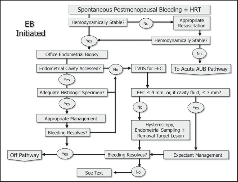
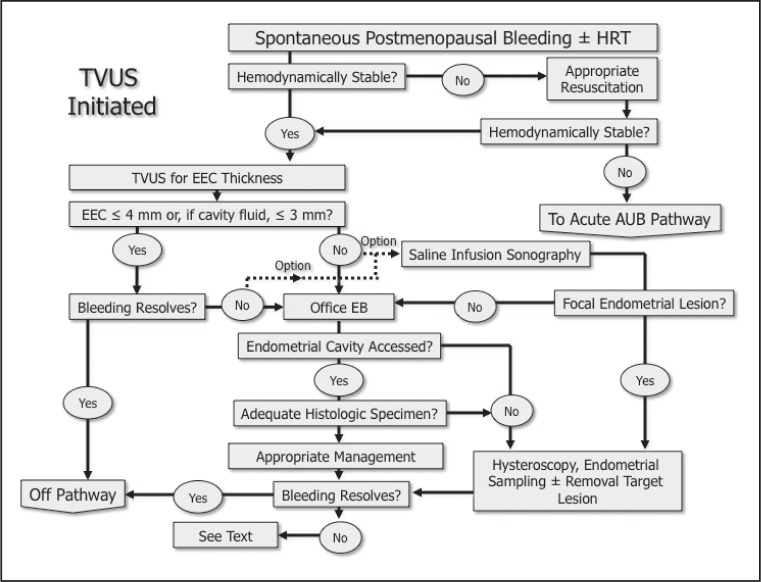
For women with spontaneous bleeding 12 or more months following the last menstrual period, the endometrium can be primarily assessed by either office-based endometrial biopsy (Figure 5) or TVUS (Figure 6). For those who undergo TVUS and have an EEC greater than 4 mm, localized thickening, or an indistinct or nonvisible EEC, endometrial sampling is a reasonable next step.
Figure 5.
Investigation of postmenopausal uterine bleeding with initial investigation of endometrial echo complex (EEC).
Entire EEC should be seen, allowing demonstration of a consistent 2-layer thickness in both the sagittal and transverse planes that is less than or equal to 4 mm. If there is clear (sonolucent) fluid, the bilaminar endometrial thickness should be less than or equal to 3 mm.
AUB = abnormal uterine bleeding; HRT = hormone replacement therapy;
TVUS = transvaginal ultrasonography; ± = with or without.
Figure 6.
Endometrial biopsy (EB) as initial investigation of post-menopausal uterine bleeding.
A satisfactory EB comprises perceptible passage of the sampling device through the cervical canal into the endometrial cavity and appropriate functioning of the aspiration mechanism. Adequacy of the specimen for histologic interpretation is determined by the pathologist.
AUB = abnormal uterine bleeding; EEC = endometrial echo complex;
HRT = hormone replacement therapy; TVUS = transvaginal ultrasonography; ± = with or without.
These approaches were compared by Weber et al100 (Class 3) from the Cleveland Clinic in a cost-modeling exercise, with primary TVUS predicted as being slightly less expensive. However, in that study, the assumptions were based on a charge model and included performance of the TVUS in a Radiology Department. In another study, Medverd and Dubinsky101 (Class 3) demonstrated that TVUS as a primary assessment modality utilized approximately 11% fewer resources than did an endometrial biopsy-originated evaluation paradigm. It is unclear whether charges or costs were considered in their analysis, but it is presumed that the imaging was performed in a Radiology Department and was interpreted by a radiologist. A cost modeling analysis from the UK, which assumed that a gynecologist performed all procedures, suggested that endometrial biopsy- and TVUS-initiated investigational paradigms were similar regarding their cost-effectiveness when 5 mm was used as the EEC threshold102 (Class 3). How this analysis translates to US fee-for-service or prepaid models is unclear, and the impact of changing the EEC threshold to 4 mm was not evaluated.
Primary Assessment of Hormone Replacement Therapy-Related Postmenopausal Bleeding (Breakthrough Bleeding)
The increased risk of endometrial cancer for women with postmenopausal bleeding who are receiving unopposed estrogen requires assessment in all circumstances. For women with postmenopausal bleeding while receiving combined estrogen-progestin HRT regimens, the approach is less clear, as it is apparent that such women are at significantly reduced risk of endometrial carcinoma compared with women not receiving HRT103 (Class 5). Furthermore, women using estrogen and cyclic progestin therapy and who have cyclic bleeding near to or following the end of the progestogenic component of the regimen require no investigation. In addition, those women who use estrogen and continuous progestin regimens will frequently experience breakthrough bleeding in the first 6 months of therapy and generally require no investigation. However, women receiving cyclic progestin regimens who have unscheduled bleeding, or for those receiving continuous progestin therapy who have breakthrough bleeding beyond 6 months and especially following a period of amenorrhea, the endometrium should be assessed. Available evidence suggests that either endometrial biopsy or TVUS can be used in a fashion similar to that recommended for spontaneous bleeding using a similar EEC threshold for endometrial sampling65 (Class 2),68 (Class 5). Although it is possible that a higher EEC threshold might apply to women receiving sequential HRT, the committee could not find convincing evidence to support such a notion. Consequently, clinicians are encouraged to perform TVUS for EEC measurement in the early phase of the “cycle” shortly after the completion of the progestin component.
Unless appropriately trained in either endometrial sampling techniques or TVUS, the primary care provider should refer any patient whose postmenopausal bleeding fits within the criteria requiring endometrial assessment. The gynecologist should determine which patients, if any, require TVUS assessment, including those who should be referred to a radiologist or other individual with specialist training in ultrasound of the uterus.
Investigation of Postmenopausal Bleeding in Women Receiving Hormone Replacement Therapy
Evaluation of women with postmenopausal bleeding while using HRT is affected by a number of factors, including the type of regimen, the dose of estrogen, and, if used, the scheduling of progestin administration (cyclic or continuous). These and other factors have an effect on the nature and timing of anticipated and unscheduled bleeding and may have a role in the timing and interpretation of ultrasound findings.
Causes of Abnormal Uterine Bleeding
Causes of abnormal bleeding in women using HRT include the following:
Poor compliance
Poor gastrointestinal absorption (for oral preparations)
Drug interactions
Coagulation defects
Liver disease
Gynecologic disorders, including but not limited to endometrial cancer, endometrial polyps, and cervical or vaginal lesions
Nonreproductive tract origins (eg, urinary tract, gastrointestinal tract).
Important Points in the Patient History
The clinician should determine if the bleeding pattern is within acceptable limits (see earlier section, Hormone Replacement Therapy). In addition, the provider should ascertain if there are any other factors or symptoms that may place the patient at increased risk of endometrial cancer. Pertinent questions to ask include the following:
When does the bleeding occur with respect to progestin administration? Women receiving cyclical progestin therapy should have bleeding near the end of or shortly after discontinuation of the progestin component of the regimen.
How long does the bleeding last? How heavy is it? Heavy bleeding, even when experienced in the context of a cyclically administered progestin, may suggest the presence of intrauterine pathology.
Was there a period of amenorrhea after HRT was started? For continuous combined regimens, breakthrough bleeding is common, especially in the first six months, but should a period of amenorrhea be established initially, bleeding, even within the first six months, suggests intrauterine abnormalities.
Is there evidence of poor compliance? The patient should understand the appropriate method for following her treatment regimen. Some women take their medications sporadically or use their progestin in an unconventional fashion.
Is there a reason to suspect poor gastrointestinal absorption? A history of nausea, vomiting, or diarrhea is a potential explanation for incomplete absorption and resultant bleeding.
Is there any evidence of hepatocellular disease? The liver is responsible for metabolizing estrogen. Should there be active or chronic hepatocellular disease, the circulating levels of estrogen may be higher than normal and abnormal bleeding may occur secondary to endometrial stimulation.
Is the patient receiving any other drugs? The intentional or inadvertent use of other gonadal steroids (estrogens with or without progestins) may explain unexpected bleeding.
Discontinuation of Hormone Replacement Therapy before Clinical Investigation
There is no convincing reason to discontinue HRT before clinical investigation. If the primary physician is able to complete the required investigative steps, this issue is moot. However, if the physician will be referring the patient for clinical assessment by a gynecologist, there may be a time period between the referral and the actual consultation appointment. In such instances, women may continue their regimen (Class 7).
Recurrent Spontaneous or Breakthrough Bleeding
Spontaneous or HRT-related, recurrent postmenopausal bleeding can occur. In a prospective study of women with postmenopausal bleeding who were not receiving HRT and whose EEC measured 4 mm or less, the incidence of recurrent bleeding was 10% at a mean of 49 weeks following the original TVUS104 (Class 5). Of these 25 patients, 8% (n = 2) had endometrial carcinoma and 4% (n = 1) had a malignant melanoma. In another retrospective study of 1536 women with postmenopausal bleeding evaluated over a period of almost 5 years with both TVUS and endometrial biopsy, the prevalence of endometrial cancer was 3% with primary assessment.105 On the other hand, the prevalence was 4% in the 126 patients with normal results of the initial evaluation who presented with persistent postmenopausal bleeding105 (Class 5).
There is no current evidence supporting any specific recommendations for reinvestigation in the face of a normal TVUS or satisfactory and normal endometrial biopsy specimen. However, considering the false-negative rates associated with either approach, the indications for reassessment should be rather liberal considering one or a combination of repeat TVUS, repeat endometrial biopsy, and contrast sonography or hysteros-copy64,106 (Class 5),49 (Class 6),107 (Class 7). Determination of the specific order and combinations of these investigations will depend on the clinical judgment of, and resources available to, the clinician.
Tamoxifen-Related Bleeding
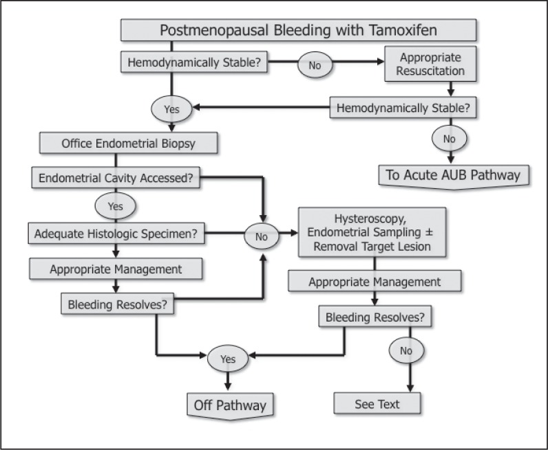
As previously described, patients receiving tamoxifen who are postmenopausal experience an increased risk of endometrial neoplasia that is greatest after four years of exposure. Whereas there is no evidence to support the use of routine biopsy of such patients, any who present with bleeding while receiving tamoxifen should undergo office-based endometrial sampling if possible (Figure 7). If not feasible, such patients should undergo hysteroscopy and curettage.
Figure 7.
Investigation of postmenopausal uterine bleeding in women receiving tamoxifen.
Transvaginal ultrasonography is not sufficient for the evaluation of women experiencing bleeding while receiving tamoxifen. Endometrial sampling is necessary.
AUB = abnormal uterine bleeding; ± = with or without.
The tamoxifen-induced impact on the endometrium frequently, if not usually, creates a thickened EEC, often with diffuse subendometrial cystic change that is in no way indicative of endometrial hyperplasia or malignancy108–113 (Classes 2–4). As a result, TVUS for evaluation of EEC thickness is not useful in the evaluation of tamoxifen-related postmenopausal bleeding.
Many patients with tamoxifen-related bleeding have endometrial polyps114,115 (Class 1b). Consequently, if bleeding persists, evaluation of the endometrial cavity is indicated, generally with hysteroscopy, with hysteroscopically guided excision of any identified polyps and curettage for reassessment of the endometrium. Contrast sonography may also be useful in this regard; however, there is evidence that saline infusion sonography is inferior to hysteroscopy in detecting focal lesions in these patients116 (Class 3).
For patients who are receiving tamoxifen, who have persistent episodes of postmenopausal bleeding, who have no evidence of focal lesions, and for whom both the cervical canal and endometrial cavity have been adequately evaluated with imaging and adequate endometrial sampling showing benign tissue, annual repeated sampling is indicated (Class 7).
Acknowledgments
Kathleen Louden, ELS, of Louden Health Communications provided editorial assistance.
Footnotes
The group also developed guidelines that apply to women with both acute and chronic abnormal uterine bleeding in the reproductive years.
Adapted from the Scottish Intercollegiate Guidelines Network73
Disclosure Statement
The author has no conflicts of interest to disclose.
More Burdened
A determining point in the history of gynecology is to be found in the fact that sex plays a more important part in the life of woman than in that of man, and that she is more burdened by her sex.
—Henry E Sigerist, 1891–1957, Swiss-born American medical historian
References
- 1.North American Menopause Society The 2012 hormone therapy position statement of The North American Menopause Society. Menopause. 2012 Mar;19(3):257–71. doi: 10.1097/gme.0b013e31824b970a. DOI: http://dx.doi.org/10.1097/gme.0b013e31824b970a. [DOI] [PMC free article] [PubMed] [Google Scholar]
- 2.Marjoribanks J, Farquhar C, Roberts H, Lethaby A. Long term hormone therapy for perimenopausal and postmenopausal women. Cochrane Database Syst Rev. 2012 Jul 11;7:CD004143. doi: 10.1002/14651858.CD004143.pub4. DOI: http://dx.doi.org/10.1002/14651858.CD004143.pub4. [DOI] [PubMed] [Google Scholar]
- 3.Moyer VA. Menopausal hormone therapy for the primary prevention of chronic conditions: US Preventive Services Task Force recommendation statement. Ann Intern Med. 2013 Jan 1;158(1):47–54. doi: 10.7326/0003-4819-158-1-201301010-00553. DOI: http://dx.doi.org/10.7326/0003-4819-158-1-201301010-00553. [DOI] [PubMed] [Google Scholar]
- 4.Research on the menopause in the 1990s. Report of a WHO Scientific Group. World Health Organ Tech Rep Ser. 1996;866:1–107. [PubMed] [Google Scholar]
- 5.Spencer CP, Cooper AJ, Whitehead MI. Management of abnormal bleeding in women receiving hormone replacement therapy. BMJ. 1997 Jul 5;315(7099):37–42. doi: 10.1136/bmj.315.7099.37. DOI: http://dx.doi.org/10.1136/bmj.315.7099.37. [DOI] [PMC free article] [PubMed] [Google Scholar]
- 6.Ettinger B, Li DK, Klein R. Unexpected vaginal bleeding and associated gynecologic care in postmenopausal women using hormone replacement therapy: comparison of cyclic versus continuous combined schedules. Fertil Steril. 1998 May;69(5):865–9. doi: 10.1016/s0015-0282(98)00047-8. DOI: http://dx.doi.org/10.1016/S0015-0282(98)00047-8. [DOI] [PubMed] [Google Scholar]
- 7.Jemal A, Bray F, Center MM, Ferlay J, Ward E, Forman D. Global cancer statistics. CA Cancer J Clin. 2011 Mar-Apr;61(2):69–90. doi: 10.3322/caac.20107. DOI: http://dx.doi.org/10.3322/caac.20107. [DOI] [PubMed] [Google Scholar]
- 8.Bokhman JV. Two pathogenetic types of endometrial carcinoma. Gynecol Oncol. 1983 Feb;15(1):10–7. doi: 10.1016/0090-8258(83)90111-7. DOI: http://dx.doi.org/10.1016/0090-8258(83)90111-7. [DOI] [PubMed] [Google Scholar]
- 9.Creasman WT, Odicino F, Maisonneuve P, et al. Carcinoma of the corpus uteri. Int J Gynaecol Obstet. 2003 Oct;83(Suppl 1):79–118. doi: 10.1016/s0020-7292(03)90116-0. DOI: http://dx.doi.org/10.1016/S0020-7292(03)90116-0. [DOI] [PubMed] [Google Scholar]
- 10.Duong LM, Wilson RJ, Ajani UA, Singh SD, Eheman CR. Trends in endometrial cancer incidence rates in the United States, 1999–2006. J Women’s Health (Larchmt) 2011 Aug;20(8):1157–63. doi: 10.1089/jwh.2010.2529. DOI: http://dx.doi.org/10.1089/jwh.2010.2529. [DOI] [PubMed] [Google Scholar]
- 11.Ferrazzi E, Torri V, Trio D, Zannoni E, Filiberto S, Dordoni D. Sonographic endometrial thickness: a useful test to predict atrophy in patients with postmenopausal bleeding. An Italian multicenter study. Ultrasound Obstet Gynecol. 1996 May;7(5):315–21. doi: 10.1046/j.1469-0705.1996.07050315.x. DOI: http://dx.doi.org/10.1046/j.1469-0705.1996.07050315.x. [DOI] [PubMed] [Google Scholar]
- 12.Gredmark T, Kvint S, Havel G, Mattsson LA. Histopathological findings in women with postmenopausal bleeding. Br J Obstet Gynaecol. 1995 Feb;102(2):133–6. doi: 10.1111/j.1471-0528.1995.tb09066.x. DOI: http://dx.doi.org/10.1111/j.1471-0528.1995.tb09066.x. [DOI] [PubMed] [Google Scholar]
- 13.Lidor A, Ismajovich B, Confino E, David MP. Histopathological findings in 226 women with postmenopausal uterine bleeding. Acta Obstet Gynecol Scand. 1986;65(1):41–3. doi: 10.3109/00016348609158227. DOI: http://dx.doi.org/10.3109/00016348609158227. [DOI] [PubMed] [Google Scholar]
- 14.Burbos N, Musonda P, Giarenis I, et al. Age-related differential diagnosis of vaginal bleeding in postmenopausal women: a series of 3047 symptomatic postmenopausal women. Menopause Int. 2010 Mar;16(1):5–8. doi: 10.1258/mi.2010.010005. DOI: http://dx.doi.org/10.1258/mi.2010.010005. [DOI] [PubMed] [Google Scholar]
- 15.Gambrell RD, Jr, Massey FM, Castaneda TA, Ugenas AJ, Ricci CA, Wright JM. Use of the progestogen challenge test to reduce the risk of endometrial cancer. Obstet Gynecol. 1980 Jun;55(6):732–8. [PubMed] [Google Scholar]
- 16.Persson I, Adami HO, Bergkvist L, et al. Risk of endometrial cancer after treatment with oestrogens alone or in conjunction with progestogens: results of a prospective study. BMJ. 1989 Jan 21;298(6667):147–51. doi: 10.1136/bmj.298.6667.147. DOI: http://dx.doi.org/10.1136/bmj.298.6667.147. [DOI] [PMC free article] [PubMed] [Google Scholar]
- 17.Weiderpass E, Adami HO, Baron JA, et al. Risk of endometrial cancer following estrogen replacement with and without progestins. J Natl Cancer Inst. 1999 Jul 7;91(13):1131–7. doi: 10.1093/jnci/91.13.1131. DOI: http://dx.doi.org/10.1093/jnci/91.13.1131. [DOI] [PubMed] [Google Scholar]
- 18.Fornander T, Rutqvist LE, Cedermark B, et al. Adjuvant tamoxifen in early breast cancer: occurrence of new primary cancers. Lancet. 1989 Jan 21;1(8360):117–20. doi: 10.1016/s0140-6736(89)91141-0. DOI: http://dx.doi.org/10.1016/S0140-6736(89)91141-0. [DOI] [PubMed] [Google Scholar]
- 19.Rutqvist LE, Johansson H, Signomklao T, Johansson U, Fornander T, Wilking N. Adjuvant tamoxifen therapy for early stage breast cancer and second primary malignancies. Stockholm Breast Cancer Study Group. J Natl Cancer Inst. 1995 May 3;87(9):645–51. doi: 10.1093/jnci/87.9.645. DOI: http://dx.doi.org/10.1093/jnci/87.9.645. [DOI] [PubMed] [Google Scholar]
- 20.Fisher B, Costantino JP, Wickerham DL, et al. Tamoxifen for prevention of breast cancer: report of the National Surgical Adjuvant Breast and Bowel Project P-1 Study. J Natl Cancer Inst. 1998 Sep 16;90(18):1371–88. doi: 10.1093/jnci/90.18.1371. DOI: http://dx.doi.org/10.1093/jnci/90.18.1371. [DOI] [PubMed] [Google Scholar]
- 21.Curtis RE, Boice JD, Jr, Shriner DA, Hankey BF, Fraumeni JF., Jr Second cancers after adjuvant tamoxifen therapy for breast cancer. J Natl Cancer Inst. 1996 Jun 19;88(12):832–4. doi: 10.1093/jnci/88.12.832. DOI: http://dx.doi.org/10.1093/jnci/88.12.832. [DOI] [PubMed] [Google Scholar]
- 22.Cancer registration statistics Scotland 1986–1995. Edinburgh, Scotland: National Health Service in Scotland, Information & Statistics Division; 1998. [Google Scholar]
- 23.Lethaby A, Suckling J, Barlow D, Farquhar CM, Jepson RG, Roberts H. Hormone replacement therapy in postmenopausal women: endometrial hyperplasia and irregular bleeding. Cochrane Database Syst Rev. 2004;(3):CD000402. doi: 10.1002/14651858.CD000402.pub2. DOI: http://dx.doi.org/10.1002/14651858.CD000402.pub2. [DOI] [PubMed] [Google Scholar]
- 24.Tahir MM, Bigrigg MA, Browning JJ, Brookes ST, Smith PA. A randomised controlled trial comparing transvaginal ultrasound, outpatient hysteroscopy and endometrial biopsy with inpatient hysteroscopy and curettage. Br J Obstet Gynaecol. 1999 Dec;106(12):1259–64. doi: 10.1111/j.1471-0528.1999.tb08179.x. DOI: http://dx.doi.org/10.1111/j.1471-0528.1999.tb08179.x. [DOI] [PubMed] [Google Scholar]
- 25.Furness S, Roberts H, Marjoribanks J, Lethaby A, Hickey M, Farquhar C. Hormone therapy in postmenopausal women and risk of endometrial hyperplasia. Cochrane Database Syst Rev. 2009 Apr 15;(2):CD000402. doi: 10.1002/14651858.CD000402.pub3. DOI: http://dx.doi.org/10.1002/14651858.CD000402.pub3. [DOI] [PubMed] [Google Scholar]
- 26.Williams DB, Voigt BJ, Fu YS, Schoenfeld MJ, Judd HL. Assessment of less than monthly progestin therapy in postmenopausal women given estrogen replacement. Obstet Gynecol. 1994 Nov;84(5):787–93. [PubMed] [Google Scholar]
- 27.Ettinger B, Selby J, Citron JT, Vangessel A, Ettinger VM, Hendrickson MR. Cyclic hormone replacement therapy using quarterly progestin. Obstet Gynecol. 1994 May;83(5 Pt 1):693–700. [PubMed] [Google Scholar]
- 28.Ettinger B, Pressman A, Van Gessel A. Low-dosage esterified estrogens opposed by progestin at 6-month intervals. Obstet Gynecol. 2001 Aug;98(2):205–11. doi: 10.1016/s0029-7844(01)01419-3. DOI: http://dx.doi.org/10.1016/S0029-7844(01)01419-3. [DOI] [PubMed] [Google Scholar]
- 29.Jaakkola S, Lyytinen HK, Dyba T, Ylikorkala O, Pukkala E. Endometrial cancer associated with various forms of postmenopausal hormone therapy: a case control study. Int J Cancer. 2011 Apr 1;128(7):1644–51. doi: 10.1002/ijc.25762. DOI: http://dx.doi.org/10.1002/ijc.25762. [DOI] [PubMed] [Google Scholar]
- 30.Ettinger B, Ensrud KE, Wallace R, et al. Effects of ultralow-dose transdermal estradiol on bone mineral density: a randomized clinical trial. Obstet Gynecol. 2004 Sep;104(3):443–51. doi: 10.1097/01.AOG.0000137833.43248.79. DOI: http://dx.doi.org/10.1097/01.AOG.0000137833.43248.79. [DOI] [PubMed] [Google Scholar]
- 31.Simon J, Nachtigall L, Ulrich LG, Eugster-Hausmann M, Gut R. Endometrial safety of ultralow-dose estradiol vaginal tablets. Obstet Gynecol. 2010 Oct;116(4):876–83. doi: 10.1097/AOG.0b013e3181f386bb. DOI: http://dx.doi.org/10.1097/AOG.0b013e3181f386bb. [DOI] [PubMed] [Google Scholar]
- 32.Barret-Connor E. Hormone replacement. Am J Geriatr Cardiol. 1993 Oct;2(5):36–7. [PubMed] [Google Scholar]
- 33.Archer DF, Pickar JH, Bottiglioni F. Bleeding patterns in postmenopausal women taking continuous combined or sequential regimens of conjugated estrogens with medroxyprogesterone acetate. Menopause Study Group. Obstet Gynecol. 1994 May;83(5 Pt 1):686–92. [PubMed] [Google Scholar]
- 34.Nagele F, O’Connor H, Baskett TF, Davies A, Mohammed H, Magos AL. Hysteroscopy in women with abnormal uterine bleeding on hormone replacement therapy: a comparison with postmenopausal bleeding. Fertil Steril. 1996 Jun;65(6):1145–50. doi: 10.1016/s0015-0282(16)58329-0. [DOI] [PubMed] [Google Scholar]
- 35.Hussain SA, Williams S, Stevens A, Rea DW. Endocrine therapy for early breast cancer. Expert Rev Anticancer Ther. 2004 Oct;4(5):877–88. doi: 10.1586/14737140.4.5.877. DOI: http://dx.doi.org/10.1586/14737140.4.5.877. [DOI] [PubMed] [Google Scholar]
- 36.Bernstein L, Deapen D, Cerhan JR, et al. Tamoxifen therapy for breast cancer and endometrial cancer risk. J Natl Cancer Inst. 1999 Oct 6;91(19):1654–62. doi: 10.1093/jnci/91.19.1654. DOI: http://dx.doi.org/10.1093/jnci/91.19.1654. [DOI] [PubMed] [Google Scholar]
- 37.van Leeuwen FE, Benraadt J, Coebergh JW, et al. Risk of endometrial cancer after tamoxifen treatment of breast cancer. Lancet. 1994 Feb 19;343(8895):448–52. doi: 10.1016/s0140-6736(94)92692-1. DOI: http://dx.doi.org/10.1016/S0140-6736(94)92692-1. [DOI] [PubMed] [Google Scholar]
- 38.Bergman L, Beelen ML, Gallee MP, Hollema H, Benraadt J, van Leeuwen FE. Risk and prognosis of endometrial cancer after tamoxifen for breast cancer. Comprehensive Cancer Centres’ ALERT Group. Lancet. 2000 Sep 9;356(9233):881–7. doi: 10.1016/s0140-6736(00)02677-5. [DOI] [PubMed] [Google Scholar]
- 39.Love CD, Muir BB, Scrimgeour JB, Leonard RC, Dillon P, Dixon JM. Investigation of endometrial abnormalities in asymptomatic women treated with tamoxifen and an evaluation of the role of endometrial screening. J Clin Oncol. 1999 Jul;17(7):2050–4. doi: 10.1200/JCO.1999.17.7.2050. [DOI] [PubMed] [Google Scholar]
- 40.Markovitch O, Tepper R, Aviram R, Fishman A, Shapira J, Cohen I. The value of sonohysterography in the prediction of endometrial pathologies in asymptomatic postmenopausal breast cancer tamoxifen-treated patients. Gynecol Oncol. 2004 Sep;94(3):754–9. doi: 10.1016/j.ygyno.2004.06.033. DOI: http://dx.doi.org/10.1016/j.ygyno.2004.06.033. [DOI] [PubMed] [Google Scholar]
- 41.Fung MF, Reid A, Faught W, et al. Prospective longitudinal study of ultrasound screening for endometrial abnormalities in women with breast cancer receiving tamoxifen. Gynecol Oncol. 2003 Oct;91(1):154–9. doi: 10.1016/s0090-8258(03)00441-4. DOI: http://dx.doi.org/10.1016/S0090-8258(03)00441-4. [DOI] [PubMed] [Google Scholar]
- 42.McGonigle KF, Shaw SL, Vasilev SA, Odom-Maryon T, Roy S, Simpson JF. Abnormalities detected on transvaginal ultrasonography in tamoxifen-treated postmenopausal breast cancer patients may represent endometrial cystic atrophy. Am J Obstet Gynecol. 1998 Jun;178(6):1145–50. doi: 10.1016/s0002-9378(98)70315-1. DOI: http://dx.doi.org/10.1016/S0002-9378(98)70315-1. [DOI] [PubMed] [Google Scholar]
- 43.Dunlop MG, Farrington SM, Carothers AD, et al. Cancer risk associated with germline DNA mismatch repair gene mutations. Hum Mol Genet. 1997 Jan;6(1):105–10. doi: 10.1093/hmg/6.1.105. DOI: http://dx.doi.org/10.1093/hmg/6.1.105. [DOI] [PubMed] [Google Scholar]
- 44.Aarnio M, Sankila R, Pukkala E, et al. Cancer risk in mutation carriers of DNA-mismatch-repair genes. Int J Cancer. 1999 Apr 12;81(2):214–8. doi: 10.1002/(sici)1097-0215(19990412)81:2<214::aid-ijc8>3.0.co;2-l. DOI: http://dx.doi.org/10.1002/(SICI)1097-0215(19990412)81:2<214::AID-IJC8>3.0.CO;2-L. [DOI] [PubMed] [Google Scholar]
- 45.Vasen HF, Wijnen JT, Menko FH, et al. Cancer risk in families with hereditary nonpolyposis colorectal cancer diagnosed by mutation analysis. Gastroenterology. 1996 Apr;110(4):1020–7. doi: 10.1053/gast.1996.v110.pm8612988. DOI: http://dx.doi.org/10.1053/gast.1996.v110.pm8612988. Erratum in: Gastroenterology 1996 Nov;111(5):1402. [DOI] [PubMed] [Google Scholar]
- 46.Feldman S, Cook EF, Harlow BL, Berkowitz RS. Predicting endometrial cancer among older women who present with abnormal vaginal bleeding. Gynecol Oncol. 1995 Mar;56(3):376–81. doi: 10.1006/gyno.1995.1066. DOI: http://dx.doi.org/10.1006/gyno.1995.1066. [DOI] [PubMed] [Google Scholar]
- 47.Haller H, Matecjcić N, Rukavina B, Krasević M, Rupcić S, Mozetic D. Transvaginal sonography and hysteroscopy in women with post-menopausal bleeding. Int J Gynaecol Obstet. 1996 Aug;54(2):155–9. doi: 10.1016/0020-7292(96)02677-x. DOI: http://dx.doi.org/10.1016/0020-7292(96)02677-X. [DOI] [PubMed] [Google Scholar]
- 48.Garuti G, Sambruni I, Cellani F, Garzia D, Alleva P, Luerti M. Hysteroscopy and transvaginal ultrasonography in postmenopausal women with uterine bleeding. International J Gynaecol Obstet. 1999 Apr;65(1):25–33. doi: 10.1016/s0020-7292(98)00224-0. DOI: http://dx.doi.org/10.1016/S0020-7292(98)00224-0. [DOI] [PubMed] [Google Scholar]
- 49.Gordon SJ, Westgate J. The incidence and management of failed Pipelle sampling in a general outpatient clinic. Aust N Z J Obstet Gynaecol. 1999 Feb;39(1):115–8. doi: 10.1111/j.1479-828x.1999.tb03460.x. DOI: http://dx.doi.org/10.1111/j.1479-828X.1999.tb03460.x. [DOI] [PubMed] [Google Scholar]
- 50.Dijkhuizen FP, Mol BW, Brölmann HA, Heintz AP. The accuracy of endometrial sampling in the diagnosis of patients with endometrial carcinoma and hyperplasia: a meta-analysis. Cancer. 2000 Oct 15;89(8):1765–72. DOI: http://dx.doi.org/10.1002/1097-0142(20001015)89:8<1765::AID-CNCR17>3.0.CO;2-F. [PubMed] [Google Scholar]
- 51.Epstein E, Skoog L, Valentin L. Comparison of Endorette and dilatation and curettage for sampling of the endometrium in women with postmenopausal bleeding. Acta Obstet Gynecol Scand. 2001 Oct;80(10):959–64. doi: 10.1034/j.1600-0412.2001.801015.x. DOI: http://dx.doi.org/10.1034/j.1600-0412.2001.801015.x. [DOI] [PubMed] [Google Scholar]
- 52.van Doorn HC, Opmeer BC, Burger CW, Duk MJ, Kooi GS, Mol BW, Dutch Study in Post-menopausal Bleeding (DUPOMEB) Inadequate office endometrial sample requires further evaluation in women with postmenopausal bleeding and abnormal ultrasound results. Int J Gynaecol Obstet. 2007 Nov;99(2):100–4. doi: 10.1016/j.ijgo.2007.05.040. DOI: http://dx.doi.org/10.1016/j.ijgo.2007.05.040. [DOI] [PubMed] [Google Scholar]
- 53.Giusa-Chiferi MG, Gonçalves WJ, Baracat EC, de Albuquerque Neto LC, Bortoletto CC, de Lima GR. Transvaginal ultrasound, uterine biopsy and hysteroscopy for post-menopausal bleeding. Int J Gynaecol Obstet. 1996 Oct;55(1):39–44. doi: 10.1016/0020-7292(96)02720-8. DOI: http://dx.doi.org/10.1016/0020-7292(96)02720-8. [DOI] [PubMed] [Google Scholar]
- 54.Naim NM, Mahdy ZA, Ahmad S, Razi ZR. The Vabra aspirator versus the Pipelle device for outpatient endometrial sampling. Aust N Z J Obstet Gynaecol. 2007 Apr;47(2):132–6. doi: 10.1111/j.1479-828X.2007.00699.x. DOI: http://dx.doi.org/10.1111/j.1479-828X.2007.00699.x. [DOI] [PubMed] [Google Scholar]
- 55.Leclair CM, Zia JK, Doom CM, Morgan TK, Edelman AB. Pain experienced using two different methods of endometrial biopsy: a randomized controlled trial. Obstet Gynecol. 2011 Mar;117(3):636–41. doi: 10.1097/AOG.0b013e31820ad45b. DOI: http://dx.doi.org/10.1097/AOG.0b013e31820ad45b. [DOI] [PubMed] [Google Scholar]
- 56.Critchley HO, Warner P, Lee AJ, Brechin S, Guise J, Graham B. Evaluation of abnormal uterine bleeding: comparison of three outpatient procedures within cohorts defined by age and menopausal status. Health Technol Assess. 2004 Sep;8(34):iii–iv. 1–139. doi: 10.3310/hta8340. [DOI] [PubMed] [Google Scholar]
- 57.Williams AR, Brechin S, Porter AJ, Warner P, Critchley HO. Factors affecting adequacy of Pipelle and Tao Brush endometrial sampling. BJOG. 2008 Jul;115(8):1028–36. doi: 10.1111/j.1471-0528.2008.01773.x. DOI: http://dx.doi.org/10.1111/j.1471-0528.2008.01773.x. [DOI] [PubMed] [Google Scholar]
- 58.Remondi C, Sesti F, Bonanno E, Pietropolli A, Piccione E. Diagnostic accuracy of liquid-based endometrial cytology in the evaluation of endometrial pathology in postmenopausal women. Cytopathology. 2012 Sep 21; doi: 10.1111/cyt.12013. DOI: http://dx.doi.org/10.1111/cyt.12013. [DOI] [PubMed] [Google Scholar]
- 59.Epstein E, Ramirez A, Skoog L, Valentin L. Dilatation and curettage fails to detect most focal lesions in the uterine cavity in women with postmenopausal bleeding. Acta Obstet Gynecol Scand. 2001 Dec;80(12):1131–6. doi: 10.1034/j.1600-0412.2001.801210.x. DOI: http://dx.doi.org/10.1034/j.1600-0412.2001.801210.x. [DOI] [PubMed] [Google Scholar]
- 60.Gebauer G, Hafner A, Siebzehnrübl E, Lang N. Role of hysteroscopy in detection and extraction of endometrial polyps: results of a prospective study. Am J Obstet Gynecol. 2001 Jan;184(2):59–63. doi: 10.1067/mob.2001.108332. DOI: http://dx.doi.org/10.1067/mob.2001.108332. [DOI] [PubMed] [Google Scholar]
- 61.Angioni S, Loddo A, Milano F, Piras B, Minerba L, Melis GB. Detection of benign intracavitary lesions in postmenopausal women with abnormal uterine bleeding: a prospective comparative study on outpatient hysteroscopy and blind biopsy. J Minim Invasive Gynecol. 2008 Jan-Feb;15(1):87–91. doi: 10.1016/j.jmig.2007.10.014. DOI: http://dx.doi.org/10.1016/j.jmig.2007.10.014. [DOI] [PubMed] [Google Scholar]
- 62.Rossi A, Forzano L, Romanello I, Fachechi G, Marchesoni D. Assessment of endometrial volume and vascularization using transvaginal 3D power Doppler angiography in women with postmenopausal bleeding. Int J Gynaecol Obstet. 2012 Oct;119(1):14–7. doi: 10.1016/j.ijgo.2012.05.023. DOI: http://dx.doi.org/10.1016/j.ijgo.2012.05.023. [DOI] [PubMed] [Google Scholar]
- 63.Wolman I, Amster R, Hartoov J, et al. Reproducibility of transvaginal ultrasonographic measurements of endometrial thickness in patients with postmenopausal bleeding. Gynecol Obstet Invest. 1998;46(3):191–4. doi: 10.1159/000010031. DOI: http://dx.doi.org/10.1159/000010031. [DOI] [PubMed] [Google Scholar]
- 64.Güner H, Tiras MB, Karabacak O, Sarikaya H, Erdem M, Yildirim M. Endometrial assessment by vaginal ultrasonography might reduce endometrial sampling in patients with postmenopausal bleeding: a prospective study. Aust N Z J Obstet Gynaecol. 1996 May;36(2):175–8. doi: 10.1111/j.1479-828x.1996.tb03280.x. DOI: http://dx.doi.org/10.1111/j.1479-828X.1996.tb03280.x. [DOI] [PubMed] [Google Scholar]
- 65.Smith-Bindman R, Kerlikowske K, Feldstein VA, et al. Endovaginal ultrasound to exclude endometrial cancer and other endometrial abnormalities. JAMA. 1998 Nov 4;280(17):1510–7. doi: 10.1001/jama.280.17.1510. DOI: http://dx.doi.org/10.1001/jama.280.17.1510. [DOI] [PubMed] [Google Scholar]
- 66.Gupta JK, Chien PF, Voit D, Clark TJ, Khan KS. Ultrasonographic endometrial thickness for diagnosing endometrial pathology in women with postmenopausal bleeding: a meta-analysis. Acta Obstet Gynecol Scand. 2002 Sep;81(9):799–816. doi: 10.1034/j.1600-0412.2001.810902.x. DOI: http://dx.doi.org/10.1034/j.1600-0412.2001.810902.x. [DOI] [PubMed] [Google Scholar]
- 67.Zhou XD, Ren XL, Zhang J, et al. Therapeutic response assessment of high intensity focused ultrasound therapy for uterine fibroid: utility of contrast-enhanced ultrasonography. Eur J Radiol. 2007 May;62(2):289–94. doi: 10.1016/j.ejrad.2006.11.039. DOI: http://dx.doi.org/10.1016/j.ejrad.2006.11.039. [DOI] [PubMed] [Google Scholar]
- 68.Granberg S, Ylöstalo P, Wikland M, Karlsson B. Endometrial sonographic and histologic findings in women with and without hormonal replacement therapy suffering from postmenopausal bleeding. Maturitas. 1997 May;27(1):35–40. doi: 10.1016/s0378-5122(97)01107-9. DOI: http://dx.doi.org/10.1016/S0378-5122(97)01107-9. [DOI] [PubMed] [Google Scholar]
- 69.Stewart LA, Parmar MK. Meta-analysis of the literature or of individual patient data: is there a difference? Lancet. 1993 Feb 13;341(8842):418–22. doi: 10.1016/0140-6736(93)93004-k. DOI: http://dx.doi.org/10.1016/0140-6736(93)93004-K. [DOI] [PubMed] [Google Scholar]
- 70.Hopewell S, McDonald S, Clarke M, Egger M. Grey literature in meta-analyses of randomized trials of health care interventions. Cochrane Database Syst Rev. 2007 Apr 18;(2):MR000010. doi: 10.1002/14651858.MR000010.pub3. DOI: http://dx.doi.org/10.1002/14651858.MR000010.pub2. [DOI] [PMC free article] [PubMed] [Google Scholar]
- 71.Tabor A, Watt HC, Wald NJ. Endometrial thickness as a test for endometrial cancer in women with postmenopausal vaginal bleeding. Obstet Gynecol. 2002 Apr;99(4):663–70. doi: 10.1016/s0029-7844(01)01771-9. DOI: http://dx.doi.org/10.1016/S0029-7844(01)01771-9. [DOI] [PubMed] [Google Scholar]
- 72.Timmermans A, Opmeer BC, Khan KS, et al. Endometrial thickness measurement for detecting endometrial cancer in women with postmenopausal bleeding: a systematic review and meta-analysis. Obstet Gynecol. 2010 Jul;116(1):160–7. doi: 10.1097/AOG.0b013e3181e3e7e8. DOI: http://dx.doi.org/10.1097/AOG.0b013e3181e3e7e8. [DOI] [PubMed] [Google Scholar]
- 73.Scottish Intercollegiate Guidelines Network (SIGN) Investigation of postmenopausal bleeding A national clinical guideline [Internet] Edinburgh, Scotland: Scottish Intercollegiate Guidelines Network (SIGN); 2002. Sep 25, [cited 2013 Sep 3]. Available from: www.guideline.gov/content.aspx?id=3456. [Google Scholar]
- 74.American Congress of Obstetricians and Gynecologists ACOG Committee Opinion No. 426: the role of transvaginal ultrasonography in the evaluation of postmenopausal bleeding. Obstet Gynecol. 2009 Feb;113(2 Pt 1):462–4. doi: 10.1097/AOG.0b013e31819930cc. DOI: http://dx.doi.org/10.1097/AOG.0b013e31819930cc. [DOI] [PubMed] [Google Scholar]
- 75.Bar-Hava I, Orvieto R, Ferber A, et al. Asymptomatic postmenopausal intrauterine fluid accumulation: characterization and significance. Climacteric. 1998 Dec;1(4):279–83. doi: 10.3109/13697139809085555. DOI: http://dx.doi.org/10.3109/13697139809085555. [DOI] [PubMed] [Google Scholar]
- 76.Goldstein SR. Postmenopausal endometrial fluid collections revisited: look at the doughnut rather than the hole. Obstet Gynecol. 1994 May;83(5 Pt 1):738–40. [PubMed] [Google Scholar]
- 77.Schmidt T, Nawroth F, Breidenbach M, Hoopmann M, Mallmann P, Valter MM. Differential indication for histological evaluation of endometrial fluid in postmenopause. Maturitas. 2005 Mar;1450(3):177–81. doi: 10.1016/j.maturitas.2004.05.016. DOI: http://dx.doi.org/10.1016/j.maturitas.2004.05.016. [DOI] [PubMed] [Google Scholar]
- 78.Debby A, Malinger G, Glezerman M, Golan A. Intra-uterine fluid collection in postmenopuasal women with cervical stenosis. Maturitas. 2006 Nov 20;55(4):334–7. doi: 10.1016/j.maturitas.2006.04.026. DOI: http://dx.doi.org/10.1016/j.maturitas.2006.04.026. [DOI] [PubMed] [Google Scholar]
- 79.Takacs P, De Santis T, Nicholas MC, Verma U, Strassberg R, Duthely L. Echogenic endometrial fluid collection in postmenopausal women is a significant risk factor for disease. J Ultrasound Med. 2005 Nov;24(11):1477–81. doi: 10.7863/jum.2005.24.11.1477. [DOI] [PubMed] [Google Scholar]
- 80.Epstein E, Valentin L. Gray-scale ultrasound morphology in the presence or absence of intrauterine fluid and vascularity as assessed by color Doppler for discrimination between benign and malignant endometrium in women with postmenopausal bleeding. Ultrasound Obstet Gynecol. 2006 Jul;28(1):89–95. doi: 10.1002/uog.2782. DOI: http://dx.doi.org/10.1002/uog.2782. [DOI] [PubMed] [Google Scholar]
- 81.Breitkopf D, Goldstein SR, Seeds JW, ACOG Committee on Gynecologic Practice ACOG technology assessment in obstetrics and gynecology. Number 3, September 2003. Saline infusion sonohysterography. Obstet Gynecol. 2003 Sep;102(3):659–62. doi: 10.1097/00006250-200309000-00058. [DOI] [PubMed] [Google Scholar]
- 82.Werbrouck E, Veldman J, Luts J, et al. Detection of endometrial pathology using saline infusion sonography versus gel instillation sonography: a prospective cohort study. Fertil Steril. 2011 Jan;95(1):285–8. doi: 10.1016/j.fertnstert.2010.04.074. DOI: http://dx.doi.org/10.1016/j.fertnstert.2010.04.074. [DOI] [PubMed] [Google Scholar]
- 83.de Kroon CD, de Bock GH, Dieben SW, Jansen FW. Saline contrast hysterosonography in abnormal uterine bleeding: a systematic review and meta-analysis. BJOG. 2003 Oct;110(10):938–47. doi: 10.1111/j.1471-0528.2003.02472.x. DOI: http://dx.doi.org/10.1111/j.1471-0528.2003.02472.x. [DOI] [PubMed] [Google Scholar]
- 84.Epstein E, Ramirez A, Skoog L, Valentin L. Transvaginal sonography, saline contrast sonohysterography and hysteroscopy for the investigation of women with postmenopausal bleeding and endometrium > 5 mm. Ultrasound Obstet Gynecol. 2001 Aug;18(2):157–62. doi: 10.1046/j.1469-0705.2001.00472.x. DOI: http://dx.doi.org/10.1046/j.1469-0705.2001.00472.x. [DOI] [PubMed] [Google Scholar]
- 85.Bingol B, Gunenc MZ, Gedikbasi A, Guner H, Tasdemir S, Tiras B. Comparison of diagnostic accuracy of saline infusion sonohysterography, transvaginal sonography and hysteroscopy in postmenopausal bleeding. Arch Gynecol Obstet. 2011 Jul;284(1):111–7. doi: 10.1007/s00404-010-1604-0. DOI: http://dx.doi.org/10.1007/s00404-010-1604-0. [DOI] [PubMed] [Google Scholar]
- 86.Kremer C, Duffy S, Moroney M. Patient satisfaction with outpatient hysteroscopy versus day case hysteroscopy: randomised controlled trial. BMJ. 2000 Jan 29;320(7230):279–82. DOI: http://dx.doi.org/10.1136/bmj.320.7230.279. [PMC free article] [PubMed] [Google Scholar]
- 87.Timmermans A, Opmeer BC, Veersema S, Mol BW. Patients’ preferences in the evaluation of postmenopausal bleeding. BJOG. 2007 Sep;114(9):1146–9. doi: 10.1111/j.1471-0528.2007.01424.x. DOI: http://dx.doi.org/10.1111/j.1471-0528.2007.01424.x. [DOI] [PubMed] [Google Scholar]
- 88.Deckardt R, Lueken RP, Gallinat A, et al. Comparison of transvaginal ultrasound, hysteroscopy, and dilatation and curettage in the diagnosis of abnormal vaginal bleeding and intrauterine pathology in perimenopausal and postmenopausal women. J Am Assoc Gynecol Laparosc. 2002 Aug;9(3):277–82. doi: 10.1016/s1074-3804(05)60404-5. DOI: http://dx.doi.org/10.1016/S1074-3804(05)60404-5. [DOI] [PubMed] [Google Scholar]
- 89.Elfayomy AK, Habib FA, Alkabalawy MA. Role of hysteroscopy in the detection of endometrial pathologies in women presenting with postmenopausal bleeding and thickened endometrium. Arch Gynecol Obstet. 2012 Mar;285(3):839–43. doi: 10.1007/s00404-011-2068-6. DOI: http://dx.doi.org/10.1007/s00404-011-2068-6. [DOI] [PubMed] [Google Scholar]
- 90.Obermair A, Geramou M, Gucer F, et al. Does hysteroscopy facilitate tumor cell dissemination? Incidence of peritoneal cytology from patients with early stage endometrial carcinoma following dilatation and curettage (D & C) versus hysteroscopy and D & C. Cancer. 2000 Jan 1;88(1):139–43. DOI: http://dx.doi.org/10.1002/(SICI)1097-0142(20000101)88:1<139::AID-CNCR19>3.0.CO;2-U. [PubMed] [Google Scholar]
- 91.Zerbe MJ, Zhang J, Bristow RE, Grumbine FC, Abularach S, Montz FJ. Retrograde seeding of malignant cells during hysteroscopy in presumed early endometrial cancer. Gynecol Oncol. 2000 Oct;79(1):55–8. doi: 10.1006/gyno.2000.5892. DOI: http://dx.doi.org/10.1006/gyno.2000.5892. [DOI] [PubMed] [Google Scholar]
- 92.Biewenga P, de Blok S, Birnie E. Does diagnostic hysteroscopy in patients with stage I endometrial carcinoma cause positive peritoneal washings? Gynecol Oncol. 2004 Apr;93(1):194–8. doi: 10.1016/j.ygyno.2004.01.005. DOI: http://dx.doi.org/10.1016/j.ygyno.2004.01.005. [DOI] [PubMed] [Google Scholar]
- 93.Cicinelli E, Tinelli R, Colafiglio G, et al. Risk of long-term pelvic recurrences after fluid mini-hysteroscopy in women with endometrial carcinoma: a controlled randomized study. Menopause. 2010 May-Jun;17(3):511–5. doi: 10.1097/gme.0b013e3181c8534d. DOI: http://dx.doi.org/10.1097/gme.0b013e3181c8534d. [DOI] [PubMed] [Google Scholar]
- 94.Gu M, Shi W, Huang J, Barakat RR, Thaler HT, Saigo PE. Association between initial diagnostic procedure and hysteroscopy and abnormal peritoneal washings in patients with endometrial carcinoma. Cancer. 2000 Jun 25;90(3):143–7. doi: 10.1002/1097-0142(20000625)90:3<143::aid-cncr2>3.0.co;2-h. DOI: http://dx.doi.org/10.1002/1097-0142(20000625)90:3<143::AID-CNCR2>3.0.CO;2-H. [DOI] [PubMed] [Google Scholar]
- 95.Polyzos NP, Mauri D, Tsioras S, Messini CI, Valachis A, Messinis IE. Intraperitoneal dissemination of endometrial cancer cells after hysteroscopy: a systematic review and meta-analysis. Int J Gynecol Cancer. 2010 Feb;20(2):261–7. doi: 10.1111/igc.0b013e3181ca2290. DOI: http://dx.doi.org/10.1111/IGC.0b013e3181ca2290. [DOI] [PubMed] [Google Scholar]
- 96.Obermair A, Geramou M, Gücer F, et al. Impact of hysteroscopy on disease-free survival in clinically stage I endometrial cancer patients. Int J Gynecol Cancer. 2000 Jul;10(4):275–9. doi: 10.1046/j.1525-1438.2000.010004275.x. DOI: http://dx.doi.org/10.1046/j.1525-1438.2000.010004275.x. [DOI] [PubMed] [Google Scholar]
- 97.Sáinz de la Cuesta R, Espinosa JA, Crespo E, Granizo JJ, Rivas F. Does fluid hysteroscopy increase the stage or worsen the prognosis in patients with endometrial cancer? A randomized controlled trial. Eur J Obstet Gynecol Reprod Biol. 2004 Aug 10;115(2):211–5. doi: 10.1016/j.ejogrb.2004.01.029. [DOI] [PubMed] [Google Scholar]
- 98.Ben-Arie A, Tamir S, Dubnik S, et al. Does hysteroscopy affect prognosis in apparent early-stage endometrial cancer? Int J Gynecol Cancer. 2008 Jul-Aug;18(4):813–9. doi: 10.1111/j.1525-1438.2007.01076.x. DOI: http://dx.doi.org/10.1111/j.1525-1438.2007.01076.x. [DOI] [PubMed] [Google Scholar]
- 99.Breijer MC, Van Doorn HC, Clark TJ, et al. Diagnostic strategies for endometrial cancer in women with postmenopausal bleeding: cost-effectiveness of individualized strategies. Eur J Obstet Gynecol Reprod Biol. 2012 Jul;163(1):91–6. doi: 10.1016/j.ejogrb.2012.03.025. DOI: http://dx.doi.org/10.1016/j.ejogrb.2012.03.025. [DOI] [PubMed] [Google Scholar]
- 100.Weber AM, Belinson JL, Bradley LD, Piedmonte MR. Vaginal ultrasonography versus endometrial biopsy in women with post-menopausal bleeding. Am J Obstet Gynecol. 1997 Oct;177(4):924–9. doi: 10.1016/s0002-9378(97)70295-3. DOI: http://dx.doi.org/10.1016/S0002-9378(97)70295-3. [DOI] [PubMed] [Google Scholar]
- 101.Medverd JR, Dubinsky TJ. Cost analysis model: US versus endometrial biopsy in evaluation of peri- and postmenopausal abnormal vaginal bleeding. Radiology. 2002 Mar;222(3):619–27. doi: 10.1148/radiol.2223001822. DOI: http://dx.doi.org/10.1148/radiol.2223001822. [DOI] [PubMed] [Google Scholar]
- 102.Clark TJ, Barton PM, Coomarasamy A, Gupta JK, Khan KS. Investigating postmenopausal bleeding for endometrial cancer: cost-effectiveness of initial diagnostic strategies. BJOG. 2006 May;113(5):502–10. doi: 10.1111/j.1471-0528.2006.00914.x. DOI: http://dx.doi.org/10.1111/j.1471-0528.2006.00914.x. [DOI] [PubMed] [Google Scholar]
- 103.Burbos N, Musonda P, Duncan TJ, Crocker SG, Nieto JJ, Morris EP. Postmenopausal vaginal bleeding in women using hormone replacement therapy. Menopause Int. 2012 Mar;18(1):5–9. doi: 10.1258/mi.2011.011111. DOI: http://dx.doi.org/10.1258/mi.2011.011111. [DOI] [PubMed] [Google Scholar]
- 104.Van Doorn HC, Timmermans A, Opmeer BC, et al. What is the recurrence rate of postmenopausal bleeding in women who have a thin endometrium during a first episode of post-menopausal bleeding? Acta Obstet Gynecol Scand. 2008;87(1):89–93. doi: 10.1080/00016340701763130. DOI: http://dx.doi.org/10.1080/00016340701763130. [DOI] [PubMed] [Google Scholar]
- 105.Ronghe R, Gaudoin M. Women with recurrent postmenopausal bleeding should be re-investigated but are not more likely to have endometrial cancer. Menopause Int. 2010 Mar;16(1):9–11. doi: 10.1258/mi.2010.010008. DOI: http://dx.doi.org/10.1258/mi.2010.010008. [DOI] [PubMed] [Google Scholar]
- 106.Tsuda H, Kawabata M, Umesaki N, Kawabata K, Ogita S. Endometrial assessment by transabdominal ultrasonography in postmenopausal women. Eur J Obstet Gynecol Reprod Biol. 1993 Dec 30;52(3):201–4. doi: 10.1016/0028-2243(93)90072-k. DOI: http://dx.doi.org/10.1016/0028-2243(93)90072-K. [DOI] [PubMed] [Google Scholar]
- 107.Grimes DA. Diagnostic dilation and curettage: a reappraisal. Am J Obstet Gynecol. 1982 Jan 1;142(1):1–6. doi: 10.1016/s0002-9378(16)32276-1. [DOI] [PubMed] [Google Scholar]
- 108.Hulka CA, Hall DA. Endometrial abnormalities associated with tamoxifen therapy for breast cancer: sonographic and pathologic correlation. AJR Am J Roentgenol. 1993 Apr;160(4):809–12. doi: 10.2214/ajr.160.4.8456669. DOI: http://dx.doi.org/10.2214/ajr.160.4.8456669. [DOI] [PubMed] [Google Scholar]
- 109.Goldstein SR. Unusual ultrasonographic appearance of the uterus in patients receiving tamoxifen. Am J Obstet Gynecol. 1994 Feb;170(2):447–51. doi: 10.1016/s0002-9378(94)70209-8. DOI: http://dx.doi.org/10.1016/S0002-9378(94)70209-8. [DOI] [PubMed] [Google Scholar]
- 110.Bornstein J, Auslender R, Pascal B, Gutterman E, Isakov D, Abramovici H. Diagnostic pitfalls of ultrasonographic uterine screening in women treated with tamoxifen. J Reprod Med. 1994 Sep;39(9):674–8. [PubMed] [Google Scholar]
- 111.McGonigle KF, Smith DD, Marx HF, et al. Uterine effects of tamoxifen: a prospective study. Int J Gynecol Cancer. 2006 Mar-Apr;16(2):814–20. doi: 10.1111/j.1525-1438.2006.00525.x. DOI: http://dx.doi.org/10.1111/j.1525-1438.2006.00525.x. [DOI] [PubMed] [Google Scholar]
- 112.Weaver J, McHugo JM, Clark TJ. Accuracy of transvaginal ultrasound in diagnosing endometrial pathology in women with post-menopausal bleeding on tamoxifen. Br J Radiol. 2005 May;78(929):394–7. doi: 10.1259/bjr/60976544. DOI: http://dx.doi.org/10.1259/bjr/60976544. [DOI] [PubMed] [Google Scholar]
- 113.Valenzano Menada M, Costantini S, Moioli M, et al. Evaluation of endometrial thickness in hormone receptor positive early stage breast cancer postmenopausal women switching from adjuvant tamoxifen treatment to anastrozole. Breast. 2008 Dec;17(6):631–6. doi: 10.1016/j.breast.2008.05.008. DOI: http://dx.doi.org/10.1016/j.breast.2008.05.008. [DOI] [PubMed] [Google Scholar]
- 114.Chalas E, Costantino JP, Wickerham DL, et al. Benign gynecologic conditions among participants in the Breast Cancer Prevention Trial. Am J Obstet Gynecol. 2005 Apr;192(4):1230–9. doi: 10.1016/j.ajog.2004.12.083. DOI: http://dx.doi.org/10.1016/j.ajog.2004.12.083. [DOI] [PubMed] [Google Scholar]
- 115.Runowicz CD, Costantino JP, Wickerham DL, et al. Gynecologic conditions in participants in the NSABP breast cancer prevention study of tamoxifen and raloxifene (STAR) Am J Obstet Gynecol. 2011 Dec;205(6):535.e1, 5. doi: 10.1016/j.ajog.2011.06.067. DOI: http://dx.doi.org/10.1016/j.ajog.2011.06.067. [DOI] [PMC free article] [PubMed] [Google Scholar]
- 116.Garuti G, Cellani F, Grossi F, Colonnelli M, Centinaio G, Luerti M. Saline infusion sonography and office hysteroscopy to assess endometrial morbidity associated with tamoxifen intake. Gynecol Oncol. 2002 Sep;86(3):323–9. doi: 10.1006/gyno.2002.6783. DOI: http://dx.doi.org/10.1006/gyno.2002.6783. [DOI] [PubMed] [Google Scholar]


