TO THE EDITOR: We report a case of atherosclerosis in the aortic valve of a seven-year-old boy with familial hypercholesterolemia. This boy, born on August 7, 1949, had familial hypercholesterolemia type IIb and asthma. At birth, he had prominent cutaneous xanthomas along the extensor surfaces of his arms and legs, gluteal folds, and Achilles tendons. His serum lipid levels were as follows: cholesterol, 951 mg per deciliter; cholesterol esters, 666 mg per deciliter; fatty acids, 1032 mg per deciliter; and phospholipids, 600 mg per deciliter. On October 31, 1956, the boy was hospitalized for the evaluation of acute cyanosis and cardiac irregularity. Sudden apnea developed, and he died. An autopsy was performed.
In 2003, we reexamined the heart to review the pathological features of the aortic valve and vascular structures. We also examined the tissues by light microscopy after hematoxylin and eosin and Masson trichrome staining to reveal evidence of atherosclerosis and the presence of collagen. This study was approved by the institutional review board of the Mayo Foundation.
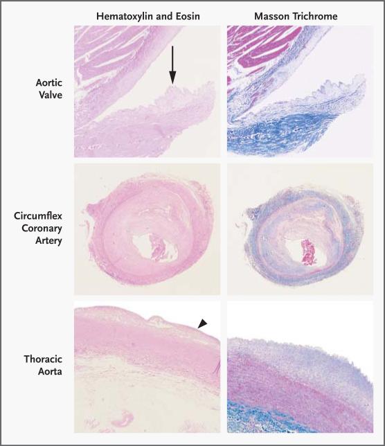
On examination, left ventricular hypertrophy was present and was concentric. The aortic valve contained atherosclerotic lesions, primarily along the aortic aspect of each cusp. Microscopical examination showed plaques rich in lipid-laden foam cells, with focal areas of collagen and scant basophilic ground substance (Fig. 1). The appearance was similar to that of vascular atherosclerosis. The circumflex coronary artery was involved by chronic atherosclerosis and acute thrombosis, and the acute thrombosis was considered the cause of death. Atherosclerotic plaques were also present throughout the thoracic aorta.
Figure 1. Photomicrographs of Tissues Obtained at Autopsy.
The aortic-valve cusp contains atheromatous plaque (arrow) along its aortic aspect (top left and right panels, ×20). The circumflex coronary artery is severely stenotic as a result of chronic atherosclerosis and acute, nonocclusive thrombosis (middle left and right panels, ×12). The thoracic aorta contains a shallow intimal plaque (arrowhead) (bottom left panel, ×12; bottom right panel, ×20).
In the United States, degenerative calcific aortic stenosis is the most common indication for aortic-valve replacement.1 The Cardiovascular Health Study has shown that the risk factors for aortic-valve stenosis are similar to those for vascular atherosclerosis and include an elevated level of low-density lipoprotein, hypertension, male sex, and smoking.2 Otto et al.3 have also reported that the presence of early, prestenotic lesions of aortic-valve sclerosis is associated with a 50 percent increase in the rate of coronary events, thereby further substantiating a relation between atherosclerotic valvular disease and vascular disease.
The current study of a case from 1956 (before the widespread use of aggressive lipid-lowering strategies) demonstrates the relation between aortic-valve disease and vascular atherosclerosis. It confirms the hypothesis that the aortic valve has an active cellular biology with the appearance of a lipid-rich plaque along the surface of the valve leaflet.
Contributor Information
Nalini M. Rajamannan, Northwestern University Chicago, IL 60611 n-rajamannan@northwestern.edu
William D. Edwards, Mayo Clinic Rochester, MN 55905
Thomas C. Spelsberg, Mayo Clinic Rochester, MN 55905
References
- 1.Edwards WD. The changing spectrum of valvular heart disease pathology. In: Braunwald E, editor. Harrison's advances in cardiology. McGraw-Hill; New York: 2002. pp. 317–23. [Google Scholar]
- 2.Stewart BF, Siscovick D, Lind BK, et al. Clinical factors associated with calcific aortic valve disease: Cardiovascular Health Study. J Am Coll Cardiol. 1997;29:630–4. doi: 10.1016/s0735-1097(96)00563-3. [DOI] [PubMed] [Google Scholar]
- 3.Otto CM, Lind BK, Kitzman DW, Gersh BJ, Siscovick DS. Association of aortic-valve sclerosis with cardiovascular mortality and morbidity in the elderly. N Engl J Med. 1999;341:142–7. doi: 10.1056/NEJM199907153410302. [DOI] [PubMed] [Google Scholar]


