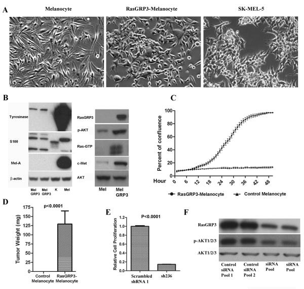
Figure 5.
Effect of RasGRP3 expression on normal melanocytes. (A) Phase constrast imaging of normal human adult melanocytes and of the RasGRP3-melanocytes. SK-MEL-5 cells are shown for comparison. Images are representative of 3 independent experiments. (B) Cell lysates of RasGRP3-melanocytes (MelGRP3), keratinocytes (K), and primary adult melanocytes (Mel) were assayed by immunoblotting as indicated (melanin A, MelA). Results are representative of 3 independent experiments. (C) Control adult melanocytes and RasGRP3-melanocytes were cultured in RPMI 1640 with 10% normal FBS for 5 days. The plates were scanned in the IncuCyte™ at one hour intervals for 60 hours. The data were analyzed by the IncuCyte™ cell proliferation assay software. Results are representative of 3 independent experiments. (D) NOD.SCID/NCr mice (7 mice for each group) were injected subcutaneously in the flanks with RasGRP3-Melanocytes or control melanocytes (plenti/V5-GW/lacZ), 5×10⁶ cells/injection. The injected mice were fed with a regular diet and sacrificed after 5 weeks. The tumors were then excised and weighed. (E) RasGRP3-melanocytes were transduced with virus containing scrambled shRNA control or sh236 respectively. Cell proliferation was determined 120 hours after transduction using the CyQuant NF cell proliferation assay. The values are normalized to that of untreated group. Values are mean ± SEM of 3 independent experiments. (F) Cells were treated as in panel E. RasGRP3 expression, Akt and p-Akt were detected by immunoblotting. Results are representative of three independent experiments.

