Abstract
Stable isotopes have greatly contributed to our understanding of nitrogen metabolism and the urea cycle. The measurement of urea flux via isotopic methods has traditionally been utilized to determine total body protein synthesis in subjects with an intact urea cycle. However, isotopic studies of nitrogen metabolism are also a useful adjunct to conventional clinical investigations in the diagnosis and management of the inherited hyperammonemias. Such studies offer a safe non-invasive method of measuring the reduction of in vivo hepatic ureagenesis, and thus may provide a more accurate measure of phenotypic severity in affected patients. In addition, isotopic methods are ideally suited to evaluate the efficacy of novel therapies to augment urea production.
Keywords: Stable isotopes, urea cycle disorders, ureagenesis, mass spectrometry
1. Introduction
Tracer methodology with stable isotopes has been immensely important in numerous studies of normal and abnormal human biochemistry and metabolism. The chemical behavior of a compound labeled with stable isotopes usually is indistinguishable from that of the parent, but the presence of the label, which is readily detectable, enables research that both measures the flux through a biochemical pathway and identifies pertinent precursor-product relationships. An important advantage of using stable isotopes is that these tracers emit no radioactivity, thereby making them safe for in vivo studies, including pediatric investigations.
Nitrogen-15 was identified and isolated in 1937 [1], only 5 years after discovery of the urea cycle [2]. This coincidence facilitated pathbreaking research in nitrogen metabolism. Indeed, Schoenheimer, Rittenberg and other luminaries of 20th Century biochemistry exploited 15N tracers to perform their seminal studies of in vivo protein turnover [3–6], thereby documenting the “dynamic” nature of body constituents [5,7].
Urea is the major end-product of mammalian nitrogen metabolism. The biochemical pathway required for urea synthesis consists of 6 enzymes and 2 membrane transporters [8]. Deficiencies in each of these proteins have been identified in humans, resulting in discrete clinical and biochemical phenotypes that have hyperammonemia as a common biochemical feature [8–11]. Severe hyperammonemia usually causes acute encephalopathy the length of which appears to correspond to the subsequent degree of learning difficulties or intellectual disability [12].
The measurement of urea production via isotopic methods is often used as an estimate of net protein catabolism in those with an intact urea cycle [3,4,13–15]. However, the application of metabolic tracers also has an important role in studying the urea cycle flux in patients with defects in the urea cycle. Such studies are not only a useful adjunct in diagnosis and management, but have also been employed to evaluate the efficacy of therapeutic interventions.
2. Measurement of isotopic enrichment
Many methods for measuring isotopic abundance are available; each has advantages and drawbacks. Initial studies employed isotope ratio mass spectrometry (IRMS), which is extremely sensitive and precise with regard to the assay of the isotopic ratio (15N/14N or 13C/12C) [16]. These instruments involve separation of gases (typically nitrogen (m/z 29/28) or carbon dioxide (m/z 45/44)) in an electromagnetic field and subsequent focusing of the labeled or unlabeled species into separate collectors, which then record the molecular abundance of each species. The ratio of labeled-to-unlabeled material then is calculated from a computerized algorithm. This technique obliges conversion of samples to a gas. It is exquisitely sensitive with respect to the measurement of isotopic abundance, but it requires that a relatively large amount of material be introduced to the mass spectrometer.
An important methodologic advance was the pairing of a gas chromatograph coupled with a mass spectrometer (GCMS), which allowed rapid separation of analytes on the GC column prior to introduction of each compound to a mass spectrometer, which assayed isotopic abundance. Most GCMS apparatuses use a quadrupole mass analyzer in order to rapidly analyze a wide array of compounds of varying molecular weight. This methodology is sensitive with respect to sample size, but it is less sensitive than IRMS for detection of isotopic abundance [17,18]. Furthermore, gas chromatography requires chemical derivatization in order to render molecules thermally volatile in the GC oven. More recently, the resolving power of GC has been coupled to IRMS. However, this necessitates an intermediate in-line combustion oven, because IRMS can only analyze simple gases. While this method obviates the need for extensive offline purification of the sample, complete separation of the analyte must occur during the chromatographic phase; whereas verification of sample purity with a quadrupole analyzer can be performed by examining its mass spectrum, when IRMS is employed it is impossible to identify the parent molecules following their combustion.
An alternative to GCMS is liquid chromatography-mass spectrometry (LCMS). The advantage of this technology is that it requires minimal sample preparation and, at least in some instances, also obviates the need for derivatization. However, with LCMS there is limited fragmentation of the parent molecule, thus restricting the ability to identify the site of isotopic labeling [16]. As with GCMS, different types of mass analyzers may be coupled with LC.
Either a GC or LC can be coupled with a tandem mass spectrometer (MS/MS), in which the fragmentation pattern of a particular molecule often allows determination of the site of the label in a large molecule [19]. However this is typically not required in the analysis of the relatively small molecules of the urea cycle. Ultimately, the choice of instrumentation depends on the analyte to be measured and the required sensitivity.
3. Stable-isotope measurement of ureagenesis in patients with inherited hyperammonemia
Stable isotope methods provide unique insight into the diagnosis and treatment of urea cycle disorders (UCDs). Such information is not accessible with traditional clinical or biochemical investigations. For instance, no standard clinical test assesses total hepatic urea production. Though this does not address any specific enzymatic defect, it accurately and non-invasively reflects the altered nitrogen metabolism, and thus may provide an objective assessment of phenotypic severity. Two studies, employing stable isotopes in different methods, not only revealed the degree of urea cycle impairment, but were sufficiently sensitive to be able to distinguish between symptomatic and asymptomatic UCD carriers [20–22].
Yudkoff and colleagues [20] evaluated a single dose of oral 15N-ammonium chloride in 14 carriers of ornithine transcarbamylase (OTC) deficiency, a hemizygous OTC-deficient male who had presented with neonatal hyperammonemia and 9 control subjects. The isotopic ammonium is directly incorporated into the urea cycle via the carbamylphosphate synthetase (CPS1) reaction, thereby generating labeled urea.
In this study, the isotopic abundance of [15N] urea recapitulated the clinical phenotypes, with the greatest synthesis of [15N] urea observed in asymptomatic OTC heterozygotes and control subjects, followed by symptomatic heterozygotes and then the hemizygous male. Interestingly, despite no observable difference in urea synthesis, 15N enrichment of glutamine clearly differentiated between the control subjects and asymptomatic heterozygotes, thus indicating that, despite the lack of symptoms, normal plasma ammonia, and normal urea production, nitrogen metabolism is nonetheless abnormal in OTC-deficient carriers [20]. In subsequent experiments, label incorporation into urea easily distinguished between male subjects with neonatal versus late-onset disease [21].
Lee and colleagues [22] expanded the subjects of study to also include ASS and ASL deficiency, and similarly evaluated affected patients, heterozygotes and unaffected controls. This experiment involved the constant infusion of both [18O] urea and [5-15N] glutamine, with subsequent measurement of both total urea and glutamine flux. Transfer of the amide-N of glutamine to the urea cycle resulted in the production of [15N] urea. The [15N] urea/[5-15N] μglutamine ratio, a measure of urea synthesized from peripheral nitrogen sources, distinguished among unaffected control subjects, asymptomatic heterozygotes, and symptomatic individuals. Among affected individuals, this ratio was lower in those with neonatal-onset versus late-onset disease [22].
Given the current accessibility of molecular genetic testing for all of the urea cycle disorders, mutational analysis is frequently requested as part of the diagnostic work-up, especially if no pathognomonic amino acids are identified in plasma. However, pathological mutations are not always identified [23,24] and even when they are found, they may not provide much prognostic information, especially in the case of heterozygotes of OTC deficiency, the most common urea cycle disorder [25]. This may result in some scenarios where the non-invasive determination of urea cycle function can greatly contribute to clinical management.
In a notable example, Scaglia and colleagues applied the above protocol to evaluate urea cycle flux in a female with suspected partial OTCD [26] who presented at 30-months of age with developmental delay and recurrent episodes of emesis. She had hyperammonemia of 246 μmol/L (normal 22–48), and a biochemical profile suggestive of partial OTCD, including low plasma citrulline, and moderately increased urine orotic acid. Hepatic OTC activity was found to be present but reduced (770 μmol/hr/g liver; control 1500–9000 μmol/hr/g liver). While these indices supported the diagnosis of partial OTC deficiency, molecular diagnostic testing was unable to identify a mutation. However, stable isotope studies demonstrated substantially reduced nitrogen transfer from glutamine to urea. This parameter was comparable to some affected OTCD males and even lower than that observed in some late-onset males. Ultimately, these results contributed to the risk-benefit assessment of potential avenues of long-term treatment and after consideration a decision was made to perform an orthotopic liver transplantation.
It may be that quantitation of flux with stable isotopes can not only diagnose the degree of urea cycle impairment, but even predict, prior to the onset of symptoms, whether medical management is necessary. This is particularly important for young OTC heterozygotes identified prospectively through affected male relatives. In fact, among 19 symptomatic and asymptomatic female OTC heterozygote carriers who presented with a positive family history of affected male relative, isotopic studies more accurately identified those who presented with clinical symptoms than an allopurinol challenge [27].
4. Evaluating the efficacy of therapeutics
Because isotopic studies may provide a holistic assessment of in-vivo ureagenesis, they may be used to evaluate the efficacy of novel therapies which impact urea production. Thus, urea flux may be measured before and after the intervention.
Our group has long been interested in the salutary effect of N-carbamylglutamate (NCG), a stable analog of N-acetylglutamate (NAG), the obligate activator of the CPS1 enzyme. Whereas oral NAG is hydrolyzed in vivo, NCG is resistant to hydrolysis. We therefore investigated the use of oral NCG as a therapy in N-acetylglutamate synthase deficiency by employing stable isotopes.
The method entails a single oral load of sodium [1-13C] acetate. Oxidation of the isotopic acetate via the tricarboxylic acid (TCA) cycle results in production of CO2 and incorporation of the label into bicarbonate. The labeled bicarbonate is then utilized by the carbamylphosphate synthetase reaction to generate 13C-carbamylphosphate, the label of which is then incorporated into urea ([13C] urea). As demonstrated in 17 control subjects [28], sequential blood measurements then document the appearance of serum [13C] urea, whose turnover is slow compared to the period of experimental observation and provides an indicator of the production of urea over time.
We performed such investigations in a patient with NAGS deficiency, before and after a 3-day trial with NCG [28]. No changes were made in the diet or medications of this otherwise well patient. Hence, any alterations in ureagenesis were due to the action of N-carbamylglutamate. In fact, a marked increase in urea production was observed after the short trial of NCG, providing direct evidence of the salutary effect of the NCG. This was corroborated by improvements in other biomarkers, such as a decrease in plasma ammonia and glutamine, and an increase in serum urea [28].
We then employed this methodology to evaluate other conditions in which a secondary NAG deficiency is thought to reduce flux through CPS1 and result in hyperammonemia. For instance, in propionic acidemia (PA), or a congenital deficiency of propionyl-CoA carboxylase, a decrease in NAG synthesis may occur either from competitive inhibition of NAGS by propionyl-CoA [29–32] or a relative depletion of hepatic acetyl-CoA or free coenzyme A [32]. Individual case reports suggest that NCG is helpful in the treatment of PA [33–37], but these are uncontrolled studies performed during an acute illness in which the effect of NCG is difficult to differentiate from that of standard care.
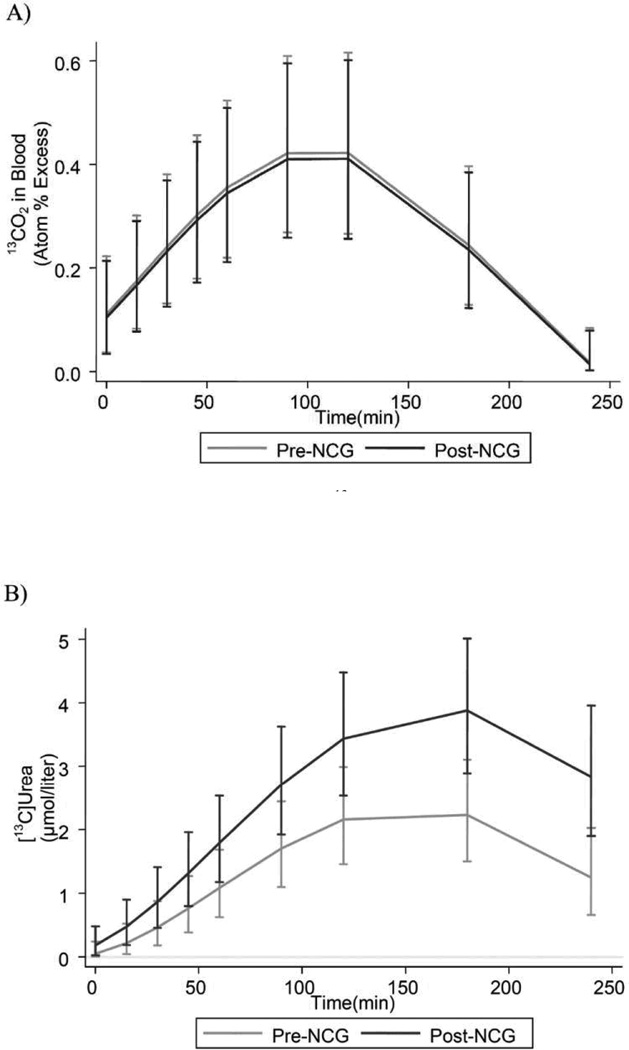
We evaluated 7 subjects with PA, performing isotopic urea turnover studies before-and-after a 3-day trial of NCG. The aggregate results (Figure 1) indicate that most administered [1-13C] acetate is converted to 13CO2, which increases very rapidly in blood and which is unaffected by NCG treatment. NCG markedly augmented [13C] urea synthesis, a phenomenon reflected by normalization of plasma ammonia levels. This not only provides direct evidence that NCG may be a useful adjunct to the treatment of hyperammonemia in PA, but additionally, offers indirect proof that insufficient activation of CPS1 may contribute to hyperammonemia in PA.
Figure 1.
Isotopic enrichment in plasma 13CO2 (A), and plasma concentrations of [13C] urea (B) in 7 patients with propionic acidemia who were administered 27.5 mg/kg of [13C] sodium acetate before and after 3d NCG therapy.
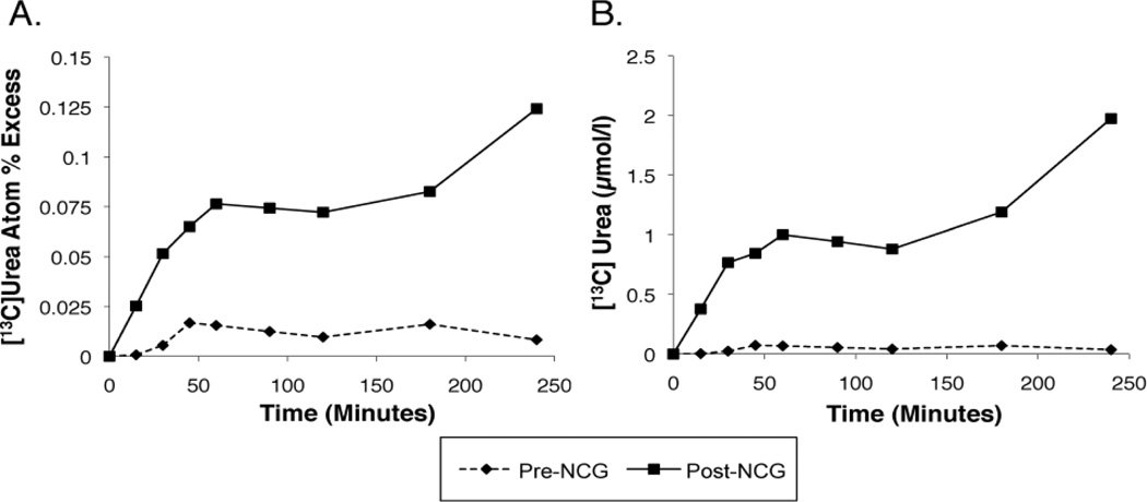
Interestingly, a marked increase in ureagenesis in response to NCG therapy was crucial in the diagnosis of a young woman with NAGS deficiency [38]. We evaluated a young patient in whom biochemical markers suggested a proximal urea cycle disorder, but in whom an allopurinol challenge was negative, and molecular diagnostic testing of NAGS, CPS1 and OTC failed to disclose a mutation. In addition, hepatic enzyme assay showed normal activity of CPS1 and OTC. The [1-13C] acetate procedure revealed a response to NCG (Figure 2) [38] which was similar to that of the NAGS deficient subject described above [28]. This made a compelling case for a diagnosis of NAGS deficiency, despite the initial negative molecular genetic testing. This prompted a search throughout the NAGS gene for a possible mutation, culminating in the discovery of a NAGS enhancer mutation. We hypothesize that this magnitude of correction to ureagenesis in response to NCG is likely unique to NAGS deficiency. Thus, this study may not only be successful in diagnosing patients in whom molecular diagnostic studies have failed, but can simultaneously validate the efficacy of the therapy.
Figure 2.
Increase over time in isotopic enrichment of [13C]urea (A) and plasma concentration of [13C]urea (B) in a patient with a suspected proximal urea-cycle disorder, who was administered 27.5 mg/kg of [1-13C]sodium acetate before and after a 3d trial of NCG. Molecular and enzymatic testing had failed to reveal a diagnosis. However, the marked augmentation in urea production following an NCG trial shown here prompted a search in the non-coding regions of the NAGS gene, and culminated in the identification of a mutation in the NAGS enhancer [38].
The rationale for developing [1-13C] acetate as a tracer is that, although the 15NH3 probe enabled effective monitoring of the response to NCG [39] the taste of solutions of NH4Cl is objectionable to most subjects and administration of ammonium salts is risky in the patient who is prone to hyperammonemia. Using [1-13C] acetate as a probe neutralizes these problems, but is not without potential complications. Thus, the method depends upon rapid intestinal absorption and hepatic metabolism of the oral 13C-acetate load, both before and after the NCG trial. Fortunately, it is possible to infer a problem with either absorption or oxidation of 13C by careful inspection of the resulting curve of 13CO2 formation in blood.
This experimental approach can demonstrate the efficacy of an intervention – in this instance, N-carbamylglutamate – but it does not disclose how rapidly the response to treatment has occurred. We therefore developed a protocol to address this issue. The procedure entailed a priming dose of H13CO3 followed by a constant infusion of this species for 5 hours. As the half-life of bicarbonate in vivo is short, this method permits the rapid attainment of a steady-state in bicarbonate enrichment. On the other hand, the half-life of urea is long – on the order of hours – thus, over time, as labeled bicarbonate is converted to urea, there is a constant linear increase in the blood [13C] urea concentration. In our study, we introduced a single oral dose of NCG after 90 minutes of isotopic bicarbonate infusion. We then observed in 5 of 6 subjects an increase (i.e., an upward inflection) in the linear rate of appearance of [13C] urea, thus demonstrating that NCG works within hours of administration.
Similar isotopic protocols may be used to evaluate novel therapies in which augmentation or restoration of ureagenesis is desired. Recent advances in gene therapy [40,41] hepatocyte transplantation [42,43] or adult hepatic stem cell [44] transplantation are examples. With each of these technologies, restoration of ureagenesis may not be uniform across the liver. Thus, liver biopsies may incorrectly determine the degree of hepatic restoration, whereas stable isotope methods presumably evaluate in vivo the rate of ureagenesis, and may provide a much more accurate estimate of any improvement in hepatic urea synthesis.
In conclusion, experiments utilizing stable isotopes have provided important insights into nitrogen metabolism and urea cycle disorders. Though underutilized clinically, the evaluation of in vivo nitrogen metabolism using tracers may provide important adjunctive information in the management of patients with urea cycle disorders. Additionally, such studies are ideally suited to evaluate the effect of novel therapies which may ameliorate nitrogen disposal via the urea cycle.
References
- 1.Urey HC, Huffman JR, Thode HG, Fox M. Concentration of N-15 by chemical methods. J Chem Phys. 1937;5(11):856–872. [Google Scholar]
- 2.Krebs HA, Henseleit K. Untersuchungen über die Harnstoffbildung im Tierkörper. Z Physiol Chem. 1932;210:33–46. [Google Scholar]
- 3.San Pietro A, Rittenberg D. A study of the rate of protein synthesis in humans. I Measurement of the urea pool and urea space. J Biol Chem. 1953;201(1):445–455. [PubMed] [Google Scholar]
- 4.San Pietro A, Rittenberg D. A study of the rate of protein synthesis in humans. II. Measurement of the metabolic pool and the rate of protein synthesis. J Biol Chem. 1953;201(1):457–473. [PubMed] [Google Scholar]
- 5.Schoenheimer R, Rittenberg D. The Application of Isotopes to the Study of Intermediary Metabolism. Science. 1938;87(2254):221–226. doi: 10.1126/science.87.2254.221. [DOI] [PubMed] [Google Scholar]
- 6.Schoenheimer R, Rittenberg D, Foster GL, Keston AS, Ratner S. The Application of the Nitrogen Isotope N15 for the Study of Protein Metabolism. Science. 1938;88(2295):599–600. doi: 10.1126/science.88.2295.599. [DOI] [PubMed] [Google Scholar]
- 7.Schoenheimer R, Clarke HT. The dynamic state of body constituents. Cambridge, Mass: Harvard university press; 1942. –3.pp. l–78.–21. [Google Scholar]
- 8.Summar M, Tuchman M. Proceedings of a consensus conference for the management of patients with urea cycle disorders. J Pediatr. 2001;138(1 Suppl):S6–S10. doi: 10.1067/mpd.2001.111831. [DOI] [PubMed] [Google Scholar]
- 9.Batshaw ML, Brusilow S, Waber L, Blom W, Brubakk AM, Burton BK, et al. Treatment of inborn errors of urea synthesis: activation of alternative pathways of waste nitrogen synthesis and excretion. N Engl J Med. 1982;306(23):1387–1392. doi: 10.1056/NEJM198206103062303. [DOI] [PubMed] [Google Scholar]
- 10.Batshaw ML, Brusilow SW. Treatment of hyperammonemic coma caused by inborn errors of urea synthesis. J Pediatr. 1980;97(6):893–900. doi: 10.1016/s0022-3476(80)80416-1. Epub 1980/12/01. [DOI] [PubMed] [Google Scholar]
- 11.Brusilow SW, Maestri NE. Urea cycle disorders: diagnosis, pathophysiology, and therapy. Adv pediatr. 1996;43:127–170. [PubMed] [Google Scholar]
- 12.Msall M, Batshaw ML, Suss R, Brusilow SW, Mellits ED. Neurologic outcome in children with inborn errors of urea synthesis. Outcome of urea-cycle enzymopathies. N Engl J Med. 1984;310(23):1500–1505. doi: 10.1056/NEJM198406073102304. [DOI] [PubMed] [Google Scholar]
- 13.Duggleby SL, Waterlow JC. The end-product method of measuring whole-body protein turnover: a review of published results and a comparison with those obtained by leucine infusion. Br J Nutr. 2005;94(2):141–153. doi: 10.1079/bjn20051460. [DOI] [PubMed] [Google Scholar]
- 14.Koletzko B, Demmelmair H, Hartl W, Kindermann A, Koletzko S, Sauerwald T, et al. The use of stable isotope techniques for nutritional and metabolic research in paediatrics. Early Hum Dev. 1998;53(Suppl):S77–S97. doi: 10.1016/s0378-3782(98)00067-x. [DOI] [PubMed] [Google Scholar]
- 15.Wolfe RR, Goodenough RD, Burke JF, Wolfe MH. Response of protein and urea kinetics in burn patients to different levels of protein intake. Ann Surg. 1983;197(2):163–171. doi: 10.1097/00000658-198302000-00007. [DOI] [PMC free article] [PubMed] [Google Scholar]
- 16.Wolfe RR, Chinkes DL. Isotope tracers in metabolic research : principles and practice of kinetic analysis. 2nd ed. Hoboken, New Jersey: Wiley-Liss; 2005. p. vii.p. 474. [Google Scholar]
- 17.Preston T, Slater C. Mass spectrometric analysis of stable-isotope-labelled amino acid tracers. Proc Nutr Soc. 1994;53(2):363–372. doi: 10.1079/pns19940042. [DOI] [PubMed] [Google Scholar]
- 18.Meier-Augenstein W. Use of gas chromatography-combustion-isotope ratio mass spectrometry in nutrition and metabolic research. Curr Opin Clin Nutr Metab Care. 1999;2(6):465–470. doi: 10.1097/00075197-199911000-00005. [DOI] [PubMed] [Google Scholar]
- 19.Meier-Augenstein W. Applied gas chromatography coupled to isotope ratio mass spectrometry. J Chromatogr A. 1999;842(1–2):351–371. doi: 10.1016/s0021-9673(98)01057-7. [DOI] [PubMed] [Google Scholar]
- 20.Yudkoff M, Daikhin Y, Nissim I, Jawad A, Wilson J, Batshaw M. In vivo nitrogen metabolism in ornithine transcarbamylase deficiency. The Journal of clinical investigation. 1996;98(9):2167–2173. doi: 10.1172/JCI119023. [DOI] [PMC free article] [PubMed] [Google Scholar]
- 21.McCullough BA, Yudkoff M, Batshaw ML, Wilson JM, Raper SE, Tuchman M. Genotype spectrum of ornithine transcarbamylase deficiency: correlation with the clinical and biochemical phenotype. Am J Med Genet. 2000;93(4):313–319. doi: 10.1002/1096-8628(20000814)93:4<313::aid-ajmg11>3.0.co;2-m. [DOI] [PubMed] [Google Scholar]
- 22.Lee B, Yu H, Jahoor F, O'Brien W, Beaudet AL, Reeds P. In vivo urea cycle flux distinguishes and correlates with phenotypic severity in disorders of the urea cycle. Proc Natl Acad Sci USA. 2000;97(14):8021–8026. doi: 10.1073/pnas.140082197. [DOI] [PMC free article] [PubMed] [Google Scholar]
- 23.Yamaguchi S, Brailey LL, Morizono H, Bale AE, Tuchman M. Mutations and polymorphisms in the human ornithine transcarbamylase (OTC) gene. Hum Mutat. 2006;27(7):626–632. doi: 10.1002/humu.20339. [DOI] [PubMed] [Google Scholar]
- 24.Häberle J, Shchelochkov OA, Wang J, Katsonis P, Hall L, Reiss S, et al. Molecular defects in human carbamoy phosphate synthetase I: mutational spectrum, diagnostic and protein structure considerations. Hum Mutat. 2011;32(6):579–589. doi: 10.1002/humu.21406. [DOI] [PMC free article] [PubMed] [Google Scholar]
- 25.Tuchman M, Lee B, Lichter-Konecki U, Summar ML, Yudkoff M, Cederbaum SD, et al. Cross-sectional multicenter study of patients with urea cycle disorders in the United States. Mol Genet Metab. 2008;94(4):397–402. doi: 10.1016/j.ymgme.2008.05.004. [DOI] [PMC free article] [PubMed] [Google Scholar]
- 26.Scaglia F, Zheng Q, O'Brien WE, Henry J, Rosenberger J, Reeds P, et al. An integrated approach to the diagnosis and prospective management of partial ornithine transcarbamylase deficiency. Pediatrics. 2002;109(1):150–152. doi: 10.1542/peds.109.1.150. [DOI] [PubMed] [Google Scholar]
- 27.Gyato K, Wray J, Huang ZJ, Yudkoff M, Batshaw ML. Metabolic and neuropsychological phenotype in women heterozygous for ornithine transcarbamylase deficiency. Ann Neurol. 2004;55(1):80–86. doi: 10.1002/ana.10794. [DOI] [PubMed] [Google Scholar]
- 28.Tuchman M, Caldovic L, Daikhin Y, Horyn O, Nissim I, Korson M, et al. Ncarbamylglutamate markedly enhances ureagenesis in N-acetylglutamate deficiency and propionic acidemia as measured by isotopic incorporation and blood biomarkers. Pediatr Res. 2008;64(2):213–217. doi: 10.1203/PDR.0b013e318179454b. [DOI] [PMC free article] [PubMed] [Google Scholar]
- 29.Coude FX, Sweetman L, Nyhan WL. Inhibition by propionyl-coenzyme A of Nacetylglutamate synthetase in rat liver mitochondria. A possible explanation for hyperammonemia in propionic and methylmalonic acidemia. J Clin Invest. 1979;64(6):1544–1551. doi: 10.1172/JCI109614. [DOI] [PMC free article] [PubMed] [Google Scholar]
- 30.Dercksen M, Ijlst L, Duran M, Mienie LJ, van Cruchten A, van der Westhuizen FH, et al. Inhibition of N-acetylglutamate synthase by various monocarboxylic and dicarboxylic short-chain coenzyme A esters and the production of alternative glutamate esters. Biochim Biophys Acta. 2013 May 2; doi: 10.1016/j.bbadis.2013.04.027. pii: S0925-4439(13)00151-8. [DOI] [PubMed] [Google Scholar]
- 31.Glasgow AM, Chase HP. Effect of propionic acid on fatty acid oxidation and ureagenesis. Pediatr Res. 1976;10(7):683–686. doi: 10.1203/00006450-197607000-00010. [DOI] [PubMed] [Google Scholar]
- 32.Stewart PM, Walser M. Failure of the normal ureagenic response to amino acids in organic acid-loaded rats. Proposed mechanism for the hyperammonemia of propionic and methylmalonic acidemia. J Clin Invest. 1980;66(3):484–492. doi: 10.1172/JCI109879. [DOI] [PMC free article] [PubMed] [Google Scholar]
- 33.Filippi L, Gozzini E, Fiorini P, Malvagia S, la Marca G, Donati MA. Ncarbamylglutamate in emergency management of hyperammonemia in neonatal acute onset propionic and methylmalonic aciduria. Neonatology. 2010;97(3):286–290. doi: 10.1159/000255168. [DOI] [PubMed] [Google Scholar]
- 34.Gebhardt B, Dittrich S, Parbel S, Vlaho S, Matsika O, Bohles H. N-carbamylglutamate protects patients with decompensated propionic aciduria from hyperammonaemia. J Inherit Metab Dis. 2005;28(2):241–244. doi: 10.1007/s10545-005-5260-7. [DOI] [PubMed] [Google Scholar]
- 35.Jones S, Reed CA, Vijay S, Walter JH, Morris AA. N-carbamylglutamate for neonatal hyperammonaemia in propionic acidaemia. J Inherit Metab Dis. 2008;31(Suppl 2):S219–S222. doi: 10.1007/s10545-008-0777-1. [DOI] [PubMed] [Google Scholar]
- 36.Schwahn BC, Pieterse L, Bisset WM, Galloway PG, Robinson PH. Biochemical efficacy of N-carbamylglutamate in neonatal severe hyperammonaemia due to propionic acidaemia. Eur J Pediatr. 2010;169(1):133–134. doi: 10.1007/s00431-009-1036-7. [DOI] [PubMed] [Google Scholar]
- 37.Levesque S, Lambert M, Karalis A, Melancon S, Russell L, Braverman N. Short-term outcome of propionic aciduria treated at presentation with N-carbamylglutamate: a retrospective review of four patients. JIMD Rep. 2012;2:97–102. doi: 10.1007/8904_2011_54. [DOI] [PMC free article] [PubMed] [Google Scholar]
- 38.Heibel SK, Ah Mew N, Caldovic L, Daikhin Y, Yudkoff M, Tuchman M. Ncarbamylglutamate enhancement of ureagenesis leads to discovery of a novel deleterious mutation in a newly defined enhancer of the NAGS gene and to effective therapy. Hum Mutat. 2011;32(10):1153–1160. doi: 10.1002/humu.21553. [DOI] [PMC free article] [PubMed] [Google Scholar]
- 39.Caldovic L, Morizono H, Daikhin Y, Nissim I, McCarter RJ, Yudkoff M, et al. Restoration of ureagenesis in N-acetylglutamate synthase deficiency by Ncarbamylglutamate. J Pediatr. 2004;145(4):552–554. doi: 10.1016/j.jpeds.2004.06.047. [DOI] [PubMed] [Google Scholar]
- 40.Moscioni D, Morizono H, McCarter RJ, Stern A, Cabrera-Luque J, Hoang A, et al. Longterm correction of ammonia metabolism and prolonged survival in ornithine transcarbamylase-deficient mice following liver-directed treatment with adenoassociated viral vectors. Mol Ther. 2006;14(1):25–33. doi: 10.1016/j.ymthe.2006.03.009. [DOI] [PubMed] [Google Scholar]
- 41.Wang L, Morizono H, Lin J, Bell P, Jones D, McMenamin D, et al. Preclinical evaluation of a clinical candidate AAV8 vector for ornithine transcarbamylase (OTC) deficiency reveals functional enzyme from each persisting vector genome. Mol Genet Metab. 2012;105(2):203–211. doi: 10.1016/j.ymgme.2011.10.020. [DOI] [PMC free article] [PubMed] [Google Scholar]
- 42.Meyburg J, Hoffmann GF. Liver, liver cell and stem cell transplantation for the treatment of urea cycle defects. Mol Genet Metab. 2010;100(Suppl 1):S77–S83. doi: 10.1016/j.ymgme.2010.01.011. [DOI] [PubMed] [Google Scholar]
- 43.Meyburg J, Schmidt J, Hoffmann GF. Liver cell transplantation in children. Clin Transplant. 2009;23(Suppl 21):75–82. doi: 10.1111/j.1399-0012.2009.01113.x. [DOI] [PubMed] [Google Scholar]
- 44.Najimi M, Khuu DN, Lysy PA, Jazouli N, Abarca J, Sempoux C, et al. Adult-derived human liver mesenchymal-like cells as a potential progenitor reservoir of hepatocytes? Cell Transplant. 2007;16(7):717–728. doi: 10.3727/000000007783465154. [DOI] [PubMed] [Google Scholar]


