Abstract
We report a case of the female patient who was admitted to the hospital because of syncope experienced while climbing stairs. Diagnostic workup raised the suspicion of a right diaphragmatic rupture that was eventually confirmed by surgery (right-sided thoracotomy). Surgery also revealed tissue protruding through the rupture site from within the retroperitoneum that was proven subsequently to be a dedifferentiated liposarcoma. Second surgery was performed to completely remove the liposarcoma tissue and repair a coincident old right lumbar region hernia. The patient recovered fully.
Spontaneous rupture of the diaphragm is rare and this is especially true for the right hemidiaphragm. We report the first case of diaphragmatic rupture caused by local infiltration by a retroperitoneal liposarcoma. This and similar reports emphasise that in cases with high clinical suspicion of diaphragmatic rupture, diagnosis should be pursued even in the absence of a preceding traumatic event.
Keywords: Diaphragm, Rupture, Liposarcoma
Case History
A 61-year-old woman was admitted to hospital because of syncope. While climbing stairs, she experienced lightheadedness and dizziness. After sitting down and calling for help, she briefly lost consciousness. She recalled having sharp pain behind her chest bone radiating to the right breast region one week ago. The pain worsened when breathing deeply. She then began experiencing exertional dyspnoea even at a slight workload. Two days before admission she started feeling a dull pain in her right lumbar region that worsened in the supine position. She reported feeling generally weak and nauseated as well as having mild abdominal discomfort. She was previously healthy except for a longstanding right lumbar region hernia. She did not smoke, drink alcohol or take any medication on a regular basis.
At the physical examination the patient was pale and her vital signs were as follows: slightly somnolent, temperature 36.6°C, blood pressure 110/60mmHg (left hand), 130/80mmHg (right hand), heart rate 64/min, respiration rate 14/min. On auscultation breathing sounds were diminished over the right lung. Heart sounds were normal. The abdomen was slightly tender in the epigastric region and under the right costal arch. A right-sided reducible lumbar hernia was present. The remainder of the physical examination was unremarkable.
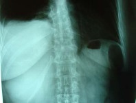
A plain abdominal x-ray showed an elevated right hemidiaphragm and apparent effusion in the right phrenicocostal sinus (Fig 1). During the x-ray examination, the patient collapsed and her blood pressure dropped to 80/40mmHg. Urgent thoracic computerised tomography was ordered because of suspected thoracic aorta dissection. This examination excluded aortic dissection but showed massive right pleural effusion. A right thoracic drain was implanted, through which 1,000ml of fresh blood was immediately evacuated and three hours later 500ml more.
Figure 1.

Plain abdominal x-ray showing elevated right hemidiaphragm and pleural effusion in right phrenicocostal sinus
A blood transfusion (610ml) was given together with fresh frozen plasma (400ml) and normal saline (2,000ml), after which the blood pressure normalised. An urgent bronchoscopic examination showed no endobronchial abnormalities.
Due to the presumed diagnosis of diaphragmatic rupture an exploratory right thoracotomy was performed through a sixth right intercostal posterolateral incision. This procedure revealed fresh and coagulated blood in the right thoracic cavity and showed diaphragmatic rupture posteriorly (lumbar part of the diaphragm) 8cm long and active arterial bleeding. Blood was evacuated and haemostasis performed. The tissue protruding through the rupture site was sampled for pathological analysis and the defect was closed by direct suturing. The patient’s postoperative recovery was excellent. Pathological analysis of the sampled tissue that protruded from within the right retroperitoneum revealed a dedifferentiated liposarcoma.
The patient was sent home and after clinical recovery she was readmitted for an elective operation to completely excise the right retroperitoneal liposarcoma tissue and perform a right-sided lumbar hernia repair. She recovered fully and at a follow-up appointment 20 months after the second operation she had no signs of disease recurrence.
Discussion
The most common causes of diaphragmatic rupture are blunt trauma and penetrating injuries. Spontaneous rupture of the diaphragm is rare, especially in the right hemidiaphragm. A thorough literature review by Losanof et al published in 2010 found only 28 detailed case reports.1
We present a case of right hemidiaphragm rupture not preceded by trauma. Even though an x-ray showed pleural effusion and elevation of the right diaphragm dome, a diagnosis of diaphragmatic rupture was not at first considered likely because history revealed no traumatic injury. An exploratory thoracotomy was the key procedure in reaching the correct diagnosis.
There have been occasional reports of spontaneous diaphragmatic rupture but they were mostly preceded by strenuous physical activity such as weightlifting,2 pilates,3 child delivery4 etc. In our case, climbing stairs could have been the trigger but we speculate that local invasion of the diaphragm by the retroperitoneal tumour was a more likely cause of this particular rupture. The only similar case in the literature of diaphragmatic infiltration leading to rupture is a report published in 2010 of an endometriosis related diaphragm rupture.5
Conclusions
This report is one of only a few published cases of spontaneous right hemidiaphragm rupture and, to the best of our knowledge, it is the first report of diaphragmatic rupture caused by local invasion of a liposarcoma (or any other tumour for that matter). This and similar reports emphasise that in cases with high clinical suspicion of diaphragmatic rupture, a diagnosis should be pursued even in the absence of a preceding traumatic event.
References
- 1.Losanoff JE, Edelman DA, Salwen WA, Basson MD. Spontaneous rupture of the diaphragm: case report and comprehensive review of the world literature. J Thorac Cardiovasc Surg. 2010:139, 127–128. doi: 10.1016/j.jtcvs.2009.05.035. [DOI] [PubMed] [Google Scholar]
- 2.Bisgaard C, Rodenberg JC, Lundgaard J. Spontaneous rupture of the diaphragm. Scand J Thorac Cardiovasc Surg. 1985;19:177–180. doi: 10.3109/14017438509102715. [DOI] [PubMed] [Google Scholar]
- 3.Yang YM, Yang HB, Park JS, et al. Spontaneous diaphragmatic rupture complicated with perforation of the stomach during Pilates. Am J Emerg Med. 2010;28:259, e1–3. doi: 10.1016/j.ajem.2009.06.012. [DOI] [PubMed] [Google Scholar]
- 4.Boufettal R, Lefriyekh MR, Boufettal H, et al. Spontaneous diaphragm rupture during delivery. Case report. J Gynecol Obstet Biol Reprod (Paris) 2008;37:93–96. doi: 10.1016/j.jgyn.2007.10.001. [DOI] [PubMed] [Google Scholar]
- 5.Triponez F, Alifano M, Bobbio A, Regnard JF. Endometriosis-related spontaneous diaphragmatic rupture. Interact Cardiovasc Thorac Surg. 2010;11:485–487. doi: 10.1510/icvts.2010.243543. [DOI] [PubMed] [Google Scholar]


