Abstract
Introduction
Pleomorphic adenoma of minor salivary glands of hard palate is a rare benign tumour. It usually presents as slow growing submucosal mass on hard palate. The purpose of this study was to collect observational data regarding age, size, symptoms, CT findings and treatment of pleomorphic adenoma of hard palate.
Material and methods
The prospective observational study was done in the Department of ENT, Department of Oral and maxillofacial surgery at SKIMS Medical College and at Peoples Care Polyclinic for a period of 2 years. Twenty cases were enrolled in the study. After clinical suspicion, computed tomography, FNAC and core needle biopsy was done in all twenty cases before any surgical intervention was done.
Results
Most common age group seen to be involved was 16–30 years. Most common symptom was submucosal slow growing mass. Core biopsy was diagnostic with 100 % efficacy. On CT scan twelve patients (60 %) were seen to have intact hard palate even without minor erosions while four patients had full thickness erosion of hard palate. Surgical wide excision was done in all cases and one patient needed total maxillectomy. All treated patients were followed for one year and none showed signs of recurrence
Conclusion
Pleomorphic adenoma of hard palate is usually seen in adults. Definitive diagnosis lies on histopathological examination. CT is necessary for ruling out any bony erosion. Treatment is by wide local excision.
Keywords: Pleomorphic adenoma, Hard palate, Excision, Palatal flap, Recurrences, Salivary gland, Rare benign tumour
Introduction
Salivary gland tumors account for <3 % of the head and neck tumors [1]. Pleomorphic adenoma (PA) is the most common salivary gland tumour, accounting for about 40–70 % of all major and minor salivary gland tumors [2]. PA is the most common benign tumor of the minor salivary gland. The most common site of this tumor in the oral cavity is the palatal area followed by the lip, buccal mucosa, floor of the mouth, tongue tonsil, pharynx, and retro molar area [3].
The majority of minor salivary gland PAs occur in the second decade of life [2]. There is slight predilection for female gender [3].
The clinical presentation of a PA of the hard palate is typically a firm or rubbery submucosal mass without ulceration or surrounding inflammation. Pain and tenderness are unusual [4].
The term pleomorphic describes the embryogenic basis of origin of these tumors, which contains both epithelial and mesenchymal tissues [5]. These tumors arise from intercalated and myoepithelial cells [6]. Differential diagnosis of the palatal lesions includes other minor salivary gland tumours, particularly mucoepidermoid carcinoma, as well as other benign and malignant mesenchymal lesions such as neurofibroma and rhabdomyosarcoma [7].
The diagnosis of PA is established on the basis of history, physical examination, cytology and histopathology. Computed tomography (CT) scan and magnetic resonance imaging (MRI) provide information of the location, size and extension of tumor to the surrounding superficial and deep structures.
The treatment of choice for PA in minor salivary gland is wide local excision with the removal of periosteum or bone if they are involved. Simple enucleation of this tumour leads to high local recurrence rate and should be avoided [4].
Materials and Methods
This prospective observational study was done in Department of ENT, Department of Oral and Maxillofacial Surgery at SKIMS Medical College and at Peoples Care Polyclinic from April 2009 to April 2011. After clinical suspicion, CT, Fine Needle Aspiration Cytology (FNAC) and core needle biopsy was done in all 20 cases before any surgical intervention was done.
Twenty patients were enrolled in the study with the following inclusion and exclusion criteria. Inclusion criteria Patients of any age group with swelling of hard palate with strong suspicion of it being PA and which on core biopsy proves to be such. Exclusion criteria Patients presenting with swelling in hard palate and FNAC suggestive of PA but core needle biopsy proved otherwise.
After confirming the diagnosis all patients underwent surgical excision of the mass followed by repair and reconstruction as appropriate to the case.
Patients without erosion of hard palate on CT scan were treated with wide surgical excision and removal of periosteum. Patients who had mild erosion of hard palate were treated with surgical excision and curettage of involved bone. Patients with full thickness erosion of hard palate were treated by removing involved portion of hard palate and in one case total maxillectomy was done.
Reconstruction was not done in cases without erosion of hard palate on CT scan. The excised wound in such cases was allowed to granulate and heal by itself. Reconstruction was done by palatal flap based on greater palatine artery and obturator in cases with full thickness erosion of the hard palate.
The surgical specimens were sent for histopathological examination.
All patients were at least followed-up for 1 year after they were treated by definitive surgical therapy while some enrolled early in the study were followed-up for 2 years
Observations and Results
Twenty patients diagnosed as PA of hard palate were seen and operated in a span of 2 years.
The most common age group involved was 16–30 years. Only two patients were seen in pediatric age group of <15. Males were seen to be involved more commonly than females (Table 1).
Table 1.
Number of patients in different age groups
| Age-group | Number of patients | |
|---|---|---|
| Males | Females | |
| 0–15 | 2 | x |
| 16–30 | 8 | 3 |
| 31–45 | 3 | 1 |
| 46–60 | 2 | x |
| 61–75 | 1 | x |
| Total | 16 | 4 |
Swelling over hard palate was the most common clinical feature. Overlying mucosa was normal in 17 patients. Ulcerations of mucosa was seen in three cases (Fig. 1). Majority of swellings (18) were smooth (Fig. 2) and only two were multilobulated (Fig. 3). The size of swelling ranged from 0 to 8 cm with maximum number of patients (12) having swelling in the range of 3–5 cm (Table 2).
Fig. 1.
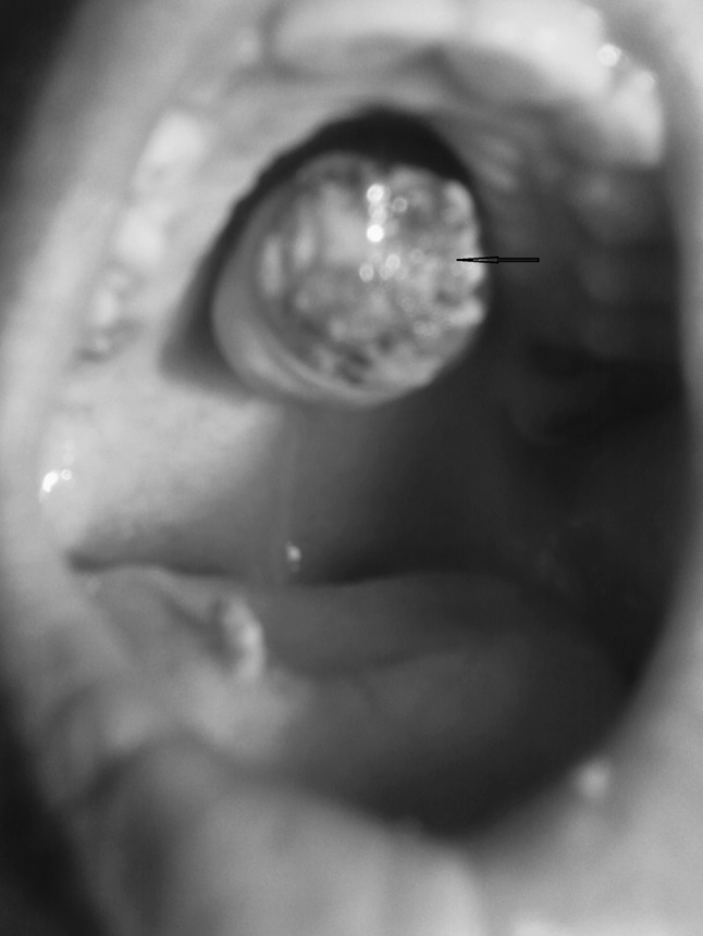
Rounded PA over hard palate with ulceration (black arrow)
Fig. 2.
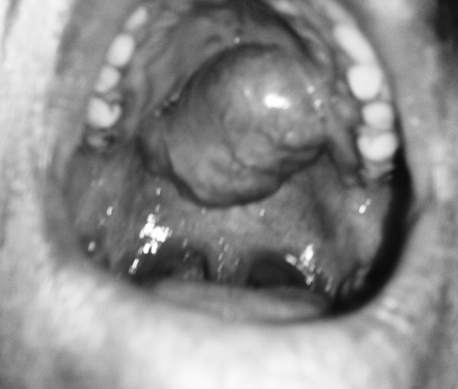
Smooth surfaced, unilobular PA over hard palate
Fig. 3.
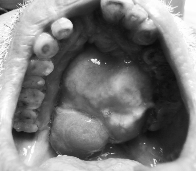
Multilobulated PA over hard palate
Table 2.
Clinical features
| Clinical features | No. of patients | Percentage |
|---|---|---|
| Pain | 3 | 15 |
| Ulceration | 3 | 15 |
| Bleeding | 3 | 15 |
| Swelling | 20 | 100 |
| Itching | 2 | 10 |
| Normal overlying mucosa | 17 | 85 |
| Smooth swelling | 18 | 90 |
| Multilobulated swelling | 2 | 10 |
| Cheek swelling | 1 | 5 |
| Size of swelling | ||
| 0–2 cm | 5 | |
| 3–5 cm | 12 | |
| 6–8 cm | 3 | |
On CT scan hard palate was intact in most of the cases (12) while only four cases had minor erosion. Full thickness erosion was seen in four patients (Fig. 4). Involvement of infratemporal fossa was seen in one patient mimicking malignancy and in the same patient anterior wall of maxilla and inferior orbital wall was eroded (Table 3).
Fig. 4.
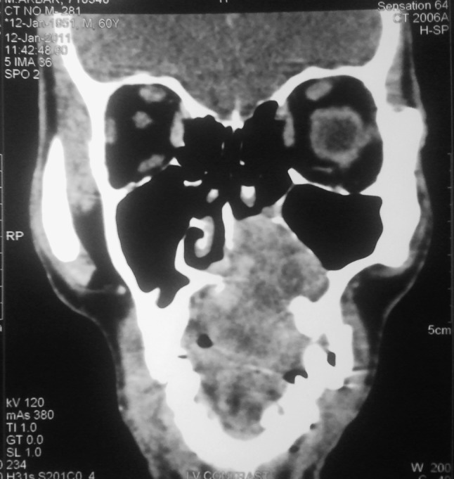
CT scan showing PA of palate eroding hard palate
Table 3.
CT findings in patients
| CT findings | No. of patients | Percentage |
|---|---|---|
| Erosion of hard palate (full thickness) | 4 | 20 |
| Erosion of maxillary sinus wall | 1 | 5 |
| Intact hard palate with no erosion | 12 | 60 |
| Intact hard palate with minor erosion | 4 | 20 |
| Involvement of infratemporal fossa | 1 | 5 |
| Involvement of floor of orbit | 1 | 5 |
The surgical wound in 12 cases without any erosion on CT scan were allowed to granulate and heal by itself (Fig. 5). Out of four cases with minor erosion of hard palate on CT scan, two cases developed full thickness defect in hard palate on curettage and in remaining two patients hard palate remained intact. These two patients who developed full thickness defect on curettage underwent reconstruction by palatal flap and remaining two patients were allowed to self granulate and heal. Out of four cases with full thickness defect on CT scan, two underwent reconstruction by palatal flap after removal of involved bone while among the other two patients, one underwent complete maxillectomy followed by reconstruction by obturator and the other underwent simple reconstruction using obturator (Table 4).
Fig. 5.
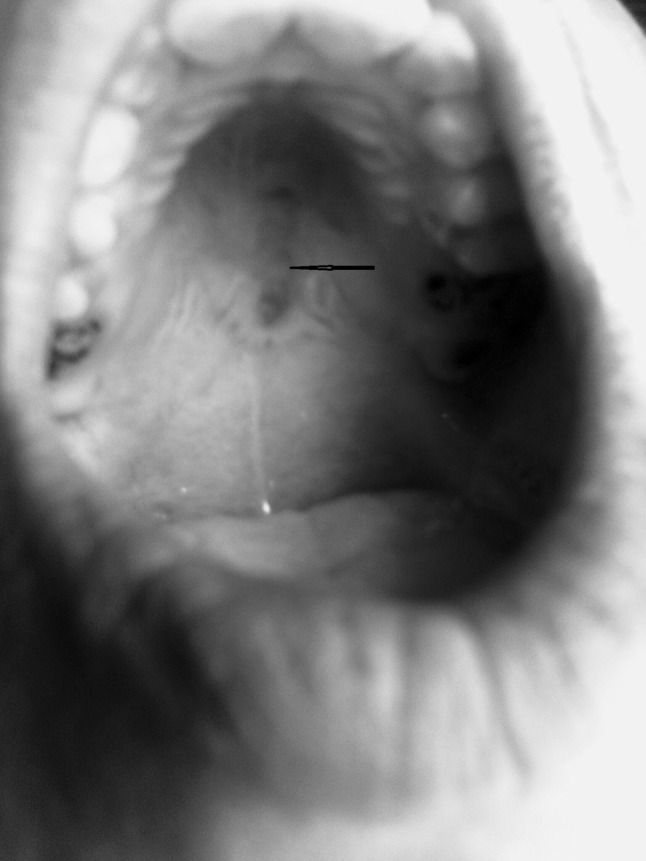
Surgical wound after 21 days showing healthy mucosa over wound area but thinner than the rest of palatal mucosa (black arrow)
Table 4.
Surgical technique and reconstruction method used
| CT findings | Surgical technique and outcome | Treatment of surgical wound/reconstruction | No. of patients |
|---|---|---|---|
| Intact hard palate bone | Excisions of mass with underlying periosteum. Surgical wound bare | Bare wound allowed to granulate and heal by itself | 12 |
| Intact hard palate with minor erosions | Excisions of mass with underlying periosteum and curettage of involved bone of hard palate | ||
| Two patients developed full thickness defect of hard palate | Reconstruction by palatal flap | 2 | |
| In two patients hard palate remained intact | Bare wound allowed to granulate and heal by itself | 2 | |
| Full thickness defect in hard palate | Excisions of the mass with underlying periosteum and bone | Reconstruction by palatal flap | 2 |
| Reconstruction by obturator | 1 | ||
| Total maxillectomy | Reconstruction by obturator | 1 |
Overall in 14 cases no reconstruction was done, in four cases palatal flap was used and in remaining two cases obturator was fitted.
There was a large interval between appearance of the first clinical feature and diagnosis, ranging from 3 months to 10 years.
Comparing with the histopathology of excised specimen diagnostic accuracy of core needle biopsy was 100 %.There were five patients in whom pathologists could not make diagnosis of PA on FNAC but results of core biopsy and final histopathology of excised specimen showed them to have PA. So the diagnostic accuracy of FNAC was found to be 75 %.
There was no recurrence in any of the cases during follow up period.
Discussion
The data from our study show predominance of PA hard palate in males over females. Other case reports and reviews contrarily reported [8, 9]. The most commonly involved age group in our study was 16–30 years. Only two cases were seen in children below 15 years. PAs of the palate in children and adolescents are rare. Byars et al. [10] first reported two cases of palatal PA in children. Since then 17 cases have been reported in English literature in paediatric age group [8]. Yamamoto et al. [9] reported 10 cases of juvenile palatal PA in patients aged 18 years and younger in Japanese literature.
The most common symptom in our study was a submucosal lump, although few cases showed ulceration (three cases), pain and bleeding which is in accordance with literature. The ulceration seen in three cases had long history of lump on hard palate and the probable reason was repeated trauma to lump due to chewing. All our patients had slow growing tumours. There was a large interval between the appearance of first symptoms and diagnosis, ranging from 3 months to 10 years. The reason for this delay is slow growth of these tumours and their relative asymptomatic nature. However few authors have described rapidly growing palatal PA [8, 11, 12]. The size of the swelling ranged from 0 to 8 cm and majority of patients had swelling in the range of 3–5 cm while in literature the size of the swelling ranges from 0.8 to 5 cm with an average of 2.6 cm [2]. Cheek swelling is an uncommon finding in such cases and was seen in one patient. It occurs in extensive cases involving the whole maxilla.
Differential diagnosis of PA include odontogenic and non-odontogenic cysts, soft tissue tumours, palatal abscess, mucoepidermoid carcinoma, adenoid cystic carcinoma and salivary gland tumors. Palatal tissues contain components of soft tissue and harbour minor salivary gland tissues. As a result, soft tissue tumors such as neurofibroma, fibroma, lipoma, neurilemmoma as well as salivary gland tumors should also be considered in the differential diagnoses for this case. Lymphoma can also present with palatal swelling in children [7].
History, physical examination and radiological examination can aid in diagnosis but the ultimate diagnosis lies on histopathology.
Histologically, it is characterized by a great variety of tissues presenting epithelial cells arranged in cord-like and duct-like cell patterns, along with areas of epidermoid metaplasia [2].
Computed tomography was done in all cases and diagnosis was verified by core biopsy. Histopathological sampling procedures include FNAC and core needle biopsy (bigger needle comparing to FNAC). FNAC operated in experienced hands, can determine whether the tumor is malignant in nature with 90 % sensitivity [6, 13] but in contrary diagnostic accuracy in our study was 75 %. FNAC can also distinguish primary salivary tumor from metastatic disease. Core needle biopsy is more invasive but is more accurate compared to FNAC with diagnostic accuracy greater than 97 % [14] and we found 100 % diagnostic accuracy of core needle biopsy. Furthermore, core needle biopsy allows more accurate histological typing of the tumor.
Different types of imaging modalities are helpful. The noninvasive diagnostic aids for salivary gland tumors include ultrasound, CT, and MRI. We only used CT in our cases. Plain X-ray and hematologic investigations play insignificant part in the diagnosis of salivary gland tumor of the palate. CT and MRI both provide important information on the location, size, and extension of the tumor into the surrounding superficial and deep tissues. CT is superior to MRI in evaluating bone, especially in diagnosing erosion and perforation of the bony palate and possible involvement of the nasal cavity or maxillary sinus [15, 16] MRI, with its high resolution for soft tissue, provides better definition of the vertical and inferior tumor extension through its multiplanar capacity and the tumor–muscle interface and more clearly indicates the degree of encapsulation [15]. These tumors are also able to invade and erode adjacent bone, causing radiolucent mottling on the X-ray of the maxilla [17].
On CT scan we found that hard palate was intact in most of the cases while minor erosion of hard palate was seen in four cases. Erosion of inferior orbital wall was seen in one patient. PA is a slow growing tumour and destruction of bony wall is seen late in the disease. Extensive erosion is seen in very rare circumstances. We found only one patient with a long history of PA with extensive erosion with minimal symptoms mimicking malignancy.
Treatment of palatal PA involves wide local excision of the tumor including its surrounding capsule, together with clear margins involving the periosteum and associated mucosa, followed by curettage of the underlying bone with a sharp spoon or bur under copious sterile normal saline irrigation, to avoid recurrence [18, 19]. We did not do curettage of bone in cases where there was no erosion of hard palate on CT scan. Total maxillectomy as seen in one of our case is done in extensive disease involving walls of maxillary sinus.
Reconstruction method of the defect has been reported differently in different reports and reviews. We did not reconstruct the area in cases where no full thickness defect was created on wide surgical excision and in cases without any preexisting erosions on CT scan. In such cases we waited for the wound to granulate and heal for 1 month. In cases where full thickness defect was preoperatively present in hard palate or was created, we reconstructed it either by obturator or by palatal flap based on greater palatine vessels. All these techniques have been reported in literature.
These tumors usually do not recur after adequate surgical excision. Recurrence if at all occurs can be attributable to inadequate surgical techniques such as simple enucleation leaving behind microscopic pseudopod-like extensions, capsular penetration, and tumor rupture with spillage of tumor cells [4]. We did not have any recurrence in 1 year of follow-up but recurrence of palatal PA in children following surgical treatment has been reported in two cases out of 16 cases from the English literature [7]. Long term follow-up is necessary as recurrences though very uncommon after proper surgical excision can be seen on long term follow up.
Conclusion
Pleomorphic adenoma of palate is a rare entity usually seen in adults. Most common symptom is slowly growing painless submucosal mass on hard palate. Definitive diagnosis lies on histopathological examination. CT is necessary for ruling out any bony erosions. Treatment is by wide local excision with removal of periosteum and curettage of bone. Reconstruction is only necessary if there is full thickness defect in the bone, otherwise excellent results are seen if wound is allowed to granulate and heal by itself. The most common way to reconstruct the defect is either by the use of obturator or palatal flap. Recurrences are uncommon but may be seen on long term follow-up.
Contributor Information
Suhail Amin Patigaroo, Email: Dr_suhail_jnmc@yahoo.co.in.
Fozia Amin Patigaroo, Email: glassglaze@yahoo.in.
Junaid Ashraf, Email: chanjack@gmail.com.
Nazia Mehfooz, Email: Dr_nazia_jnmc@yahoo.co.in.
Mohd Shakeel, Email: drmshakeel@yahoo.com.
Nazir A. Khan, Email: skims.nkhan@gmail.com
Masood H. Kirmani, Email: masoodkirmani@gmail.com
References
- 1.Luna MA, Batsakis JG, El-Naggar AK. Salivary gland tumors in children. Ann Otol Rhinol Laryngol. 1991;100:869–871. doi: 10.1177/000348949110001016. [DOI] [PubMed] [Google Scholar]
- 2.Jorge J, Pires FR, Alves FA, Perez DEC, Kowalski LP, Lopes MA. Juvenile intraoral pleomorphic adenoma: report of five cases and review of the literature. Int J Oral Maxillofac Surg. 2002;31:273–275. doi: 10.1054/ijom.2002.0206. [DOI] [PubMed] [Google Scholar]
- 3.Sanjay Byakodi, Shivayogi Charanthimath, Santosh Hiremath 4, Kashalikar JJ (2011) Pleomorphic adenoma of palate: a case report. Int J Dent Case Reports 1:36–40
- 4.Mubeen K, Vijayalakshmi KR, Patil AR, Giraddi GB, Singh C. Benign pleomorphic adenoma of minor salivary gland of palate. J Dent Oral Hyg. 2011;3:82–88. [Google Scholar]
- 5.Batsakis JG (1981) Neoplasms of the minor and ‘lesser’ major salivary glands. In: Tumors of the head and neck. The Williams and Wilkins Company, Baltimore, p 38–47
- 6.Suen JY, Synderman NL. Benign neoplasms of the salivary glands. In: Cummings CW, Fredrickson JM, Harker LA, Krause CJ, Schuller DE, editors. Otolaryngology—head and neck surgery. St. Louis: Mosby Year Book; 1993. pp. 1029–1042. [Google Scholar]
- 7.Dhanuthai K, Sappayatosok K, Kongin K. Pleomorphic adenoma of the palate in a child: a case report. Med Oral Patol Oral Cir Bucal. 2009;14:E73–E75. [PubMed] [Google Scholar]
- 8.Moubayed SP, AlSaab F, Daniel SJ. Rapidly progressing palatal pleomorphic adenoma in an adolescent. Int J Pediatr Otorhinolaryngol Extra. 2010;5:141–143. doi: 10.1016/j.pedex.2009.08.001. [DOI] [Google Scholar]
- 9.Yamamoto H, Fukumoto M, Yamaguchi F, Sakata K, Oikawa T. Pleomorphic adenoma of the buccal gland in a child. Int J Oral Maxillofac Surg. 1986;15:474–477. doi: 10.1016/S0300-9785(86)80041-2. [DOI] [PubMed] [Google Scholar]
- 10.Byars LT, Ackerman LV, Peacock E (1957) Tumors of salivary gland origin in children: a clinical pathologic appraisal of 24 cases. Ann Surg 146:40–51 [DOI] [PMC free article] [PubMed]
- 11.Lopez-Cedrun JL, Gonzalez-Landa G, Birichinaga B. Pleomorphic adenoma of the palate in children: report of a case. Int J Oral Maxillofac Surg. 1996;25(3):206–207. doi: 10.1016/S0901-5027(96)80031-2. [DOI] [PubMed] [Google Scholar]
- 12.Shaaban H, Bruce J, Davenport PJ. Recurrent pleomorphic adenoma of the palate in a child. Br J Plast Surg. 2001;54:245–247. doi: 10.1054/bjps.2000.3536. [DOI] [PubMed] [Google Scholar]
- 13.Feinmesser R, Gay I. Pleomorphic adenoma of the hard palate: an invasive tumour? J Laryngol Otol. 1983;97:1169–1171. doi: 10.1017/S002221510009616X. [DOI] [PubMed] [Google Scholar]
- 14.Clauser L, Mandrioli S, Dallera V, Sarti E, Galiè M, Cavazzini L. Pleomorphic adenoma of the palate. J Craniofac Surg. 2004;15:1026–1029. doi: 10.1097/00001665-200411000-00029. [DOI] [PubMed] [Google Scholar]
- 15.Pogrel MA. The management of salivary gland tumors of the palate. J Oral Maxillofac Surg. 1994;52:454. doi: 10.1016/0278-2391(94)90339-5. [DOI] [PubMed] [Google Scholar]
- 16.Noghreyan A, Gatot A, Maor E, et al. Palatal pleomorphic adenoma in a child. J Laryngol Otol. 1995;109:343. doi: 10.1017/S0022215100130105. [DOI] [PubMed] [Google Scholar]
- 17.Weber AL. Pleomorphic adenoma of the hard palate. Ann Otol Rhinol Laryngol. 1981;90:192–194. doi: 10.1177/000348948109000222. [DOI] [PubMed] [Google Scholar]
- 18.De Courten A, Lombardi T, Samson J. Pleomorphic adenoma in a child: 9-year follow-up. Int J Oral Maxillofac Surg. 1996;25:293–295. doi: 10.1016/S0901-5027(06)80060-3. [DOI] [PubMed] [Google Scholar]
- 19.Lopez-Cedrum JL, Gonzalez-Landa G, Birichinaga B. Pleomorphic adenoma of the palate in children: report of a case. Int J Oral Maxillofac Surg. 1996;25:206. doi: 10.1016/S0901-5027(96)80031-2. [DOI] [PubMed] [Google Scholar]


