Abstract
Reconstruction of full-thickness buccal defect is challenging as two linings need to be addressed. Either two different flaps or double-paddle for one free flaps are necessary for this defect. The prolonged operation might not be tolerated by patients because of advanced age or medical comorbidity. A 77-year-old gentleman, with significant medical comorbidity, presented with a 4.0 × 4.5 cm ulcerative mass due to squamous cell carcinoma arising from the left buccal mucosa. The tumor extended to the left cheek skin. There was no palpable neck node. CT scan did not show any bony erosion or suspicious neck node. Full-thickness resection of the tumour was undertaken. For the full-thickness buccal defect, a bi-paddled pedicled submental flap after de-epithelialization of the flap skin was used for both the cutaneous and mucosal resurfacing. The flap survived completely and patient recovered smoothly. The surgery is simple and operation time is much shorter than free flap reconstruction. This modified utilization of submental flap simplifies the closure of complicated oro-facial wound.
Keywords: Bipaddled, Buccal, Composite defect, Pedicled flap, Submental flap
Introduction
Reconstruction of composite oro-facial defect is challenging. Free flaps are often necessary for re-surfacing the inner mucosal and cutaneous linings. However, not all patients can benefit from free flap reconstruction due to various reasons, such as severe comorbidity or old age, which are relative contraindications for prolonged free flap operation. Reliable pedicled flaps remain important backups in such circumstances.
Submental flap was first reported by Martin et al. [1] in 1993. The advantages peculiar to this flap comprise good colour or texture match to facial skin, suppleness and concealed donor site. The operative time and hospital stay are shorter than using the gold standard radial forearm free flap [2]. The usage of submental flap for aggressive oro-facial cancer has been gaining attention in recent years [3–7]. However, these reports mainly described the application of submental flap for two-dimensional defect of the oral cavity. Recently, one of our patients with through-and-through buccal defect after cancer extirpation of the oral cavity was successfully repaired by a bipaddled pedicled submental flap.
Case Report
A 77-year-old gentleman complained of a left buccal mass since 3 weeks. He was a chronic smoker and ex-drinker. His past health was significant: hypertension, hyperlipidaemia and history of transient ischaemic stroke in 2008 and was taking aspirin.
On physical examination, a 4.0 × 4.5 cm ulcerative mass arising from the left buccal mucosal was present and extended to the left cheek skin. There was no palpable neck node. CT scan did not show any bony erosion or suspicious neck node. Biopsy of the mass confirmed the diagnosis of squamous cell carcinoma. The tumour stage was cT4N0M0. Due to patient’s advanced age and co-morbidity, prolonged free flap operation was deemed risky. Eventually, full-thickness tumor resection, left neck dissection and reconstruction with bipaddled submental flap was chosen as the surgical treatment.
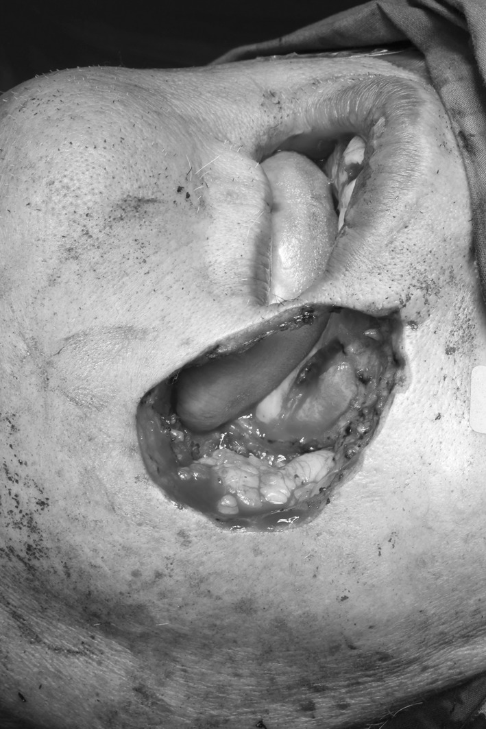
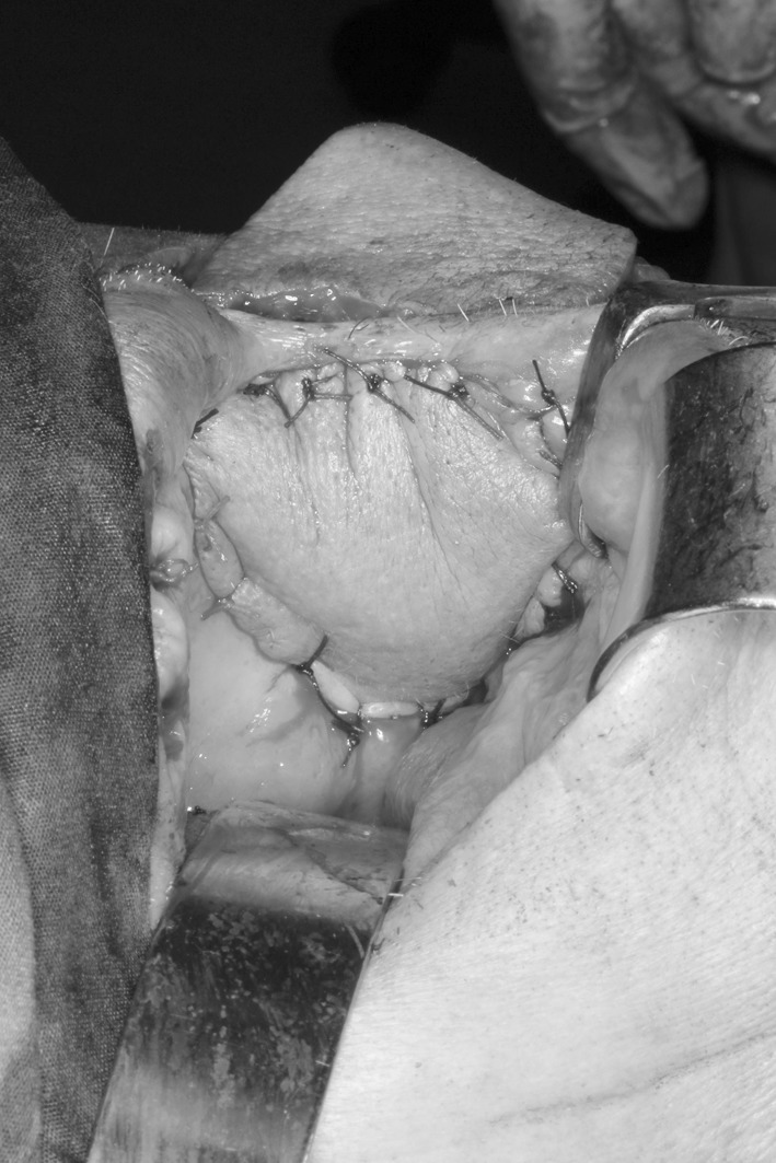
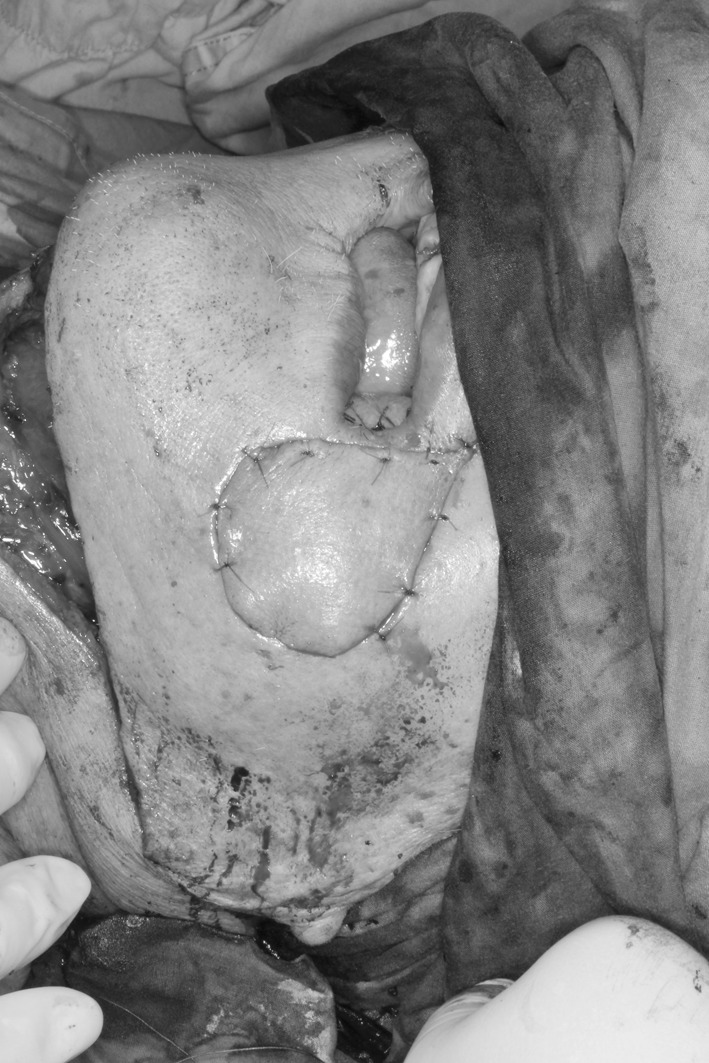
The left buccal tumour was removed, including the orifice of the left Stensen’s duct and cheek skin, with 1 cm margins. The left commissure of the mouth could just be preserved without compromising the oncologic clearance. The cosmetic outcome and continence function is expected to be better by keeping the commissure. Resection margins were proven to be free of tumour on frozen section examination. The full-thickness defect is shown (Fig. 1). Submental flap spanning between the mandible angles was designed as an elliptical skin paddle with width of 6 cm. After the incision was made, the flap was dissected just beneath the platysma muscle. The left submental vein was identified at the superior border of submandibular gland and submental artery was deep to the gland. The artery originates from the facial artery while the vein drains to the anterior facial vein. The vascular branches to the gland were meticulously controlled and divided. The flap pedicle vessels were followed and dissected toward the midline. The anterior belly of the left digastric muscle and the mylohyoid muscle were detached and included in the flap in order to protect the perforators [8] which are located between these two muscles. Left neck node dissection was carried out when the flap harvest was complete. The facial vein and facial artery were preserved. The flap was finally transferred to the oral cavity via a subcutaneous tunnel. De-epithelialization for 1 cm flap skin was done. This divided the flap into two skin paddles: the bigger paddle distal to the perforators was utilized for mucosal re-surfacing (Fig. 2) while the smaller paddle proximal to the perforators for skin coverage (Fig. 3). The whole operation lasted about 6 h.
Fig. 1.

The defect after the full-thickness resection of left buccal carcinoma
Fig. 2.

The bigger skin paddle of submental flap resurfacing the buccal mucosa. The submental flap was de-epithelialized near the commissure of lip
Fig. 3.

The smaller skin paddle of submental flap for cheek skin coverage
The flap survived uneventfully and the patient recovered smoothly. Currently he is taking soft diet and his speech is normal.
Discussion
Full-thickness cheek reconstruction is technically difficult. Free flap e.g. anterolateral thigh flap or radial forearm flap designed with two skin paddles are commonly used for this sophisticated defect. However, not every patient is a good candidate for free flap reconstruction. Moreover, some patients would just decline it due to various reasons. Pedicled flaps would then remain the useful alternatives under such circumstances. Submental flap is one of these alternatives that provide supple skin conformable to the three dimensional defect after tumor resection. As of the time we operated on this patient, to our knowledge, submental flap for full-thickness cheek defect has not been reported.
After the surgery, we searched the literature with the Medline System and only one case of submental flap for reconstructing full-thickness buccal wound after cancer extirpation was reported by Ramkumar et al. [9] in 2012. Our case represents the second reported case. The technique of flap design is slightly different for these two cases. For the patient managed by Ramkumar et al., the two skin paddles were made by completely incising the flap skin to the platysma muscle. The flap pedicle vessel distal to this incision should be prudentially identified and protected. In our case, only the flap skin was de-epithelialised down to the subcutaneous flap to create the two cutaneous paddles. Therefore, the flap pedicle vessels would not be damaged so easily. In both cases, the bigger distal paddles were utilized to re-surface the mucosal wound while the smaller proximal paddles for the cheek cutaneous coverage. To ensure the integrity of flap pedicle and perforators of this bipaddled flap, mylohyoid muscle as well as the anterior belly of digastric muscle should be harvested in the flap.
Bipaddled design is an innovative modification of submental flap to expand the armamentarium of pedicled flap for reconstruction of the challenging composite cheek defect after tumour resection. The surgery is simple and operation time is much shorter. Bipaddled submental flap simplifies the closure of complicated oro-facial wound.
Conflict of interest
This work has not been submitted for publication, and none of the authors have conflict of interest using any of the products.
References
- 1.Martin D, Pascal JF, Baudet J, Mondie JM, Farhat JB, Athoum A, Le Gaillard P, Peri G. The submental island flap: a new donor site. Anatomy and clinical applications as a free or pedicled flap. Plast Reconstr Surg. 1993;92:867–873. doi: 10.1097/00006534-199392050-00013. [DOI] [PubMed] [Google Scholar]
- 2.Paydarfar JA, Patel UA. Submental island pedicled flap vs radial forearm free flap for oral reconstruction. Arch Otolaryngol Head Neck Surg. 2011;137:82–87. doi: 10.1001/archoto.2010.204. [DOI] [PubMed] [Google Scholar]
- 3.Chow TL, Chan TTF, Chow TK, Fung SC, Lam SH. Reconstruction with submental flap for aggressive orofacial cancer. Plast Reconstr Surg. 2007;120:431–436. doi: 10.1097/01.prs.0000267343.10982.dc. [DOI] [PubMed] [Google Scholar]
- 4.Sebastian P, Thomas S, Varghese BT, Iype EM, Balagopal PG, Mathew PC. The submental island flap for reconstruction of defects in oral cancer patients. Oral Oncol. 2008;44:1014–1018. doi: 10.1016/j.oraloncology.2008.02.013. [DOI] [PubMed] [Google Scholar]
- 5.Uppin SB, Ahmad QG, Yadav P, Shetty K. Use of the submental island flap in orofacial reconstruction—a review of 20 cases. J Plast Reconstr Aesthet Surg. 2009;62:514–519. doi: 10.1016/j.bjps.2007.11.023. [DOI] [PubMed] [Google Scholar]
- 6.Taghinia AH, Movassaghi K, Wang AX, Pribaz JJ. Reconstruction of the upper aerodigestive tract with the submental artery flap. Plast Reconstr Surg. 2009;123:562–570. doi: 10.1097/PRS.0b013e3181977fe4. [DOI] [PubMed] [Google Scholar]
- 7.Amin AA, Sakkary MA, Khalil AA, Rifaat MA, Zayed SB. The submental flap for oral cavity reconstruction: extended indications and technical refinements. Head Neck Oncol. 2011;3:51–57. doi: 10.1186/1758-3284-3-51. [DOI] [PMC free article] [PubMed] [Google Scholar]
- 8.Patel UA, Bayles SW, Hayden E. The submental flap: a modified technique for resident training. Laryngoscope. 2007;117:186–189. doi: 10.1097/01.mlg.0000246519.77156.a4. [DOI] [PubMed] [Google Scholar]
- 9.Ramkumar A, Francis NJ, Senthil Kumar R, Dinesh Kumar S. Bipaddled submental artery flap. Oral Maxillofac Surg. 2012;41:458–460. doi: 10.1016/j.ijom.2011.12.030. [DOI] [PubMed] [Google Scholar]


