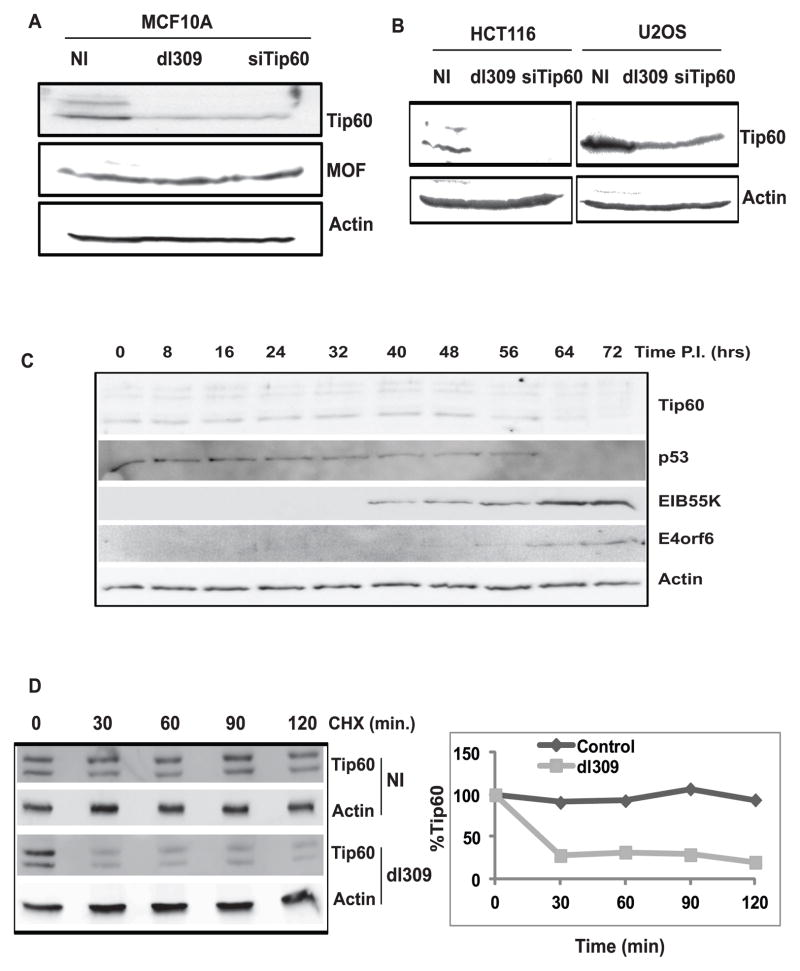
Figure 1. Tip60 is destabilized in adenovirus-infected cells.
(A) Immunoblot with indicated antibodies of MCF10A cells (NI- no infection; dl309- infected with dl309 adenovirus; siTip60- transfected with siTip60). Cells were harvested 68 hrs post infection. Knockdown of Tip60 confirms the identity of the Tip60 bands.
(B) Destabilization of Tip60 by adenovirus in other cell lines. HCT116 and U2OS cells were lysed 40 hours after infection or 72 hr after siRNA transfection prior to immunoblotting.
(C) Tip60 destabilization is concurrent with p53 degradation in adenovirus-infected cells. dl309 infected MCF10A cells were harvested at different time points as indicated after infection and lysates were used to probe with indicated antibodies.
(D) Tip60 protein half life decreases in adenovirus-infected cells. dl309-infected and uninfected MCF10A cells were treated with cycloheximide (added at 68hrs post infection) for different time points as indicated and lysates were resolved for Western blot by indicated antibodies. (Graph) Levels of Tip60 were quantitated using Gene Tool Software (Syngene). Tip60 levels in both 0 hours lanes are taken as 100%.

