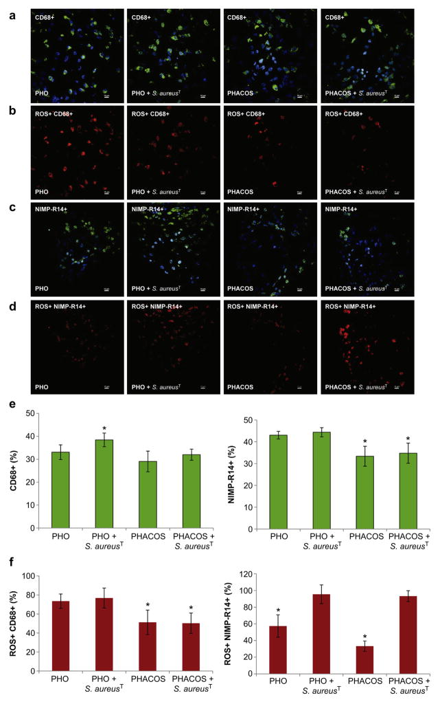
Fig. 7.
Immunohistochemical staining for macrophages and neutrophils in implant-associated inflammation. (a) Representative co-localization images of macrophages (CD68+; green) and nuclei (DAPI; blue) in 14-d: sterile PHO; S. aureusT precolonized PHO; sterile PHACOS; S. aureus precolonized PHACOS implants (from left to right). (b) Intracellular ROS (H-Cy5+, red) co-localizing with macrophages in: sterile PHO; S. aureusT pre-colonized PHO; sterile PHACOS; S. aureusT pre-colonized PHACOS implants (from left to right). (c) Neutrophils (NIMP-R14+; green) with DAPI in 14-d: sterile PHO; S. aureus precolonized PHO; sterile PHACOS; S. aureusT precolonized PHACOS implants. (d) Intracellular ROS (H-Cy5+, red) co-localizing with neutrophils in: sterile PHO; S. aureusT pre-colonized PHO; sterile PHACOS; S. aureusT pre-colonized PHACOS implants (from left to right). (e) Quantification of CD68+ (left) and NIMP-R14 cells (right) stained positive for CD68+ (macrophages) and for NIMP-R14+ cells (neutrophiles) (n = 3 mice/time point; *P < 0.05 between macrophage recruitment to S. aureusT pre-colonized PHO and S. aureusT pre-colonized PHACOS implant; neutrophil recruitment to sterile PHO and sterile PHACOS implants; neutrophil recruitment to S. aureusT pre-colonized PHO and S. aureusT pre-colonized PHACOS). (f) Quantification of CD68+ and ROS+ (left) and NIMP-R14 and ROS+ (right) cells co-localization (n = 3 mice/time point; *P < 0.05 between ROS active macrophage on sterile PHO and sterile PHACOS implant; ROS active macrophage on S. aureusT pre-colonized PHO and S. aureusT pre-colonized PHACOS implant; ROS active neutrophil on sterile PHO and S. aureusT pre-colonized PHO; ROS active neutrophil on sterile PHACOS and S. aureusT pre-colonized PHACOS implant).

