Abstract
BACKGROUND
Electrocardiogram (ECG) manifestations of takotsubo cardiomyopathy (TC) produce ST-segment elevation or T-wave inversion, mimicking acute coronary syndrome (ACS). We describe the ECG manifestation of TC, including ECG evolution, and its different points from ACS.
METHODS
We studied 37 consecutive patients (age 67 ± 15 years, range 23–89, M:F = 12:25) from March 2004 to November 2012 with a diagnosis of TC who were proven to have apical ballooning on echocardiography or left ventricular angiography and normal coronary artery. We analyzed their standard 12-lead ECGs, including rate, PR interval, QRS duration, corrected QT (QTc) interval, ECG evolutions, and arrhythmia events.
RESULTS
Two common ECG findings in TC were ST-segment elevation (n = 13, 35%) and T inversion (n = 24, 65%), mostly in the precordial leads. After ST-segment resolution, in a few days (3.5 days), diffuse and often deep T-wave inversion developed. Eight patients (22%) had transient Q-waves lasting a few days in precordial leads. No reciprocal ST-segment depression was noted. T-wave inversion continued for several months. QT prolongation (<440 milliseconds) was observed in 37 patients (97%). There were no significant life-threatening arrhythmias except atrial fibrillation (n = 6, 16%).
CONCLUSION
There are distinct differences between the ECGs of TC and ACS. These differences will help to differentiate TC from ACS.
KEYWORDS: takotsubo cardiomyopathy, electrocardiography
Introduction
Takotsubo cardiomyopathy (TC) is a cardiomyopathy that shows distinctive clinical conditions. It is also called apical ballooning because it shows wide-ranging acute akinesia in the apex or mid-portion of the left ventricle of the heart.1–5 Unusual forms of abnormalities in myocardial motion without coronary arterial stenosis are noted, and the appearance of the electrocardiogram (ECG) may cause confusion in initial diagnosis because it is similar to that of acute coronary syndrome (ACS). Therefore, this study analyzed changes in the ECG of TC patients over time and the differences in the ECG of TC patients from those of ACS patients to examine if the results can help distinguish between TC and ACS.
Materials and Methods
Study subjects
Between March 2004 and November 2012, 37 consecutive patients with TC (12 men, 25 women) were retrospectively enrolled in this study. The study was approved by the institutional review board of Inje University Ilsan Paik Hospital. Informed consent was waived by the board. Based on previous reports, the inclusion criteria were as follows: (1) transient akinesia or dyskinesia beyond a single major coronary-artery vascular distribution; (2) absence of significant coronary-artery disease as assessed by angiograms (diameter stenosis <50% by visual estimation) or angiographic evidence of acute plaque rupture, (3) electrocardiographic changes (ST-segment elevation, T-wave inversion, or ST-segment change), and (4) evidence of complete recovery from apical ballooning proven by echocardiography.6
Clinical approach
All patients’ medical histories and risk factors for coronary-artery diseases were identified through their medical records to conduct the study. Factors that would have induced mental or physical stress to patients suspected of this disease were checked. The patients were either hospitalized to the emergency room because of the onset of acute symptoms or their acute symptoms occurred while they were in the hospital for various diseases. When an acute symptom occurred, cardiac enzymes (creatinine kinase [CK-MB] and troponin-T) were measured and the ECG was continuously monitored over time. ECGs were also conducted at the time symptoms occurred and at the time of recovery from the symptoms in all patients to follow-up left ventricle functions and regional wall motion abnormality. The existence of coronary-artery diseases was checked in all patients through coronary angiography or coronary CT angiography, and the coronary angiography was conducted together with LV angiography.
ECG analysis
In all patients, a 12-lead ECG was continuously measured at a paper speed of 25 mm/second and an amplification of 10 mm/mV at least once a day over time from the time when symptoms began during their hospitalization period. Basic parameters (ventricular rate, PR interval, QRS duration, and QT interval) were measured in all cases of ECG and corrected QT (QTc) intervals were measured using Bazett’s formula . In addition, changes in the ST segment and T waves, the existence of reciprocal changes in limb leads, and arrhythmia events were checked.
Statistical analysis
Statistical analysis and application of descriptive statistics were done using MedCalc® for Windows software (version 9; MedCalc Software, Broekstraat 52, B-9030 Mariakerke, Belgium). P < 0.05 was considered to be statistically significant.
Results
Clinical characteristics
In this retrospective study, a total of 37 patients met our inclusion criteria and were enrolled into the study. Of them, 25 (67.6%) were female and 12 (32.4%) were male. The mean age of the patients was 67.2 ± 15 years (range 23–89 years). The baseline clinical characteristics of the study patients are shown in Table 1. The patients had more physical stress than mental stress as they had 3 mental stress-inducing factors and 31 physical stress-inducing factors.
Table 1.
Baseline characteristics of the study patients.
| STUDY PATIENTS | 37 |
|---|---|
| Age, years (range) | 67 (23–89) |
| Gender, n | M:F = 12:25 |
| Risk factors | |
| Diabetes | 10 |
| History of hypertension | 16 |
| Dyslipidemia | 3 |
| Current smoker | 4 |
| Systolic BP, mmHg | 113 |
| Diastolic BP, mmHg | 68 |
| Heart rate, bpm | 98 |
| CK, U/L | 573 |
| CK-MB, ng/mL | 20.45 |
| Troponin-T, μg/L | 1.0 |
| LVEF | 44 |
| Underlying disease | |
| Bronchial asthma or COPD | 4 |
| Cancer | 2 |
| CRF | 1 |
| CVA | 4 |
| Dementia | 1 |
| Drug intoxication | 1 |
| No underlying disease | |
| Precipitating stress | 26 |
| Emotional | 3 |
| Physical | 31 |
| Clinical presentations | |
| Chest pain | 7 |
| Dyspnea | 16 |
| Cardiogenic shock | 2 |
| Mental change | 6 |
| Abdominal pain | 2 |
| Fever | 2 |
| Back pain | 1 |
| General weakness | 1 |
Abbreviations: CK, creatinine kinase; CK-MB, creatinine kinase-MB; LVEF, left ventricular ejection fraction; CRF, chronic renal failure; CVA, cerebrovascular accident.
Findings on the standard 12-lead ECG
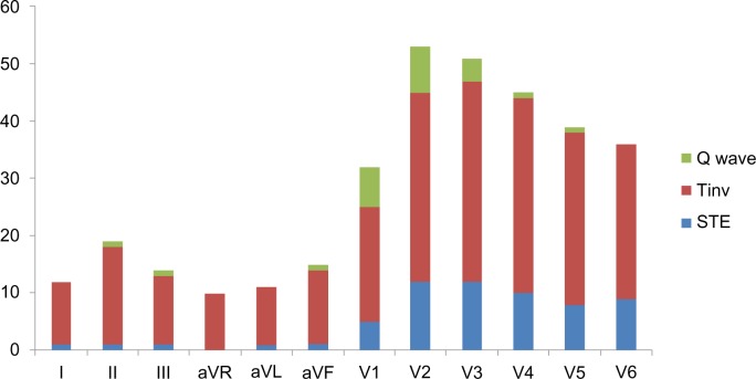
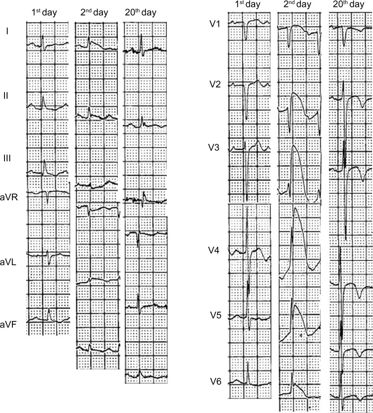
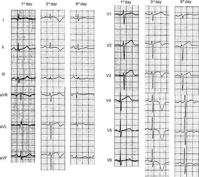
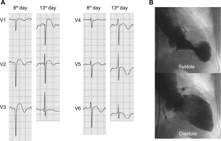
Changes in ECG because of TC mainly occurred in precordial leads, and T inversion occurred most frequently between T inversion, ST elevation, and Q-waves (Fig. 1). Representative changes in ECG appear in two forms: ST-segment elevation and T-wave inversion (Table 2). The number of cases where T inversion appeared from the beginning without ST elevation was 24; this was higher than the number of cases where ST elevation appeared first, which was 13. In cases where ST elevation appeared first, T inversion appeared when the ST segment had subsided. ST elevation persisted for one to three days and subsided thereafter (average 1.2 ± 1.1 days, 1.5 hours to 3.5 days) (Fig. 2). T inversion persisted for 1 to 38 days (average 10.6 ± 7.4 days, 1 to 38 days). The peak of T inversion was 2.3 ± 2.0 days (2.4 hours to 7.6 days). T inversion persisted during the periods of hospitalization for most patients and for several months thereafter, even after discharge from the hospital (Fig. 3). QTc prolonged during these periods (537 ± 64 milliseconds, 415 to 699 milliseconds). Q-waves occurred in eight (22%) patients, but disappeared over time as these were reversible (Fig. 4). Unlike the ECG for ACS, there was no reciprocal change in limb leads (Fig. 2). The regional wall motion abnormalities appeared first in the echocardiography in most cases, and changes in the ECG appeared approximately on day later. Even after recovery from abnormal myocardial motion, changes in the ECG and in T inversion, in particular, persisted. Arrhythmia occurred rarely and was benign in cases when it did occur. The most frequently occurring symptom of arrhythmia was atrial fibrillation (Table 2). Atrioventricular block did not occur.
Figure 1.
Prevalence of ST elevation, T-wave inversion, and Q-wave in patients with TC.
Table 2.
Electrocardiographic findings of the study patients.
| ST ELEVATION AND T INVERSION (N = 13) |
T INVERSION ONLY (N = 24) |
P | |
|---|---|---|---|
| Ventricular rate | 116 ± 21 (72, 155) | 87 ± 18 (58, 119) | 0.294 |
| PR interval | 148 ± 27 (120, 216) | 168 ± 38 (120, 276) | 0.509 |
| QRS duration | 87 ± 16 (66, 130) | 89 ± 14 (68, 128) | 0.379 |
| QTc interval | 462 ± 73 (324, 586) | 512 ± 61 (402, 636) | 0.332 |
| Arrhythmia event | 5 | 8 | 0.515 |
| AF | 4 | 2 | |
| PAC | 1 | 1 | |
| PVC | 0 | 4 | |
| SVT | 0 | 1 |
Abbreviations: AF, atrial fibrillation; PAC, premature atrial contraction; PVC, premature ventricular contraction; SVT, supraventricular tachycardia.
Figure 2.
Representative 12-lead ECG change in a patient with TC. On the second day, marked ST elevation was seen in I, II, aVF, and V3 to V6. Note the absence of reciprocal ST-segment depression in limb leads on the 20th day; ST elevation disappeared and T inversion was on the same leads.
Figure 3.
Representative T-wave changes of 12-lead ECG in a patient with TC. On the third day, deep T-wave inversion was seen in I, II, aVF, and V3 to V6. On the sixth day, T-wave inversion disappeared.
Figure 4.
(A) Representative reversible Q-wave change in a patient with TC. On the eighth day, Q wave was seen in V1 to V3. On the 13th day, Q wave disappeared on the same leads. (B) The left ventriculogram in the same patient with TC showed apical ballooning during systole.
Discussion
The main findings in this study are as follows: (1) there were two forms of representative changes in ECG: ST elevation and T inversion; (2) in the form of ST elevation, T inversion appeared when ST elevation had subsided; (3) when T inversion appeared, QT prolongation occurred; (4) there was no reciprocal change in limb leads; (5) Q-waves were reversible; (6) there was no atrioventricular block; and (7) there was no significant arrhythmia event.
ECGs for TC were generally in two forms: ST elevation and T inversion. In the case of the former form, ST elevation appeared first at the beginning, followed by T inversion, which persisted from several days to several weeks and disappeared thereafter. There were cases where T inversion occurred from the beginning and persisted without ST elevation. QT intervals prolonged, whereas T inversion persisted. Although the mechanism of it is not clear, it is assumed to occur because of disorders in repolarization because of the effects of catecholamine.7–8 Similar changes were observed when there were stressful events. QT prolongation was observed when epinephrine was intravenously injected rapidly.9
According to the results of the analysis of the ECGs, there were distinctive differences in ECGs for general myocardial infarction; there were cases where Q-waves occurred, although the Q-waves were reversible and disappeared over time. This differs from a study conducted by Ogura et al. indicating that there was no expression of abnormal Q-waves.10 The reason why Q-waves disappear is assumed to be related to the recovery of myocardial motion in the case of TC because TC is not true myocardial necrosis unlike ACS. The fact that there is no reciprocal change in limb leads is also a difference from myocardial infarction. Although the mechanism of the appearance of reciprocal ECG changes in ACS has not been completely disclosed, it is generally known to be an electrophysiological phenomenon reflecting the myocardial necrosis or ischemia caused by acute infarction.11–13 ST depression occurring early in the acute phase of myocardial infarction is likely to be a reflection of electrophysiological changes taking place at the site of the infarction that is manifested in the contralateral surface of the heart. In anterior myocardial infarction patients, high lateral wall infarction which is supplied with blood streams from the proximal left anterior descending coronary artery is known to induce reciprocal changes in inferior leads.14–17 The reason why there is no reciprocal ST depression in TC is because of the fact that myocardial dysfunctions in TC are limited to the cardiac apex.
T inversion and ST elevation mainly appear in precordial leads and more rarely in limb leads. The duration of ST elevation is short, whereas that of T inversion is long. In some cases, T inversion appears for a while after ST elevation begins, disappears, and then begins to appear again. Differences between these two forms require prospective studies with more patients.
When abnormalities in the ECG were expressed, sinus tachycardia was found in many cases. This is thought to be attributable to increases in catecholamine because of the rise of sympathetic tone in TC. The expression of an ECG abnormality generally occurs in precordial leads rather than in limb leads, and the reason for this is thought to be the fact that abnormal myocardial changes in TC mainly occur in the cardiac apex. Pant et al.18 reported that arrhythmias are present in about one-quarter of patients with TC and worsen the outcome. In this study, arrhythmia rarely occurred and those that did were most frequently atrial fibrillation, a supraventricular tachycardia, and not of ventricular origin. Atrial fibrillation is the most common form of arrhythmia despite the TC is of ventricular origin. The exact pathogenesis of inducing atrial fibrillation in TC is unknown. But it is presumed that increased catecholamine or transient myocardial ischemia is responsible for its occurrence. No atrioventricular block occurred. The reason for this is assumed to be the fact that myocardial changes mainly occurred in the cardiac apex, and thus did not affect the conduction system.
Limitations
This study has limitations as a retrospective study and may have errors in standardizing ECG changes as a result of TC because the number of subject patients was small. In addition, this study may have other limitations because it was a nonrandomized study in which the subject patients were not directly compared to ACS patients.
Conclusions
Through this study, it was determined that although the appearance of the ECG for TC is similar to that of ACS, which is infarct or ischemia because of coronary occlusion, it has distinctive differences from that of ACS because TC as a reversible myocardial change was identified. Because the ECG for TC shows similar to that of ACS, which would bring about confusion in clinical studies at the beginning of diagnosis, based on the results drawn in this study, analysis of ECGs over time is considered helpful in the diagnosis and treatment of TC.
Footnotes
ACADEMIC EDITOR: Thomas E. Vanhecke, Editor in Chief
FUNDING: Author discloses no funding sources.
COMPETING INTERESTS: Author discloses no potential conflicts of interest.
Author Contributions
JN was responsible for conception and design, acquisition, analysis and interpretation of data, and draft of the manuscript. The author reviewed and approved of the final manuscript.
DISCLOSURES AND ETHICS
As a requirement of publication the author has provided signed confirmation of compliance with ethical and legal obligations including but not limited to compliance with ICMJE authorship and competing interests guidelines, that the article is neither under consideration for publication nor published elsewhere, of their compliance with legal and ethical guidelines concerning human and animal research participants (if applicable), and that permission has been obtained for reproduction of any copyrighted material. This article was subject to blind, independent, expert peer review. The reviewers reported no competing interests.
REFERENCES
- 1.Tsuchihashi K, Ueshima K, Uchida T, et al. Transient left ventricular apical ballooning without coronary artery stenosis: a novel heart syndrome mimicking acute myocardial infarction. Angina pectoris-myocardial infarction investigations in Japan. J Am Coll Cardiol. 2001;38(1):11–8. doi: 10.1016/s0735-1097(01)01316-x. [DOI] [PubMed] [Google Scholar]
- 2.Kurisu S, Sato H, Kawagoe T, et al. Tako-tsubo-like left ventricular dysfunction with ST-segment elevation: a novel cardiac syndrome mimicking acute myocardial infarction. Am Heart J. 2002;143(3):448–55. doi: 10.1067/mhj.2002.120403. [DOI] [PubMed] [Google Scholar]
- 3.Bybee KA, Kara T, Prasad A, et al. Systematic review: transient left ventricular apical ballooning: a syndrome that mimics ST-segment elevation myocardial infarction. Ann Intern Med. 2004;141(11):858–65. doi: 10.7326/0003-4819-141-11-200412070-00010. [DOI] [PubMed] [Google Scholar]
- 4.Sharkey SW, Lesser JR, Zenovich AG, et al. Acute and reversible cardiomyopathy provoked by stress in women from the United States. Circulation. 2005;111(4):472–9. doi: 10.1161/01.CIR.0000153801.51470.EB. [DOI] [PubMed] [Google Scholar]
- 5.Gianni M, Dentali F, Grandi AM, Sumner G, Hiralal R, Lonn E. Apical ballooning syndrome or Takotsubo cardiomyopathy: a systematic review. Eur Heart J. 2006;27(13):1523–9. doi: 10.1093/eurheartj/ehl032. [DOI] [PubMed] [Google Scholar]
- 6.Hahn JY, Gwon HC, Park SW, et al. The clinical features of transient left ventricular nonapical ballooning syndrome: comparison with apical ballooning syndrome. Am Heart J. 2007;154:1166–73. doi: 10.1016/j.ahj.2007.08.003. [DOI] [PubMed] [Google Scholar]
- 7.Obayashi T, Tokunaga T, Iiizumi T, Shiigai T, Hiroe M, Marumo F. Transient QT interval prolongation with inverted T waves indicates myocardial salvage on dual radionuclide single-photon emission computed tomography in acute anterior myocardial infarction. Jpn Circ J. 2001;65:7–10. doi: 10.1253/jcj.65.7. [DOI] [PubMed] [Google Scholar]
- 8.Bonnemeier H, Hartmann F, Wiegand UK, Bode F, Katus HA, Richardt G. Course and prognostic implications of QT interval and QT interval variability after primary coronary angioplasty in acute myocardial infarction. J Am Coll Cardiol. 2001;37:44–50. doi: 10.1016/s0735-1097(00)01061-5. [DOI] [PubMed] [Google Scholar]
- 9.Abildskov JA. Adrenergic effects on the QT interval of the electrocardiogram. Am Heart J. 1976;92:210–6. doi: 10.1016/s0002-8703(76)80256-6. [DOI] [PubMed] [Google Scholar]
- 10.Ogura R, Hiasa Y, Takahashi T, et al. Specific findings of the standard 12-lead ECG in patients with ‘Takotsubo’ cardiomyopathy: comparison with the findings of acute anterior myocardial infarction. Circ J. 2003;67:687–90. doi: 10.1253/circj.67.687. [DOI] [PubMed] [Google Scholar]
- 11.Katz R, Conroy RM, Robinson K, Mulcahy R. The aetiology and prognostic implications of reciprocal electrocardiographic changes in acute myocardial infarction. Br Heart J. 1986;55:423–7. doi: 10.1136/hrt.55.5.423. [DOI] [PMC free article] [PubMed] [Google Scholar]
- 12.Greenwood JP, Younger JF, Ridgway JP, Sivananthan MU, Ball SG, Plein S. Safety and diagnostic accuracy of stress cardiac magnetic resonance imaging vs. exercise tolerance testing early after acute ST elevation myocardial infarction. Heart. 2007;93:1363–8. doi: 10.1136/hrt.2006.106427. [DOI] [PMC free article] [PubMed] [Google Scholar]
- 13.Stevenson RN, Ranjadayalan K, Umachandran V, Timmis AD. Significance of reciprocal ST depression in acute myocardial infarction: a study of 258 patients treated by thrombolysis. Br Heart J. 1993;69:211–4. doi: 10.1136/hrt.69.3.211. [DOI] [PMC free article] [PubMed] [Google Scholar]
- 14.Sapin PM, Musselman DR, Dehmer GJ, Cascio WE. Implications of inferior ST-segment elevation accompanying anterior wall acute myocardial infarction for the angiographic morphology of the left anterior descending coronary artery morphology and site of occlusion. Am J Cardiol. 1992;69:860–5. doi: 10.1016/0002-9149(92)90783-u. [DOI] [PubMed] [Google Scholar]
- 15.Birnbaum Y, Solodky A, Herz I, et al. Implications of inferior ST-segment depression in anterior acute myocardial infarction: Electrocardiographic and angiographic correlation. Am Heart J. 1994;127:1467–73. doi: 10.1016/0002-8703(94)90372-7. [DOI] [PubMed] [Google Scholar]
- 16.Tamura A, Kataoka H, Nagase K, Mikuriya Y, Nasu M. Clinical significance of inferior ST elevation during acute anterior myocardial infarction. Br Heart J. 1995;74:611–4. doi: 10.1136/hrt.74.6.611. [DOI] [PMC free article] [PubMed] [Google Scholar]
- 17.Sasaki K, Yotsukura M, Sakata K, Yoshino H, Ishikawa K. Relation of ST-segment changes in inferior leads during anterior wall acute myocardial infarction to length and occlusion site of the left anterior descending coronary artery. Am J Cardiol. 2001;87:1340–5. doi: 10.1016/s0002-9149(01)01549-1. [DOI] [PubMed] [Google Scholar]
- 18.Pant S, Deshmukh A, Mehta K, et al. Burden of arrhythmias in patients with Takotsubo Cardiomyopathy (apical ballooning syndrome) Int J Cardiol. 2013;170(1):64–8. doi: 10.1016/j.ijcard.2013.10.041. [DOI] [PubMed] [Google Scholar]


