Abstract
In order to better characterize the Bacillus anthracis typing phage AP50c, we designed a genetic screen to identify its bacterial receptor. Insertions of the transposon mariner or targeted deletions of the structural gene for the S-layer protein Sap and the sporulation genes spo0A, spo0B, and spo0F in B. anthracis Sterne resulted in phage resistance with concomitant defects in phage adsorption and infectivity. Electron microscopy of bacteria incubated with AP50c revealed phage particles associated with the surface of bacilli of the Sterne strain but not with the surfaces of Δsap, Δspo0A, Δspo0B, or Δspo0F mutants. The amount of Sap in the S layer of each of the spo0 mutant strains was substantially reduced compared to that of the parent strain, and incubation of AP50c with purified recombinant Sap led to a substantial reduction in phage activity. Phylogenetic analysis based on whole-genome sequences of B. cereus sensu lato strains revealed several closely related B. cereus and B. thuringiensis strains that carry sap genes with very high similarities to the sap gene of B. anthracis. Complementation of the Δsap mutant in trans with the wild-type B. anthracis sap or the sap gene from either of two different B. cereus strains that are sensitive to AP50c infection restored phage sensitivity, and electron microscopy confirmed attachment of phage particles to the surface of each of the complemented strains. Based on these data, we postulate that Sap is involved in AP50c infectivity, most likely acting as the phage receptor, and that the spo0 genes may regulate synthesis of Sap and/or formation of the S layer.
INTRODUCTION
Bacteriophages are the most abundant entities on Earth; according to some estimates, there are approximately 10-fold more phages than bacteria (1–3). Bacteriophages exhibit very high specificity to the bacteria that they infect, most often at the species level and sometimes even at the strain level. This specificity can be attributed to the interaction of a phage-encoded protein or a structure present on the bacteriophage with a receptor on the bacterial surface (4), and it is comparable to the specificity of the antigen-antibody interaction, making phages highly suitable for bacterial diagnosis/detection (5–8). Although reports of the use of phage therapy in Western medicine are rare (9), phages have been used to differentiate bacterial species and strains (10–13). Bacterium-phage specificity has been exploited for various bacterial typing schemes for decades; examples include Salmonella, Listeria, Vibrio cholerae, and Mycobacterium (14–17). Phages have been used for developing many rapid diagnostic tools for bacterial detection, and it is also conceivable to design universal systems by modifying the specificities on the surface of the phage (18–20). While the high specificity exhibited by phages to their bacteria is desirable for diagnostics, broad-spectrum countermeasures are desired for therapeutics.
Bacteria and phages are engaged in a constant evolutionary arms race in which they have developed resistance and counterresistance mechanisms over millions of years (21). An informed decision about using phages for diagnostic/therapeutic applications requires knowledge of the phage receptors and resistance mechanisms of the bacteria. This information is valuable in the intelligent design of phage cocktails that contain phages that use different bacterial receptors, so that one can minimize the possibility of emergence of resistance during phage therapy applications.
Bacillus anthracis, a member of the B. cereus sensu lato group, is a Gram-positive bacterium that is the etiologic agent of anthrax illness. Because it readily forms spores that can be aerosolized, it is considered a bioterror threat. B. anthracis has been the subject of a great deal of research interest since a series of letters containing anthrax spores was mailed in the United States in 2001, leading to the deaths of five individuals (22).
Spores are the virulent form of B. anthracis, and a number of studies have focused on understanding the mechanisms of sporulation in the genus Bacillus in general, especially in B. subtilis. Although the factors involved in sporulation of B. anthracis have not been fully characterized, in B. subtilis, external signals for sporulation cause phosphorylation of spo0F, which phosphorylates spo0B, which in turn phosphorylates spo0A, and phosphorylated Spo0A becomes an active transcription factor (23). The high degree of conservation of these proteins suggests that this cascade operates in the same way in all members of the B. cereus sensu lato group (24).
Previously, we characterized a B. anthracis-specific phage of the Tectiviridae family, AP50, isolated in 1972 (25, 26). We extended the genetic characterization of AP50 and determined its genome sequence, which we found to be highly similar to that of other Gram-positive Tectiviridae phages in genetic organization and encoded proteins (27). AP50 exhibits a narrow host range, infecting B. anthracis exclusively, although one B. thuringiensis strain (ATCC 35646) was shown to possess an AP50-like element. We identified mutations in the AP50 genome responsible for lytic/lysogeny control and isolated a clear plaque mutant of AP50 (suggesting that the mutant phage is obligatorily lytic), designated AP50c, that differs from AP50 by two nucleotides. Spontaneous bacterial mutants resistant to killing by AP50c produce a polysaccharide material that coats the cell surface (27).
We were interested in identifying the bacterial receptor of phage AP50c and characterizing phage resistance mechanisms. Two parallel approaches were used to achieve this goal. Initially, spontaneous phage-resistant mutants were isolated, and a whole-genome sequencing approach was used to identify relevant mutations. In this screen, all the spontaneous phage-resistant mutants were found to have mutations in the csaB gene (28). CsaB is a protein involved in cell surface anchoring of various proteins, including the S-layer protein Sap (29). Therefore, we hypothesized that Sap (or the S layer composed of Sap) may be the receptor of AP50c. In our second approach, described in the present study, we obtained evidence supporting this hypothesis.
MATERIALS AND METHODS
Media and chemicals.
BGGM is brain heart infusion (BHI) containing 10% glycerol, 0.4% glucose, and 10 mM MgCl2. Phage buffer was 10 mM Tris-HCl, pH 8.0, 10 mM MgCl2, 1% ammonium acetate. Electroporation competence buffer was 1 mM HEPES, 10% glycerol, pH 7.0.
Plasmids, bacterial strains, and phages.
Plasmids and bacterial strains are listed in Table 1. The transposon insertion vector pJZ037 has been previously described (30). It is temperature sensitive for replication and contains a chloramphenicol resistance gene for Gram-positive organisms, as well as a gene encoding the C9 variant of the Himar1 transposase and an erythromycin resistance gene, located between two inverted repeats that are transposed by the transposase.
TABLE 1.
Bacterial strains and plasmids used in this study
| Strain or plasmida | Description | Source or reference |
|---|---|---|
| Strains | ||
| Escherichia coli | ||
| GM119 | dam dcm mutant | 36 |
| C2925 | dam dcm mutant | New England Biolabs |
| B. anthracis | ||
| 34F2 | Sterne/pXO1+/pXO2− | P. C. Hanna lab |
| 7702 | Sterne/pXO1+/pXO2− | T. M. Koehler lab |
| JB220 | 7702 ΔBAS2245 | This study |
| BAP350 | 7702 ΔcsaB | This study |
| BA749 | 7702 ΔBAS0566 | This study |
| BA750 | 7702 Δsap | This study |
| BA751 | 7702 Δeag | This study |
| BA752 | 7702 ΔBAS1792 | This study |
| BA753 | 7702 ΔBAS3559 | This study |
| BA754 | 7702 Δspo0A | This study |
| BA755 | 7702 Δspo0F | This study |
| DP-B-5747 | JB220 Δspo0B | This study |
| B. cereus | ||
| RS110 | ATCC 4342 | 60 |
| RS112 | RTS100 (B. anthracis-like) | |
| RS415 | ATCC 25621 | 61 |
| RS416 | ATCC 43881 | 62 |
| RS417 | ATCC 14893 | 63 |
| RS431 | ATCC 27877 | 64 |
| RS432 | ATCC 7064 (NRS201) | 60 |
| RS436 | B. cereus transition state strain CDC13100 | 47 |
| RS437 | B. cereus transition state strain CDC13140 | 47 |
| RS438 | B. cereus transition state strain CDC32805 | 47 |
| RS710 | B. cereus var. Saigon BGSC 6A6 | Simon Cutting (unpublished data) |
| RS711 | BGSC 6E1 (GP7)/pBC15/pBC16, Tc | 65 |
| RS741 | F4431/73 (FRI-30) | 66 |
| RS756 | E33L ZK (Zebra Killer) | 48 |
| B. megaterium | ||
| RS712 | BGSC 7A1 strain 899 | 67 |
| RS713 | BGSC 7A16 (QM B1551) | 68 |
| RS721 | ATCC 8245 (γ phage sensitive) | 69 |
| B. thuringiensis | ||
| RS763 | HD682 | |
| RS766 | 97-27 (pathogen) | 70 |
| RS781 | Al Hakam (Iraq) | 71 |
| BGSC 4CC1 | B. thuringiensis serovar pulsiensis | Bacillus Genetic Stock Center |
| BGSC 4BA1 | B. thuringiensis serovar pondicheriensis | Bacillus Genetic Stock Center |
| BGSC 4AJ1 | B. thuringiensis serovar monterrey | Bacillus Genetic Stock Center |
| BGSC 4AW1 | B. thuringiensis serovar andalousiensis | Bacillus Genetic Stock Center |
| Plasmids | ||
| pJZ037 | Temperature-sensitive transposon vector | 30 |
| pKSV7 | Allelic exchange vector | 33 |
| pRP1028 | Mutant construction | 28 |
| pSS4332 | Mutant construction | 28 |
| pSW4 | E. coli-B. anthracis shuttle vector | 40 |
| pET15b Sap ΔSLH | Sap expression plasmid | O. Schneewind |
| pNL9164 | Source of cadC promoter | Sigma-Aldrich |
| pNBGD1001 | pSW4 with pagA promoter with NotI-NdeI sites | 28 |
| pNBGD1004 | pSW4 with cadC promoter with NotI-NdeI sites | This study |
| pNBGD1005 | pNBGD1004::oriT | This study |
| pNBGD1006 | pNBGD1005 carrying sap from 34F2 | This study |
| pNBGD1007 | pNBGD1005 carrying sap from RS438 | This study |
| pNBGD1008 | pNBGD1005 carrying sap from RS756 | This study |
| pNBGD1009 | pNBGD1001 carrying sap from 34F2 | This study |
| pNBGD1010 | pNBGD1001 carrying sap from RS438 | This study |
| pNBGD1011 | pNBGD1001 carrying sap from RS756 | This study |
Strains with the prefix RS were obtained from Raymond Schuch (formerly of Rockefeller University).
B. anthracis typing phage AP50c has been previously described (27). It was grown as a plate stock on the B. anthracis Sterne strain, using PA medium (31). The phage inoculum was 10,000 PFU. After overnight growth at 30°C, 5 ml of phage buffer was poured on each plate, and the soft agar layer was scraped off. After removal of the agar by low-speed centrifugation, the titer was ∼7 × 10⁸ PFU/ml.
Expression and purification of recombinant Sap.
An expression plasmid for recombinant histidine-tagged C-terminal Sap was kindly provided by Olaf Schneewind. Plasmid pET15b Sap ΔSLH was transformed into Escherichia coli C43(DE3). A single colony was inoculated into 3 ml LB containing 100 μg/ml ampicillin (LB-amp), and the culture was grown at 37°C to an A600 of 0.5. The full preculture was transferred to a 100-ml volume of LB-amp and incubated at 37°C to an A600 of 0.5. Isopropyl-beta-d-1-thiogalactopyranoside was added to a final concentration of 1 mM, and the culture was incubated for an additional 4 h. The full volume was centrifuged, the pellet was resuspended in 8 ml of lysis buffer (50 mM NaH2PO4, pH 8.0, 0.5 M NaCl, containing 8 mg/ml lysozyme), and the mixture was incubated for 30 min on ice. Following sonication on ice, the preparation was centrifuged for 15 min at 4,000 × g, and the supernatant was retained. The lysate was applied to a His-Pur nickel-nitrilotriacetic acid column (Thermo Scientific, Rockford, IL) and rotated for 30 min at room temperature. The column was washed four times with wash buffer (50 mM NaH2PO4, pH 8.0, 0.5 M NaCl, 20 mM imidazole), and the protein was eluted with elution buffer (50 mM NaH2PO4, pH 8.0, 0.5 M NaCl, 250 mM imidazole). Aliquots were analyzed by SDS-PAGE and Coomassie staining. Eluates were combined, and the protein was concentrated and buffer exchanged using an Amicon Ultra-15 concentrator with a 30,000-Da-molecular-mass cutoff (Thermo Scientific) into 25 mM Tris-HCl, pH 7.5.
Assessment of effect of recombinant Sap on AP50c activity.
One microliter of AP50c phage stock was incubated for 45 min at 24°C with a 9-μl volume of purified recombinant C-terminal Sap (140 μg/ml), with 9 μl of bovine serum albumin at the same concentration in the same buffer, or with 9 μl of buffer alone (25 mM Tris-HCl, pH 7.5). The mixtures were then used in a soft agar plaque assay, using strain JB220 on BHI agar plates and incubating overnight at 24°C. For a control sample, 1 μl of phage stock was used. The percent reduction in activity of each phage mixture was calculated by subtracting the number of plaques on the experimental plate from the number of plaques on the control plate and dividing the result by the number of plaques on the control plate. This experiment was performed three times, and the mean and standard deviation were calculated.
Construction of a B. anthracis Sterne strain with deletion of the gene for chloramphenicol resistance.
A construct for the deletion of BAS2245, a chloramphenicol resistance gene, from the Sterne strain 7702 was engineered using splicing by overlap extension PCR (SOE-PCR) (32). Primers are listed in Table 2. Primers 2245-A and 2245-B were used to amplify the upstream flanking region. Primers 2245-C and 2245-D amplified the downstream flanking region. The fragment obtained with primers 2245-A and 2245-B (the AB fragment) and the fragment obtained with primers 2245-C and 2245-D (the CD fragment) were amplified by PCR with Sterne DNA as the template, and SOE-PCR was used to generate the final product obtained with primers 2245-A and 2245-D (the AD product). This fragment was cloned into the Topo TA plasmid (Invitrogen, Grand Island, NY), and the EcoRI sites that flank the Topo TA cloning site were used to excise the AD fragment, which was then cloned into the EcoRI site of the pKSV7 allelic exchange vector (33). The pKSV7ΔBAS2245 construct was introduced into the Sterne strain, and allelic exchange (34) was used to generate strain JB220. The existence of the deletion was confirmed by PCR assays and Southern blot analysis.
TABLE 2.
Oligonucleotide primers used in this study
| Primer | Sequence (5′ to 3′)a | Restriction site(s) | Application |
|---|---|---|---|
| 1004 | GGGCCCCATATGGGATCCCGGGGGGTAAG | NdeI | pSW PA promoter |
| 1005 | GGGCCCGCGGCCGCTTATGTTTAATAGGATGAATCCGAACCTCATTACAC | NotI | |
| 1064 | GGGCCCGCGGCCGCCGCTTGCCCTCATCTGTTAC | NotI | oriT |
| 1065 | GGGCCCGCGGCCGCCTCTCGCCTGTCCCCTCAGT | NotI | |
| 2484 | TATGGTCTCCGGCCGCTTGCATACTACGAACCGTACCAGTG | BsaI, EagI | Create ΔBAS0566 mutant |
| 2485 | TATGGTCTCCCCGGGTAATGTTTGTTGCATTGTCCTCCTC | BsaI, XmaI | |
| 2486 | TATGGTCTCCCCGGGAATGATAAGGAGGAATAAAAATGAAAAAGATGG | BsaI, XmaI | |
| 2487 | TATGGTCTCGGATCCTCTGCTTTCTTACCAGTTGCTTCAG | BsaI, BamHI | |
| 2488 | TATGGTCTCCGGCCGCAACATTAATATATAAGGAATGTTGG | BsaI, EagI | Create ΔBAS0841 mutant |
| 2489 | TATGGTCTCCCCGGGGTTAGTCTTTGCCATTTTATATATTTCCTC | BsaI, XmaI | |
| 2490 | TATGGTCTCCCCGGGAAACCTGCAACAAAATAATTAAAAATC | BsaI, XmaI | |
| 2491 | TATGGTCTCGGATCCACTTTCTCAAAAGAAGATTCGACAC | BsaI, BamHI | |
| 2492 | TATGGTCTCCGGCCGCGTAATAATTTTAGTCGAATGTAGTCTG | BsaI, EagI | Create ΔBAS0842 mutant |
| 2493 | TATGGTCTCCCCGGGGTTAGTCTTTGCCATTTTATAAATTTCCTC | BsaI, XmaI | |
| 2494 | TATGGTCTCCCCGGGAATAACCCAAATCTATAAGTCGATTATAG | BsaI, XmaI | |
| 2495 | TATGGTCTCGGATCCGGTGAAAAATTGTGCTACTACACAC | BsaI, BamHI | |
| 2496 | TATGGTCTCCGGCCGCCGTTAAGAAAAAGTGGATATACTCC | BsaI, EagI | Create ΔBAS1792 mutant |
| 2497 | TATGGTCTCCCCGGGACCAGTAGCCATCATTCTTAGCCATTTACG | BsaI, XmaI | |
| 2498 | TATGGTCTCCCCGGGCCAGTTCAAAGTAAATAGACTGAATTTTCTGTTG | BsaI, XmaI | |
| 2499 | TATGGTCTCGGATCCTAATACTGTTTTTAAGAAACGTCTC | BsaI, BamHI | |
| 2500 | TATGGTCTCCGGCCGCTCATGCAAATAATGTGCGTCGTGTC | BsaI, EagI | Create ΔBAS3559 mutant |
| 2501 | TATGGTCTCCCCGGGGATTGATTGCTTCATGTCGAATCCTC | BsaI, XmaI | |
| 2502 | TATGGTCTCCCCGGGTCAATTGAATTAGAGTAGTAATGAATTATTGTAC | BsaI, XmaI | |
| 2503 | TATGGTCTCGGATCCCCTTCTGGACCTAGCTTCCCTGTAG | BsaI, BamHI | |
| 2504 | TATGGTCTCCGGCCGCAAGGCATGGTAATAAAAGTAACAGAC | BsaI, EagI | Create ΔBAS4076 mutant |
| 2505 | TATGGTCTCCCCGGGTTTAATTTTCTCCACAGCTTTTCCTC | BsaI, XmaI | |
| 2506 | TATGGTCTCCCCGGGGAACATAAGGCTAGCTGAGAATAGAAGAAG | BsaI, XmaI | |
| 2507 | TATGGTCTCGGATCCGAAGAGCGCAAATAAAAGCAACGTC | BsaI, BamHI | |
| 2512 | TATGGTCTCCGGCCGCATTGCTACAACTTCTTCATCTGTTG | BsaI, EagI | Create ΔBAS5185 mutant |
| 2513 | TATGGTCTCCCCGGGCCCTTCCATAACTCCCAACCTTTCTGTG | BsaI, XmaI | |
| 2514 | TATGGTCTCCCCGGGAATGAGCTCGCTGTAGAGGCTTAAAAATG | BsaI, XmaI | |
| 2515 | TATGGTCTCGGATCCATTTTTCGAAGCTTGAACCATGGTC | BsaI, BamHI | |
| 2714 | GCTTCACCAAGCGGTGCATCAACAC | None | Confirm ΔBAS0566 mutant |
| 2715 | CACTTTGAAAGAATCTTGCGCACTC | None | |
| 2716 | CTCGTAAAAGCTTGTAAACTTATTCTC | None | Confirm ΔBAS0841 mutant |
| 2717 | CACATAAAAAAATGATACAAACCGTTAGATG | None | |
| 2718 | GCTTTCAAAAATAGAAGAATACCTC | None | Confirm ΔBAS0842 mutant |
| 2719 | CAACCAACTATACCAACTCATCGATG | None | |
| 2720 | CCTAATAGTATGAAAGAACGTCTAGAC | None | Confirm ΔBAS1792 mutant |
| 2721 | CGCTAATACATGTTCTTTCTCCTG | None | |
| 2722 | GAAGTAGACCCAGAAAGCGCTGAG | None | Confirm ΔBAS3559 mutant |
| 2723 | GGTATAGGAGTTACCGGACCTACTG | None | |
| 2724 | GATATTGAAGTTGTTAGTACAGTAC | None | Confirm ΔBAS4076 mutant |
| 2725 | CATTATTTCCATCGTCAAAATGATGATG | None | |
| 2728 | GATGCCGACAATAGAGTGTCCATCTG | None | Confirm ΔBAS5185 mutant |
| 2729 | GATGTGAAACCTGCATCGATTGCTTC | None | |
| 2245-A | GTATTCAAAGATGCTGGGATTAGTA | None | Create ΔBAS2245 mutant |
| 2245-B | AGCCACTCGTTGCAACTATTACTTACCCTATCAATTACGTGAAACTTCAT | None | |
| 2245-C | ATGAAGTTTCACGTAATTGATAGGGTAAGTAATAGTTGCAACGAGTGGCT | None | |
| 2245-D | GCTTGGATATTCCGCTTCTATTAAT | None | |
| 4339-A | AAAGTCGACAAAGTTCTTTTCGTTGGTGGC | SalI | Create ΔBAS4339 mutant |
| 4339-B | CCTGATCTATAAACATTATTTCACTGTCCATTTTTTATTCATTTAGATA | None | |
| 4339-C | TATCTAAATGAATAAAAAATGGACAGTGAAATAATGTTTATAGATCAGG | None | |
| 4339-D | TTTCTGCAGTTTCGTTACAGCTGAGATTGG | PstI | |
| 4339-F1 | AAGACAGGTTCCCTAAAGGC | None | Confirm ΔBAS4339 mutant |
| 4339-F2 | TCAGGTGGTTCAATTCTTTAC | None | |
| 4339-R1 | TATCAAATTTAACACCTTCAGTC | None | |
| 4339-R2 | TGTCCATGCGTTACTAAATCC | None | |
| Sap_NdeI_F | ACCTCGATCGCCATATGGCAAAGACTAACTCTTAC | NdeI | sap complementation |
| Sap_BamHI_R | CCTATCGATTGGATCCTTATTTTGTTGCAGGTTTTGC | BamHI |
Restriction sites are underlined.
Construction of a B. anthracis Sterne strain with deletion of BAS4339 (spo0B).
The procedure for construction of a B. anthracis Sterne strain with deletion of BAS4339 (spo0B) was the same as that used for deletion of BAS2245. Additionally, primers 4339-A and -D included restriction sites for SalI and PstI, respectively. These sites were used to clone the final AD product into pKSV7. This plasmid, pKSV7ΔBAS4339, was introduced into JB220 as described above, generating DP-B-5747 (ΔBAS2245, ΔBAS4339). The existence of the deletion was confirmed by PCR.
Construction of other B. anthracis Sterne strain deletions.
Primers used to delete other genes and to confirm the deletions by PCR are listed in Table 2. Markerless, in-frame deletions were engineered as previously described (28, 35). Briefly, approximately 500 bp of DNA on each side of the gene to be deleted was amplified from B. anthracis. PCR products were digested with BsaI and cloned into BsaI (EagI 5′ overhang)/BamHI-digested pRP1028 in a three-fragment ligation reaction. Plasmids were transferred to B. anthracis and integrated into the chromosome following a temperature shift. Plasmid pSS4332 was transferred to each integrant, after which I-SceI expression stimulated the second crossover event. Mutations were confirmed by PCR and sequencing.
Construction of transposon insertion libraries.
pJZ037 was passaged through the dam dcm mutant Escherichia coli strain GM119 (36) to generate unmethylated DNA. B. anthracis Sterne strain derivative JB220 was grown in BHI to an A600 of 0.5. Cells were collected by filtration on a Millipore 0.45-μm-pore-size HA filter and then washed twice with electroporation competence buffer. Cells were resuspended in electroporation buffer and divided into 200-μl samples for electroporation. One microgram of unmethylated plasmid DNA was added to each sample and set on ice for 15 min. Cells were pulsed at 25 μF, 200 Ω, and 2.5 kV, and 1 ml of BGGM was immediately added. The mixture was transferred to a tube and incubated for 1.5 h at 37°C with shaking. Cells were plated on BHI agar and incubated at 30°C overnight. Cells were also serially diluted and plated on BHI agar for enumeration of the library size. On the following day, all plates were replica plated onto BHI agar containing 1 μg/ml erythromycin and 25 μg/ml lincomycin (Erm-Linc). The cells were incubated overnight at 30°C. On the following day, all plates were replicated onto BHI agar containing the same antibiotics and incubated at 42°C to select against replication of the plasmid and for transposon insertion mutants. Colonies were counted, scraped, and resuspended in BHI with 40% glycerol for storage at −80°C.
To test for plasmid retention, a series of 10-fold dilutions was prepared from the frozen stocks of the library. An aliquot of each dilution was plated on BHI plates with either 10 μg/ml chloramphenicol (to test for the presence of the plasmid) or 1 μg/ml erythromycin (to test for the presence of the transposon). Fewer than 1 cell in 1,000 cells retained the plasmid.
Preparation of libraries for AP50 resistance assay.
Dilutions of the library were plated on BHI Erm-Linc agar to obtain single colonies. Individual colonies were inoculated into 100 μl of BHI Erm-Linc broth in 51 plates, each containing 96 wells. The first two wells were empty, wells 3 to 6 contained medium alone, wells 7 and 8 contained E. coli, and wells 9 and 10 contained the Sterne strain. Each plate was inoculated into fresh BHI medium and grown overnight at 37°C with no shaking. On the following day, plates were back diluted in triplicate into BHI and incubated at 37°C for 6 h. For two of the three plates, the AP50c phage stock was diluted 1:10,000 into BHI, 10 μl was added to each well, and the contents were mixed well. The third plate remained uninfected with phage. Plates were incubated overnight at 37°C without shaking. On the following day, the A600 of each plate was measured. Most cultures were sensitive to AP50c and had an A600 of about 0.08. AP50-resistant cultures had an A600 of 0.15 to 0.25. Forty-two of the 4,284 B. anthracis cultures tested had approximately the same A600 on all three plates.
Sequence analysis to locate the transposon insertions.
The transposon insertion sites that resulted in AP50 resistance were sequenced using a strategy described previously (37). DNA cleavage and primers were described previously (30).
Tests of transposon insertion mutants for adsorption of AP50.
Erm-Linc-resistant transductants were tested for the ability to produce plaques of AP50c. Those transductants that failed to produce AP50 plaques were tested for adsorption as follows: cells were grown overnight at room temperature with shaking in PA medium, giving a cell density of 2 × 10⁸/ml. AP50c at 7,000 PFU was mixed with 108 cells in 0.5 ml PA medium, and the mixture was incubated at room temperature for 45 min. The mixture was filtered through a 0.45-μm-pore-size filter, and 50 μl of the filtrate was tested for plaque formation. When all of the PFU passed through the filter, the strain was considered unable to adsorb AP50.
Transduction of inserted transposons.
In order to eliminate strains with secondary mutations that are unlinked to the transposons, we transduced them into their parental strain, JB220, using bacteriophage CP-51 (38, 39). Plate stocks of CP-51were made on the donor strains using PA medium (38). The JB220 recipient was grown in BHI supplemented with 0.5% glycerol to mid-log phase. A total of 2.5 ml of culture was infected at a multiplicity of infection (MOI) of 0.3 phage particle per bacterium. The mixture was allowed to sit at room temperature for 30 min and was then incubated with aeration for 2 h at room temperature. A total of 0.1 ml of the mixture was spread onto BHI Erm-Linc agar to select for transductants. Samples of phage and bacteria alone were also plated as controls.
Cloning of sap genes and complementation of B. anthracis Δsap mutant.
The starting plasmid for recombinant Sap plasmids was pSW4 (40), which was modified by inverse PCR to include a NotI-NdeI site on either side of the pagA promoter using primers 1004 and 1005. The cadC promoter was amplified from pNL9164 (Sigma-Aldrich, St. Louis, MO), which is based on pNL9162 (41), a derivative of pCN37 (42), and fragments were digested with NotI and NdeI and cloned, giving rise to pNBGD1004. Plasmid pNBGD1005 was derived from pNBGD1004 by introducing an origin of transfer, oriT, as a NotI fragment at the NotI site. The oriT fragment was PCR amplified from pIN297, which is a derivative of pPL2 (43), using primers 1064 and 1065. The sap genes were PCR amplified using DNA of strains 34F2, RS438, and RS756 (E33L) as the template and primers Sap_NdeI_F and Sap_BamHI_R. Plasmids pNBGD1006 through pNBGD1008 were derived from pNBGD1005 by cloning NdeI-BamHI-digested PCR fragments carrying the sap gene sequences from strains 34F2, RS438, and RS756 (E33L), respectively, into NdeI- and BamHI-digested pNBGD1005. Plasmids pNBGD1009 through pNBGD1011 were derived from plasmids pNBGD1006 through pNBGD1008, respectively, by subcloning the NdeI-BamHI fragments carrying the sap genes into NdeI-BamHI-digested pNBGD1001. Plasmids pNBGD1009 through pNBGD1011 were transformed into competent cells of the dam dcm mutant E. coli strain NEB C2925, selecting for ampicillin resistance. The plasmids were isolated from NEB C2925 and then transformed into competent cells of the Δsap strain, selecting for kanamycin resistance.
S-layer fractionation.
Separation of B. anthracis cultures into medium, S-layer, and cellular fractions was conducted as previously described (44, 45), with some modifications. Briefly, overnight cultures of the Sterne strain and derivatives were grown at 37°C in BHI broth supplemented with sodium bicarbonate (0.8%). The A600 was measured, and a volume of culture of each strain equivalent to an absorbance reading of 1.4 was centrifuged at 16,000 × g for 3 min. The supernatants (medium fractions) were subjected to trichloroacetic acid (TCA) precipitation and acetone drying, and the precipitates were resuspended in 4× lithium dodecyl sulfate sample buffer (NuPAGE; Life Technologies, Grand Island, NY) containing 2% beta-mercaptoethanol. The cell pellets were washed twice in phosphate-buffered saline (PBS), 100 μl of 3 M urea in PBS was added, and the samples were heated to 95°C for 10 min and centrifuged. The supernatants (S-layer fractions) were removed to a separate tube, and sample buffer was added. The pellets were washed twice in PBS, resuspended in 1 ml of PBS, and added to FastPrep microcentrifuge tubes containing Matrix B (MP Biomedicals, Solon, OH). Tubes were processed in a FastPrep-24 instrument (MP Biomedicals) using five 1-min cycles at 6.5 m/s and centrifuged, and the supernatants (cellular fractions) were subjected to TCA precipitation, acetone drying, and resuspension in sample buffer, as described above. Aliquots were subjected to SDS-PAGE (4 to 12% Novex NuPAGE bis-Tris gels; Life Technologies). Gels were Coomassie G-250 stained (SimplyBlue SafeStain; Life Technologies), or proteins were transferred to nitrocellulose (iBlot; Life Technologies). For immunoblotting, the membrane was first probed with a rabbit polyclonal antibody to Sap (45) and horseradish peroxidase-conjugated goat anti-rabbit IgG secondary antibody (Bio-Rad Laboratories, Hercules, CA). Following detection of chemiluminescence (ECL Select; GE Healthcare, Buckinghamshire, United Kingdom), the membrane was washed, incubated for 10 min in Restore PLUS Western blot stripping buffer (Thermo Scientific), and reprobed with a mouse monoclonal antibody to protective antigen (catalog number 18726; QED Bioscience, San Diego, CA) and goat anti-mouse IgG secondary antibody (KPL, Gaithersburg, MD).
SEM and TEM.
Standard methods of sample preparation were employed for transmission electron microscopy (TEM) and scanning electron microscopy (SEM). Briefly, for TEM and SEM analysis, the bacterial cells with phage (MOI, 1) and without phage were fixed in 4% paraformaldehyde with 1% glutaraldehyde in 0.1 M sodium cacodylate buffer. After fixation, the samples were washed three times, and a portion was sterility tested to ensure inactivation. The samples were then postfixed with 1% osmium tetroxide in buffer and 0.5% uranyl acetate in water. The samples were then dehydrated through an ethanol series, and a portion of the suspension was saved for SEM processing. The TEM samples were dehydrated in propylene oxide, infiltrated with epoxy resin, and cured at 60°C for 48 h. The blocks were sectioned and counterstained with uranyl acetate and lead citrate. The SEM samples were critically point dried and coated with gold-palladium prior to imaging. The TEM sections were imaged in an FEI T12 Spirit TEM at 100 kV, and the SEM samples were imaged in an FEI Quanta 200 FEG SEM at 5 kV.
RESULTS
Transposon mutagenesis to identify genes that are needed for AP50c adsorption to B. anthracis vegetative cells.
In order to identify the bacterial receptor of phage AP50c and any additional genes that may be involved in adsorption of phage AP50c to B. anthracis vegetative cells, we screened a mariner transposon insertion library of B. anthracis Sterne strain 7702. We used a temperature-sensitive plasmid carrying a Himar1-mariner-based transposon (pJZ037 [30]) to insert an erythromycin resistance gene into nonessential genes of B. anthracis. Because we planned to screen for the presence of pJZ037 by chloramphenicol resistance, we used a derivative of B. anthracis Sterne (JB220) in which a cryptic chloramphenicol resistance gene (BAS2245) was deleted. Plasmid pJZ037 was introduced into JB220 by electroporation, selecting for erythromycin resistance at 30°C. The cells were then incubated at 37°C to eliminate the plasmid, and loss of the plasmid was recognized by chloramphenicol sensitivity. A high-throughput microtiter plate assay for phage infection and growth was used to screen for insertion mutants that were defective in supporting AP50c growth. In order to test for resistance to AP50c, we incubated 4,284 independent erythromycin-resistant, chloramphenicol-sensitive colonies in replica wells in the presence of AP50c. Transposon insertion loci in 42 survivors of phage infection were mapped by an inverse PCR strategy, followed by sequence analysis of the insertion junctions, as described in Materials and Methods. The insertions were located in 19 genes. For each of these genes, one transposon insertion strain was tested for the ability to adsorb AP50c by incubation with phage, followed by filtration. If all of the added phage passed through a 0.45-μm-pore-size filter, then the strain was judged to be unable to adsorb AP50c. Two of the strains with insertions that blocked AP50c growth still adsorbed phage, and these genes were not studied further. Presumably, these two mutants had defects in steps of the phage life cycle postadsorption. For each of the 17 remaining genes of interest, the erythromycin resistance of one strain with a transposon insertion in the gene was transduced into the parental strain, in order to eliminate unlinked, secondary mutations. This procedure reduced the genes of interest to seven, which are listed in Table 3. Mutants with transposon insertions in any of these genes were defective for phage adsorption, as 70 to 100% of the phage remained in the supernatant in phage adsorption assays. In the remaining 10 mutants, the transposon-encoded erythromycin resistance did not cotransduce with phage resistance; hence, these strains were not characterized further.
TABLE 3.
Genes identified from transposon library and targeted deletion to be involved in phage AP50c adsorption/infection
| Gene/protein identifier based on transposon insertion junction sequence data | ORF in Sterne | No. of transposon insertionsa | % phage left in supernatant | Cotransduction | Targeted deletion (strain) | Phage resistance | Sporulation |
|---|---|---|---|---|---|---|---|
| Transcriptional regulator/TPR domain protein | BAS0566 | 1 | 100 | Yes | Yes (BA749) | No | NDb |
| S-layer protein (sap) | BAS0841 | 3 | 100 | Yes | Yes (BA750) | Yes | Yes |
| Branched-chain amino acid ABC transporter | BAS1792 | 1 | 88 | Yes | Yes (BA752) | No | ND |
| RNA binding protein Hfq | BAS3559 | 1 | 70 | Yes | Yes (BA753) | No | ND |
| spo0A | BAS4076 | 10 | 88–100 | Yes | Yes (BA754) | Yes | No |
| GTPase ObgEc | BAS4338 | 9 | 96–100 | Yes | Nod | ND | ND |
| spo0F | BAS5185 | 3 | 92–97 | Yes | Yes (BA755) | Yes | No |
| spo0Be | BAS4339 | 0 | ND | NAf | Yes (DP-B-5747) | Yes | No |
| eage | BAS0842 | 0 | ND | NA | Yes (BA751) | No | Yes |
| csaBe | BAS0840 | 0 | 62–100 | NA | Yes (BAP350) | Yes | Yes |
Out of a total of 41 strains with transposon insertions.
ND, not determined.
A putative sporulation initiation phosphotransferase.
Two attempts failed.
Targeted deletion.
NA, not applicable.
Targeted deletion of genes to verify their role in AP50c infection.
Because the transducing phage CP-51 carries 150 kb of DNA, there was still the possibility that our transposon insertion strains carried secondary mutations that were located very close to the original insertion site. Therefore, using a markerless allelic exchange method described earlier (28, 35), we attempted to construct targeted in-frame deletions of the seven genes of interest to check their effect on AP50c growth (Table 3). We succeeded in engineering strains with deletions of each of six of the seven genes, with the exception being BAS4338. Of the six strains, only mutants with deletion of BAS0841 (sap), BAS4076 (spo0A), or BAS5185 (spo0F) were resistant to phage infection. This difference could be due to a polar effect of the original transposon insertion on downstream genes or due to multiple transposon insertions and/or unlinked mutations elsewhere on the chromosome of the original insertion. We also constructed targeted deletions of csaB and eag, which are located in the same chromosomal region as sap. Deletion of csaB resulted in phage resistance, as was reported previously (28), whereas deletion of eag did not affect phage sensitivity (Table 3).
Construction of a mutant with a targeted deletion of BAS4338 (GTPase ObgE, a putative sporulation initiation phosphotransferase) failed after multiple attempts. We surmised that this failure might be due to the essentiality of the gene (as demonstrated in B. subtilis [46]) and that the original insertion may not have completely inactivated the gene. It was also possible that the original transposon insertion had a polar effect on spo0B, which is located downstream of BAS4338. In order to determine whether spo0B is involved in AP50c infectivity, we constructed a mutant with a targeted deletion of this gene (BAS4339) and found that the Δspo0B mutant indeed exhibited phage resistance.
Other phenotypes of phage AP50c-resistant mutants.
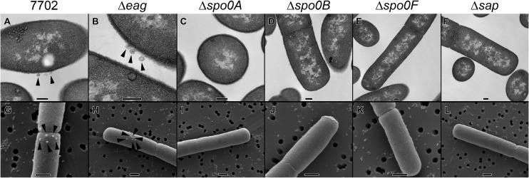
Besides their defects in phage adsorption and infectivity, strains with mutations in the sporulation genes were defective for sporulation, as expected, whereas the strains with individual sap, csaB, and eag mutations sporulated normally (data not shown). Additionally, we conducted scanning and transmission electron microscopy of bacilli that had been incubated with AP50c. In SEM and TEM of the wild type and various mutants (Fig. 1), after examining numerous fields of view of cells, we did not observe any phage particles associated with the cell surface of the Δsap mutant (Fig. 1F and L) or of the Δspo0A (Fig. 1C and I), Δspo0B (Fig. 1D and J), or Δspo0F (Fig. 1E and K) mutants, whereas phage particles were associated with the surfaces of the Sterne 7702 (Fig. 1A and G) and Δeag (Fig. 1B and H) strains.
FIG 1.
Electron microscopic analysis of B. anthracis Sterne and mutant derivatives in the presence of AP50c. Bacterial strains were prepared for TEM (A to F) and SEM (G to L), as described in Materials and Methods, in the presence of AP50c phage at an MOI of 1. Phage particles are visible in association with the surfaces of strains Sterne (7702; A and G) and BA751 (Δeag; B and H) but not with the surface of strain BA754 (Δspo0A; C and I), DP-B-5747 (Δspo0B; D and J), BA755 (Δspo0F; E and K), or BA750 (Δsap; F and L). Bars, 0.2 μm (A to F) and 0.5 μm (G to L). Arrowheads, AP50c phage particles associated with the bacterial surface.
Effect of targeted deletion of spo0 genes on Sap expression and localization.
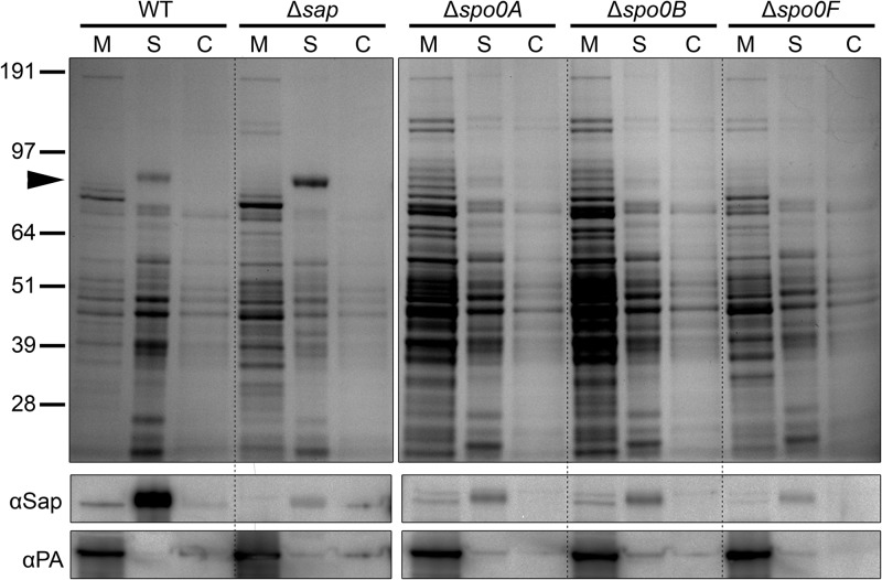
Considering that strains with targeted deletions of spo0A, spo0B, or spo0F were resistant to AP50c, we sought to assess the expression level and subcellular localization of Sap in these strains. We grew broth cultures of the various strains and performed fractionation into medium, S-layer, and cell fractions. Coomassie staining of SDS-polyacrylamide gels revealed a difference in the overall pattern of protein expression in the spo0 strains compared to the parent strain (Fig. 2). Replicate gels were transferred to nitrocellulose and probed with a rabbit polyclonal antibody to Sap. In the S layer of the sap deletion strain BA750, the anti-Sap antibody detected a protein or proteins migrating at approximately the same molecular mass seen in the S-layer fraction of the Sterne strain, although the intensity of the band was substantially reduced (Fig. 2); the presence of a very low intensity band of approximately the same molecular mass as Sap could reflect cross-reactivity of the rabbit polyclonal anti-Sap antibody with EA1 (with which Sap shares substantial homology). In the S-layer fraction of each of the strains with targeted deletion of spo0A, spo0B, or spo0F, there was a reduction in the amount of protein reacting with the anti-Sap antibody in comparison to that in the S-layer fraction of the Sterne strain. Additionally, in the medium fractions of the spo0 deletion strains, a doublet of bands was apparent, whereas in the medium fraction of the Sterne strain, only a single band was seen; whether the additional band represents a modified form of Sap or EA1 is unknown. Taken together, the resistance to AP50c and the reduction in the amount of Sap in the S-layer fractions of these strains are evidence that the presence of Sap in the S layer is important for AP50c activity.
FIG 2.
Effect of deletion of spo0 genes on Sap expression. Broth cultures of Sterne (wild type [WT]), BA750 (Δsap), BA754 (Δspo0A), DP-B-5747 (Δspo0B), and BA755 (Δspo0F) were fractionated into medium (M), S-layer (S), and cell (C) fractions, and aliquots of each fraction were separated by SDS-PAGE. Replicate gels were either Coomassie stained or transferred to nitrocellulose membranes. (Top) Coomassie stain; numbers along the left side mark the migration points of molecular mass markers (in kDa); arrowhead, migration point of Sap and/or EA1; (middle) membranes were probed with a rabbit polyclonal antibody to Sap (αSap); (bottom) membranes were stripped and probed with a mouse monoclonal antibody to protective antigen (αPA).
Reduction of AP50c activity following incubation with purified Sap.
We reasoned that if Sap is the receptor for AP50c, then incubation of phage with purified Sap should reduce the number of infective particles (PFU) in the phage preparation. When an AP50c phage lysate was incubated with purified recombinant C-terminal Sap (lacking the S-layer homology domain), the activity of the phage was substantially reduced. Specifically, incubation of phage with bovine serum albumin reduced the titer by 2% (±2.0%), incubation with buffer alone reduced the titer by 17% (±2.0%), and incubation with purified recombinant C-terminal Sap reduced the titer by 83% (±8.1%). These results provide evidence that Sap is indeed the receptor of AP50c.
Identification of B. cereus strains that are sensitive to AP50c infection.
We also took a genetic approach to test whether the Sap present in the S layer is the AP50c phage receptor. We hypothesized that B. cereus strains that are sensitive to AP50c infection might produce an S layer that is similar to the B. anthracis S layer. We screened a collection of B. cereus sensu lato strains for susceptibility to AP50c infection, including strains that are closely related to B. anthracis on the basis of multilocus sequence typing analysis (47). Of the 20 B. cereus strains screened, 2 were sensitive to AP50c. For B. cereus RS438 (CDC2000032805) (47), the efficiency of plating (EOP) of AP50c was 60% of that on JB220. For B. cereus RS756 (E33L ZK; Zebra Killer [48]), the EOP was 68% of that on JB220.
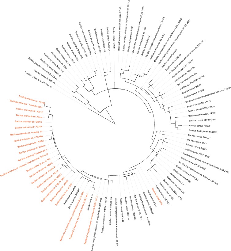
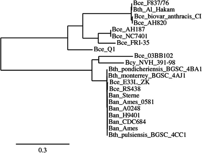
Furthermore, we generated a phylogenetic tree on the basis of whole-genome sequence data for all the B. cereus sensu lato strains available in the PATRIC database (49) and mapped the strains that carry Sap protein similarities with a cutoff of 95% over 95% of the protein sequence. This analysis revealed that one of the two B. cereus strains (RS756 [E33L]) and three B. thuringiensis strains (B. thuringiensis serovar pondicheriensis BGSC 4BA1, B. thuringiensis serovar monterrey BGSC 4AJ1, and B. thuringiensis serovar pulsiensis BGSC 4CC1) possess Sap sequences similar to the Sap sequence of B. anthracis (Fig. 3). Because the whole-genome sequence of RS438 was not available, the sap gene from this strain was PCR amplified and sequenced by the Sanger method. Sequence analysis indicated that the sap genes of RS438 and RS756 (E33L) encode proteins whose lengths are similar to the length of B. anthracis Sap but that differ from B. anthracis Sap at three positions each (RS438, S405A, T571K, and K654E; RS756, K57T, T190S, and T571K; Table 4). The Sap proteins of RS438 and RS756 (E33L) are distinctly different from those of strains outside the group that includes B. anthracis and its close neighbors (Fig. 4). B. thuringiensis strain BGSC 4CC1 encodes a Sap protein identical to that of B. anthracis, whereas the BGSC 4AJ1 and BGSC 4BA1 Sap proteins differ from the Sap protein of B. anthracis by one (V421L) and two (V421L, T586K) amino acid changes, respectively. Although the three B. thuringiensis strains possess Sap proteins similar to the Sap protein of B. anthracis, AP50c did not infect these strains, even at high phage concentrations, and they adsorbed AP50c to various degrees (JB220, 99%; BGSC 4CC1, 90%; BGSC 4BA1, 0%; and BGSC 4AJ1, 69%; Table 4). Comparison of the amino acid sequences of the Sap proteins of the five strains (RS438, RS756, BGSC 4CC1, BGSC 4AJ1, and BGSC 4BA1) indicated that the amino acid residues V421 and T586 may be critical for adsorption, because combined changes in these two positions completely abolished adsorption (BGSC 4BA1), whereas a change in one of these positions (BGSC 4AJ1) partially abolished adsorption (Table 4).
FIG 3.
Distribution of Sap homologs in B. cereus genomes. Phylogenetic tree based on whole-sequence data for all the B. cereus sensu lato strains available in the PATRIC database. B. cereus genomes that encode predicted Sap proteins with greater than 95% sequence identity to the Sap protein of B. anthracis Ames using BLASTp (57) are highlighted in red. BLAST analysis was performed using the PATRIC web server (49). The whole-genome phylogeny of B. cereus group genomes was downloaded from PATRIC in March 2013.
TABLE 4.
Amino acid differences in select positions among selected B. anthracis, B. cereus, and B. thuringiensis Sap proteins
| Strain | Amino acid at positiona: |
AP50c adsorptionb | AP50c plating | ||||||
|---|---|---|---|---|---|---|---|---|---|
| 57 | 190 | 405 | 421 | 571 | 586 | 654 | |||
| B. anthracis Ames | K | T | S | V | T | T | K | +++ | Yes |
| B. thuringiensis serovar pulsiensis BGSC 4CC1 | K | T | S | V | T | T | K | +++ | No |
| B. cereus RS438 CDC2000032805 | K | T | A | V | K | T | E | +++ | Yes |
| B. cereus RS756 (E33L ZK) | T | S | S | V | K | T | K | +++ | Yes |
| B. thuringiensis serovar monterrey BGSC 4AJ1 | K | T | S | L | T | T | K | ++ | No |
| B. thuringiensis serovar pondicheriensis BGSC 4BA1 | K | T | S | L | T | K | K | − | No |
| B. cytotoxicus NVH 391–98c | K | S | S | V | T | K | K | NT | NT |
| B. cereus 03BB102c | E | S | A | L | T | T | K | NT | NT |
| B. cereus AH187c | A | S | V | L | K | E | A | NT | NT |
| B. cereus NC7401c | A | S | V | L | K | E | A | NT | NT |
| B. cereus FRI-35c | A | S | V | L | K | N | A | NT | NT |
| B. cereus Q1c | A | S | V | L | K | K | D | NT | NT |
| B. cereus biovar anthracis CIc | E | S | V | L | K | E | A | NT | NT |
| B. cereus AH820c | E | S | V | L | K | E | A | NT | NT |
| B. cereus F837/76c | E | S | V | L | K | E | A | NT | NT |
| B. thuringiensis Al Hakamc | E | S | V | L | K | E | A | NT | NT |
Position of the amino acid residue in the 814-amino-acid Sap protein of B. anthracis Ames; residues in bold are those that differ from the residues of Sap of B. anthracis Ames.
+++, >66 to 100% adsorption; ++, >34 to 66% adsorption; −, 0% adsorption; NT, not tested.
Additional differences in the Sap proteins of these strains are not listed.
FIG 4.
Phylogenetic tree based on Sap amino acid alignment of a subset of the strains in Fig. 3. The maximum-likelihood tree is based on amino acid alignment of the Sap protein sequences from 21 strains belonging to the B. anthracis (Ban), B. cereus (Bce), and B. thuringiensis (Bth) groups obtained using the Phylogeny Fr website (http://www.phylogeny.fr/version2_cgi/index.cgi). The phylogeny was calculated using the PhyML program (58), and the tree was generated by the TreeDyn program (59).
Intra- and interspecies complementation of the sap deletion strain with wild-type sap genes restores phage sensitivity.
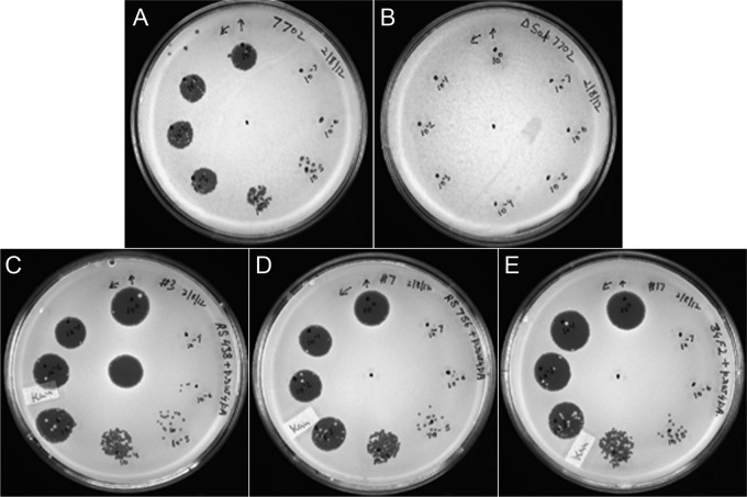
We tested whether complementation of the Δsap mutant with a wild-type sap gene from B. anthracis Sterne 34F2, B. cereus RS438, or B. cereus RS756 (E33L) would restore AP50c sensitivity. The respective sap genes were cloned into a pSW4 derivative as described in Materials and Methods. A Δsap mutant, BA750, was transformed with each of the recombinant plasmids, and the transformants were tested for AP50c infection. The results clearly showed that the mutants carrying the sap plasmids were completely sensitive to AP50c and that the B. cereus sap genes could restore infectivity in B. anthracis (Fig. 5). The complemented strains plated AP50c with EOPs approximately 10 times higher than the EOP of Sterne 7702 (data not shown), perhaps because of overexpression of Sap from the plasmid.
FIG 5.
In trans complementation of the Δsap mutant with B. anthracis and B. cereus sap genes. The recombinant plasmids carrying the sap genes from B. anthracis Sterne and B. cereus strains RS438 and RS756 (E33L) were transformed into BA750 (Δsap), and the resulting transformants were tested for AP50c sensitivity by spot tests. Ten microliters of 10× serial dilutions of phage was spotted on a lawn of each strain. Clearing in the spots after overnight incubation indicates lysis of the bacteria and phage sensitivity. (A) 7702; (B) BA750 (Δsap); (C) BA750 complemented with RS438 sap; (D) BA750 complemented with RS756 (E33L) sap; (E) BA750 complemented with 34F2 sap. In panel C, undiluted AP50c phage stock was spotted both in the center of the plate and at the 12 o'clock position; in the other panels, undiluted phage stock was spotted only at the 12 o'clock position.
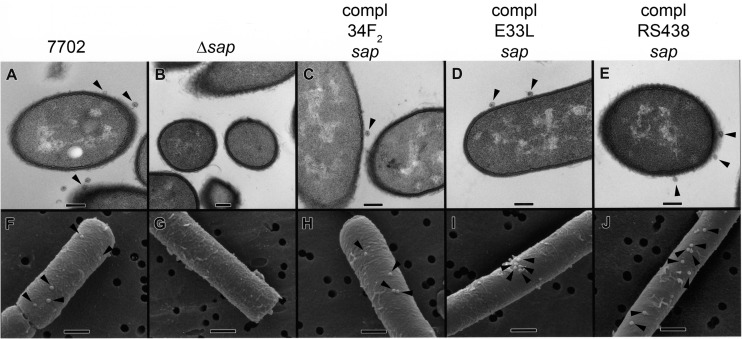
In order to visualize the bacterial cell surface and the association of phage particles with bacilli, we incubated strains with AP50c and conducted scanning and transmission electron microscopy (Fig. 6). Phage particles were visible in association with the bacterial surface of 7702 (Fig. 6A and F) and the complemented strains (Fig. 6C to E and H to J) but not with that of the Δsap strain (Fig. 6B and G). These data provide further evidence that Sap acts as the receptor of phage AP50c.
FIG 6.
Electron microscopic analysis of B. anthracis Sterne, BA750 (Δsap), and complemented (compl) strains in the presence of AP50c. Bacterial strains were prepared for TEM (A to E) and SEM (F to J), as described in Materials and Methods, in the presence of AP50c phage at an MOI of 1. Phage particles are visible in association with the surface of Sterne (7702; A and F) but not with the surface of BA750 (Δsap; B and G). Complementation of BA750 with sap of 34F2 (C and H), RS756 (E33L; D and I), or RS438 (E and J) restored the ability of AP50c to bind to the bacterial surface. Bars, 0.2 μm (A to E) and 0.5 μm (F to J). Arrowheads, AP50c phage particles associated with the bacterial surface.
DISCUSSION
In this study, we strove to identify the B. anthracis receptor for phage AP50c using a genetic/genomic approach. Our results strongly suggest that the S-layer protein Sap or the S layer composed of Sap is the receptor. Although we show that purified Sap can reduce AP50c infectivity, further clarification of whether the active form of phage receptor is Sap itself or the S layer containing Sap awaits future study.
In the chromosome of B. anthracis, sap is located upstream of eag, which encodes EA1, the other S-layer protein (50). Upstream of sap is an operon that includes csaB, which encodes the pyruvyl transferase responsible for anchoring S-layer homology-containing proteins (including Sap and EA1) to the bacterial surface (29). In a previous study, we found that spontaneous and targeted mutations in csaB, leading to changes in the CsaB protein, or targeted deletion of the entire gene resulted in AP50c resistance (28). We reasoned that phage resistance in these mutants could be the result of a failure to display the receptor on the surface. The random mutagenesis approach that we employed in this study led us to focus on the S-layer homology-containing protein Sap. We noted, however, that Sap and EA1 are very similar in their first 200 residues (66% identity, 74% similarity) (51). We therefore engineered a markerless, in-frame deletion strain of eag; this strain remained sensitive to AP50c, demonstrating the specificity of the phage-receptor interaction. We note that in these two studies, spontaneous phage-resistant mutants arose with alterations in csaB, whereas random transposon mutagenesis and screening for phage resistance identified changes in sap. The reason for this mutation spectrum bias is not clear, although differences in the mechanisms of spontaneous mutagenesis (replication errors) versus transposon mutagenesis (target specificity) could account for such a bias.
Two B. cereus and three B. thuringiensis strains with high degrees of sap homology to B. anthracis were identified in this study. The placement of these strains on the whole-genome phylogenetic tree indicates that the sap genes of the B. thuringiensis strains and B. anthracis were most likely acquired by a common ancestor, whereas the sap gene of RS756 (E33L) probably represents an independent acquisition.
A closer look at the B. cereus and B. thuringiensis strains carrying sap genes similar to the sap gene of B. anthracis shows that they also have the neighboring genes in the csa operon intact, yet B. thuringiensis strain BGSC 4CC1, which has a Sap protein identical to that of B. anthracis, did not support AP50c growth, although adsorption was normal. We speculate that the failure of the B. thuringiensis strains to support phage growth may be due to defects in steps subsequent to phage adsorption. There may be inherent differences in factors that are needed for the replication, morphogenesis, or lysis/release of progeny particles of AP50c, or these factors may be absent in these hosts.
Although the S layer of B. anthracis can be composed of the two proteins Sap and EA1, previous studies have shown that the composition of the S layer varies with growth phase. Specifically, during exponential growth, the S layer is mainly composed of Sap, but during stationary phase, it mainly consists of EA1 (52, 53). Our finding that Sap is the AP50c receptor suggests that susceptibility to AP50c infection may vary with growth phase, although we have not yet tested this experimentally. Moreover, some in vitro studies in the presence of bicarbonate (to mimic conditions found in vivo) suggest that Sap may not be expressed during infection (54); if this prediction were found to be true in vivo, then AP50c may therefore not be a good candidate for use in bacteriophage therapy.
We found that deletion of any of the sporulation-related genes spo0A, spo0B, or spo0F led to a reduction in the amount of Sap in the S layer and to resistance to AP50c. It is likely that the transcription factor Spo0A (which, of these three proteins, is the furthest downstream in the phosphorelay system [23]) controls Sap levels in some way. The effect may be on transcription, translation, Sap protein stability, posttranslational modification, targeting of Sap to the surface, or assembly of the Sap S layer.
The sap promoter is thought to be under the control of RNA polymerase containing the alternative sigma factor σA (52). However, the repressor PagR can bind to a sequence that overlaps the σA binding site, preventing sap transcription (54). The master regulator AtxA activates PagR (55) and therefore negatively regulates Sap expression (54). Moving up the pathway, we note that AtxA is negatively regulated by AbrB, which is negatively regulated by phosphorylated Spo0A (56). Considering this entire cascade of events, deletion of Spo0A (or deletion of any protein upstream of Spo0A in the phosphorelay system) would be expected to lead to an increase in Sap expression rather than a decrease. Our interpretation is that if the connection between the spo0 genes and the presence of Sap in the S layer does indeed involve AtxA, then the system must be more sophisticated than the current scientific literature suggests, perhaps with the involvement of unknown factors. Although our results strongly suggest a connection between the Spo0 phosphorelay cascade and the amount of Sap in the S layer, further study is clearly needed to elucidate the mechanism(s) involved.
ACKNOWLEDGMENTS
This work was supported by funds from the Defense Threat Reduction Agency, U.S. Department of Defense, of the U.S. government (proposal no. 8.10084_08_NM_B) to S. Sozhamannan. R. K. Pope and R. M. Hannah were funded under agreement no. HSHQDC-07-C-00020 awarded by the U.S. Department of Homeland Security (DHS) for the management and operation of the National Biodefense Analysis and Countermeasures Center (NBACC), a federally funded research and development center.
The views expressed in this article are those of the authors and do not necessarily reflect the official policy or position, expressed or implied, of the Department of the Navy, JPEO-CBD-MCS-CRP, the U.S. Department of Defense, DHS, or the U.S. government. In no event shall DHS, NBACC, or the Battelle National Biodefense Institute (BNBI) have any responsibility or liability for any use, misuse, inability to use, or reliance upon the information contained herein. DHS does not endorse any products or commercial services mentioned in this publication.
V. Mokashi is an employee of the U.S. government. This work was prepared as part of his official duties. Title 17 U.S.C. §101 defines U.S. government work as work prepared by a military service member or employee of the U.S. government as part of that person's official duties.
We thank Raymond Schuch for providing several Bacillus strains used in this study. We profusely thank Sao-Mai Nguyen-Mau for the kind gifts of rabbit polyclonal anti-Sap antibody and the Sap expression plasmid pET15b Sap ΔSLH.
Footnotes
Published ahead of print 20 December 2013
REFERENCES
- 1.Wommack KE, Colwell RR. 2000. Virioplankton: viruses in aquatic ecosystems. Microbiol. Mol. Biol. Rev. 64:69–114. 10.1128/MMBR.64.1.69-114.2000 [DOI] [PMC free article] [PubMed] [Google Scholar]
- 2.Brussow H, Hendrix RW. 2002. Phage genomics: small is beautiful. Cell 108:13–16. 10.1016/S0092-8674(01)00637-7 [DOI] [PubMed] [Google Scholar]
- 3.Rohwer F. 2003. Global phage diversity. Cell 113:141. 10.1016/S0092-8674(03)00276-9 [DOI] [PubMed] [Google Scholar]
- 4.Rakhuba DV, Kolomiets EI, Dey ES, Novik GI. 2010. Bacteriophage receptors, mechanisms of phage adsorption and penetration into host cell. Pol. J. Microbiol. 59:145–155 [PubMed] [Google Scholar]
- 5.Shao Y, Wang IN. 2008. Bacteriophage adsorption rate and optimal lysis time. Genetics 180:471–482. 10.1534/genetics.108.090100 [DOI] [PMC free article] [PubMed] [Google Scholar]
- 6.Moldovan R, Chapman-McQuiston E, Wu XL. 2007. On kinetics of phage adsorption. Biophys. J. 93:303–315. 10.1529/biophysj.106.102962 [DOI] [PMC free article] [PubMed] [Google Scholar]
- 7.Hyman P, Abedon ST. 2009. Practical methods for determining phage growth parameters. Methods Mol. Biol. 501:175–202. 10.1007/978-1-60327-164-6_18 [DOI] [PubMed] [Google Scholar]
- 8.Reverberi R, Reverberi L. 2007. Factors affecting the antigen-antibody reaction. Blood Transfus. 5:227–240. 10.2450/2007.0047-07 [DOI] [PMC free article] [PubMed] [Google Scholar]
- 9.Sulakvelidze A, Alavidze Z, Morris JG., Jr 2001. Bacteriophage therapy. Antimicrob. Agents Chemother. 45:649–659. 10.1128/AAC.45.3.649-659.2001 [DOI] [PMC free article] [PubMed] [Google Scholar]
- 10.Hagens S, Loessner MJ. 2007. Application of bacteriophages for detection and control of foodborne pathogens. Appl. Microbiol. Biotechnol. 76:513–519. 10.1007/s00253-007-1031-8 [DOI] [PubMed] [Google Scholar]
- 11.McAuliffe ORR, Fitzgerald GF. 2007. The new phage biology: from genomics to applications. Caister Academic Press, Norfolk, United Kingdom [Google Scholar]
- 12.Petty NK, Evans TJ, Fineran PC, Salmond GP. 2007. Biotechnological exploitation of bacteriophage research. Trends Biotechnol. 25:7–15. 10.1016/j.tibtech.2006.11.003 [DOI] [PubMed] [Google Scholar]
- 13.McKinstry M, Edgar R. 2005. Use of phages in therapy and bacterial detection, p 430–440 In Waldor MK, Friedman DI, Adhya SL. (ed), Phages: their role in bacterial pathogenesis and biotechnology. ASM Press, Washington, DC [Google Scholar]
- 14.Loessner MJ, Busse M. 1990. Bacteriophage typing of Listeria species. Appl. Environ. Microbiol. 56:1912–1918 [DOI] [PMC free article] [PubMed] [Google Scholar]
- 15.Basu S, Mukerjee S. 1968. Bacteriophage typing of Vibrio ElTor. Experientia 24:299–300. 10.1007/BF02152832 [DOI] [PubMed] [Google Scholar]
- 16.Rado TA, Bates JH, Engel HW, Mankiewicz E, Murohashi T, Mizuguchi Y, Sula L. 1975. World Health Organization studies on bacteriophage typing of mycobacteria. Subdivision of the species Mycobacterium tuberculosis. Am. Rev. Respir. Dis. 111:459–468 [DOI] [PubMed] [Google Scholar]
- 17.Sonnenschein C. 1928. Bakteriendiagnose mit Bakteriophagen. Dtsch. Med. Wochenschr. 54:1034–1036. 10.1055/s-0028-1125381 [DOI] [Google Scholar]
- 18.Edgar R, McKinstry M, Hwang J, Oppenheim AB, Fekete RA, Giulian G, Merril C, Nagashima K, Adhya S. 2006. High-sensitivity bacterial detection using biotin-tagged phage and quantum-dot nanocomplexes. Proc. Natl. Acad. Sci. U. S. A. 103:4841–4845. 10.1073/pnas.0601211103 [DOI] [PMC free article] [PubMed] [Google Scholar]
- 19.Yim PB, Clarke ML, McKinstry M, De Paoli Lacerda SH, Pease LF, III, Dobrovolskaia MA, Kang H, Read TD, Sozhamannan S, Hwang J. 2009. Quantitative characterization of quantum dot-labeled lambda phage for Escherichia coli detection. Biotechnol. Bioeng. 104:1059–1067. 10.1002/bit.22488 [DOI] [PubMed] [Google Scholar]
- 20.Brigati J, Williams DD, Sorokulova IB, Nanduri V, Chen IH, Turnbough CL, Jr, Petrenko VA. 2004. Diagnostic probes for Bacillus anthracis spores selected from a landscape phage library. Clin. Chem. 50:1899–1906. 10.1373/clinchem.2004.038018 [DOI] [PubMed] [Google Scholar]
- 21.Stern A, Sorek R. 2011. The phage-host arms race: shaping the evolution of microbes. Bioessays 33:43–51. 10.1002/bies.201000071 [DOI] [PMC free article] [PubMed] [Google Scholar]
- 22.Jernigan DB, Raghunathan PL, Bell BP, Brechner R, Bresnitz EA, Butler JC, Cetron M, Cohen M, Doyle T, Fischer M, Greene C, Griffith KS, Guarner J, Hadler JL, Hayslett JA, Meyer R, Petersen LR, Phillips M, Pinner R, Popovic T, Quinn CP, Reefhuis J, Reissman D, Rosenstein N, Schuchat A, Shieh WJ, Siegal L, Swerdlow DL, Tenover FC, Traeger M, Ward JW, Weisfuse I, Wiersma S, Yeskey K, Zaki S, Ashford DA, Perkins BA, Ostroff S, Hughes J, Fleming D, Koplan JP, Gerberding JL. 2002. Investigation of bioterrorism-related anthrax, United States, 2001: epidemiologic findings. Emerg. Infect. Dis. 8:1019–1028. 10.3201/eid0810.020353 [DOI] [PMC free article] [PubMed] [Google Scholar]
- 23.Burbulys D, Trach KA, Hoch JA. 1991. Initiation of sporulation in B. subtilis is controlled by a multicomponent phosphorelay. Cell 64:545–552. 10.1016/0092-8674(91)90238-T [DOI] [PubMed] [Google Scholar]
- 24.Stephenson K, Hoch JA. 2002. Evolution of signalling in the sporulation phosphorelay. Mol. Microbiol. 46:297–304. 10.1046/j.1365-2958.2002.03186.x [DOI] [PubMed] [Google Scholar]
- 25.Nagy E. 1974. A highly specific phage attacking Bacillus anthracis strain Sterne. Acta Microbiol. Acad. Sci. Hung. 21:257–263 [PubMed] [Google Scholar]
- 26.Ackermann HW, Roy R, Martin M, Murthy MR, Smirnoff WA. 1978. Partial characterization of a cubic Bacillus phage. Can. J. Microbiol. 24:986–993. 10.1139/m78-162 [DOI] [PubMed] [Google Scholar]
- 27.Sozhamannan S, McKinstry M, Lentz SM, Jalasvuori M, McAfee F, Smith A, Dabbs J, Ackermann HW, Bamford JKH, Mateczun A, Read TD. 2008. Molecular characterization of a variant of Bacillus anthracis-specific phage AP50 with improved bacteriolytic activity. Appl. Environ. Microbiol. 74:6792–6796. 10.1128/AEM.01124-08 [DOI] [PMC free article] [PubMed] [Google Scholar]
- 28.Bishop-Lilly KA, Plaut RD, Chen PE, Akmal A, Willner KM, Butani A, Dorsey S, Mokashi V, Mateczun AJ, Chapman C, George M, Luu T, Read TD, Calendar R, Stibitz S, Sozhamannan S. 2012. Whole genome sequencing of phage resistant Bacillus anthracis mutants reveals an essential role for cell surface anchoring protein CsaB in phage AP50c adsorption. Virol. J. 9:246. 10.1186/1743-422X-9-246 [DOI] [PMC free article] [PubMed] [Google Scholar]
- 29.Mesnage S, Fontaine T, Mignot T, Delepierre M, Mock M, Fouet A. 2000. Bacterial SLH domain proteins are non-covalently anchored to the cell surface via a conserved mechanism involving wall polysaccharide pyruvylation. EMBO J. 19:4473–4484. 10.1093/emboj/19.17.4473 [DOI] [PMC free article] [PubMed] [Google Scholar]
- 30.Zemansky J, Kline BC, Woodward JJ, Leber JH, Marquis H, Portnoy DA. 2009. Development of a mariner-based transposon and identification of Listeria monocytogenes determinants, including the peptidyl-prolyl isomerase PrsA2, that contribute to its hemolytic phenotype. J. Bacteriol. 191:3950–3964. 10.1128/JB.00016-09 [DOI] [PMC free article] [PubMed] [Google Scholar]
- 31.Green BD, Battisti L, Koehler TM, Thorne CB, Ivins BE. 1985. Demonstration of a capsule plasmid in Bacillus anthracis. Infect. Immun. 49:291–297 [DOI] [PMC free article] [PubMed] [Google Scholar]
- 32.Horton RM, Cai ZL, Ho SN, Pease LR. 1990. Gene splicing by overlap extension: tailor-made genes using the polymerase chain reaction. Biotechniques 8:528–535 [PubMed] [Google Scholar]
- 33.Smith K, Youngman P. 1992. Use of a new integrational vector to investigate compartment-specific expression of the Bacillus subtilis spoIIM gene. Biochimie 74:705–711. 10.1016/0300-9084(92)90143-3 [DOI] [PubMed] [Google Scholar]
- 34.Camilli A, Tilney LG, Portnoy DA. 1993. Dual roles of plcA in Listeria monocytogenes pathogenesis. Mol. Microbiol. 8:143–157. 10.1111/j.1365-2958.1993.tb01211.x [DOI] [PMC free article] [PubMed] [Google Scholar]
- 35.Janes BK, Stibitz S. 2006. Routine markerless gene replacement in Bacillus anthracis. Infect. Immun. 74:1949–1953. 10.1128/IAI.74.3.1949-1953.2006 [DOI] [PMC free article] [PubMed] [Google Scholar]
- 36.Arraj JA, Marinus MG. 1983. Phenotypic reversal in dam mutants of Escherichia coli K-12 by a recombinant plasmid containing the dam+ gene. J. Bacteriol. 153:562–565 [DOI] [PMC free article] [PubMed] [Google Scholar]
- 37.Tavazoie S, Church GM. 1998. Quantitative whole-genome analysis of DNA-protein interactions by in vivo methylase protection in E. coli. Nat. Biotechnol. 16:566–571. 10.1038/nbt0698-566 [DOI] [PubMed] [Google Scholar]
- 38.Thorne CB. 1968. Transduction in Bacillus cereus and Bacillus anthracis. Bacteriol. Rev. 32:358–361 [PMC free article] [PubMed] [Google Scholar]
- 39.Giorno R, Mallozzi M, Bozue J, Moody KS, Slack A, Qiu D, Wang R, Friedlander A, Welkos S, Driks A. 2009. Localization and assembly of proteins comprising the outer structures of the Bacillus anthracis spore. Microbiology 155:1133–1145. 10.1099/mic.0.023333-0 [DOI] [PMC free article] [PubMed] [Google Scholar]
- 40.Pomerantsev AP, Kalnin KV, Osorio M, Leppla SH. 2003. Phosphatidylcholine-specific phospholipase C and sphingomyelinase activities in bacteria of the Bacillus cereus group. Infect. Immun. 71:6591–6606. 10.1128/IAI.71.11.6591-6606.2003 [DOI] [PMC free article] [PubMed] [Google Scholar]
- 41.Yao J, Zhong J, Fang Y, Geisinger E, Novick RP, Lambowitz AM. 2006. Use of targetrons to disrupt essential and nonessential genes in Staphylococcus aureus reveals temperature sensitivity of Ll.LtrB group II intron splicing. RNA 12:1271–1281. 10.1261/rna.68706 [DOI] [PMC free article] [PubMed] [Google Scholar]
- 42.Charpentier E, Anton AI, Barry P, Alfonso B, Fang Y, Novick RP. 2004. Novel cassette-based shuttle vector system for Gram-positive bacteria. Appl. Environ. Microbiol. 70:6076–6085. 10.1128/AEM.70.10.6076-6085.2004 [DOI] [PMC free article] [PubMed] [Google Scholar]
- 43.Lauer P, Chow MY, Loessner MJ, Portnoy DA, Calendar R. 2002. Construction, characterization, and use of two Listeria monocytogenes site-specific phage integration vectors. J. Bacteriol. 184:4177–4186. 10.1128/JB.184.15.4177-4186.2002 [DOI] [PMC free article] [PubMed] [Google Scholar]
- 44.Lunderberg JM, Nguyen-Mau SM, Richter GS, Wang YT, Dworkin J, Missiakas DM, Schneewind O. 2013. Bacillus anthracis acetyltransferases PatA1 and PatA2 modify the secondary cell wall polysaccharide and affect the assembly of S-layer proteins. J. Bacteriol. 195:977–989. 10.1128/JB.01274-12 [DOI] [PMC free article] [PubMed] [Google Scholar]
- 45.Nguyen-Mau SM, Oh SY, Kern VJ, Missiakas DM, Schneewind O. 2012. Secretion genes as determinants of Bacillus anthracis chain length. J. Bacteriol. 194:3841–3850. 10.1128/JB.00384-12 [DOI] [PMC free article] [PubMed] [Google Scholar]
- 46.Trach K, Hoch JA. 1989. The Bacillus subtilis spo0B stage 0 sporulation operon encodes an essential GTP-binding protein. J. Bacteriol. 171:1362–1371 [DOI] [PMC free article] [PubMed] [Google Scholar]
- 47.Marston CK, Gee JE, Popovic T, Hoffmaster AR. 2006. Molecular approaches to identify and differentiate Bacillus anthracis from phenotypically similar Bacillus species isolates. BMC Microbiol. 6:22. 10.1186/1471-2180-6-22 [DOI] [PMC free article] [PubMed] [Google Scholar]
- 48.Han CS, Xie G, Challacombe JF, Altherr MR, Bhotika SS, Brown N, Bruce D, Campbell CS, Campbell ML, Chen J, Chertkov O, Cleland C, Dimitrijevic M, Doggett NA, Fawcett JJ, Glavina T, Goodwin LA, Green LD, Hill KK, Hitchcock P, Jackson PJ, Keim P, Kewalramani AR, Longmire J, Lucas S, Malfatti S, McMurry K, Meincke LJ, Misra M, Moseman BL, Mundt M, Munk AC, Okinaka RT, Parson-Quintana B, Reilly LP, Richardson P, Robinson DL, Rubin E, Saunders E, Tapia R, Tesmer JG, Thayer N, Thompson LS, Tice H, Ticknor LO, Wills PL, Brettin TS, Gilna P. 2006. Pathogenomic sequence analysis of Bacillus cereus and Bacillus thuringiensis isolates closely related to Bacillus anthracis. J. Bacteriol. 188:3382–3390. 10.1128/JB.188.9.3382-3390.2006 [DOI] [PMC free article] [PubMed] [Google Scholar]
- 49.Gillespie JJ, Wattam AR, Cammer SA, Gabbard JL, Shukla MP, Dalay O, Driscoll T, Hix D, Mane SP, Mao C, Nordberg EK, Scott M, Schulman JR, Snyder EE, Sullivan DE, Wang C, Warren A, Williams KP, Xue T, Yoo HS, Zhang C, Zhang Y, Will R, Kenyon RW, Sobral BW. 2011. PATRIC: the comprehensive bacterial bioinformatics resource with a focus on human pathogenic species. Infect. Immun. 79:4286–4298. 10.1128/IAI.00207-11 [DOI] [PMC free article] [PubMed] [Google Scholar]
- 50.Mesnage S, Tosi-Couture E, Mock M, Gounon P, Fouet A. 1997. Molecular characterization of the Bacillus anthracis main S-layer component: evidence that it is the major cell-associated antigen. Mol. Microbiol. 23:1147–1155. 10.1046/j.1365-2958.1997.2941659.x [DOI] [PubMed] [Google Scholar]
- 51.Fouet A, Mesnage S. 2002. Bacillus anthracis cell envelope components, p 87–113 In Koehler T. (ed), Anthrax, vol 271 Springer, Berlin, Germany: [DOI] [PubMed] [Google Scholar]
- 52.Mignot T, Mesnage S, Couture-Tosi E, Mock M, Fouet A. 2002. Developmental switch of S-layer protein synthesis in Bacillus anthracis. Mol. Microbiol. 43:1615–1627. 10.1046/j.1365-2958.2002.02852.x [DOI] [PubMed] [Google Scholar]
- 53.Kern VJ, Kern JW, Theriot JA, Schneewind O, Missiakas D. 2012. Surface-layer (S-layer) proteins Sap and EA1 govern the binding of the S-layer-associated protein BslO at the cell septa of Bacillus anthracis. J. Bacteriol. 194:3833–3840. 10.1128/JB.00402-12 [DOI] [PMC free article] [PubMed] [Google Scholar]
- 54.Mignot T, Mock M, Fouet A. 2003. A plasmid-encoded regulator couples the synthesis of toxins and surface structures in Bacillus anthracis. Mol. Microbiol. 47:917–927. 10.1046/j.1365-2958.2003.03345.x [DOI] [PubMed] [Google Scholar]
- 55.Hoffmaster AR, Koehler TM. 1999. Autogenous regulation of the Bacillus anthracis pag operon. J. Bacteriol. 181:4485–4492 [DOI] [PMC free article] [PubMed] [Google Scholar]
- 56.Saile E, Koehler TM. 2002. Control of anthrax toxin gene expression by the transition state regulator abrB. J. Bacteriol. 184:370–380. 10.1128/JB.184.2.370-380.2002 [DOI] [PMC free article] [PubMed] [Google Scholar]
- 57.Altschul SF, Gish W, Miller W, Myers EW, Lipman DJ. 1990. Basic local alignment search tool. J. Mol. Biol. 215:403–410 [DOI] [PubMed] [Google Scholar]
- 58.Guindon S, Dufayard JF, Lefort V, Anisimova M, Hordijk W, Gascuel O. 2010. New algorithms and methods to estimate maximum-likelihood phylogenies: assessing the performance of PhyML 3.0. Syst. Biol. 59:307–321. 10.1093/sysbio/syq010 [DOI] [PubMed] [Google Scholar]
- 59.Dereeper A, Guignon V, Blanc G, Audic S, Buffet S, Chevenet F, Dufayard JF, Guindon S, Lefort V, Lescot M, Claverie JM, Gascuel O. 2008. Phylogeny.fr: robust phylogenetic analysis for the non-specialist. Nucleic Acids Res. 36:W465–W469. 10.1093/nar/gkn180 [DOI] [PMC free article] [PubMed] [Google Scholar]
- 60.Smith NR, Gordon RE, Clark FE. 1952. Aerobic sporeforming bacteria. U.S. Department of Agriculture, Washington, DC [Google Scholar]
- 61.Delaporte B. 1969. Description of Bacillus medusa n. sp. C. R. Hebd. Seances Acad. Sci. D 269:1129–1131 (In French.) [PubMed] [Google Scholar]
- 62.Rosenberg H, La Nauze JM. 1967. The metabolism of phosphonates by microorganisms. The transport of aminoethylphosphonic acid in Bacillus cereus. Biochim. Biophys. Acta 141:79–90 [DOI] [PubMed] [Google Scholar]
- 63.Work E. 1959. The action of a lytic enzyme from spores of a bacillus on various species of bacteria. Ann. Inst. Pasteur (Paris) 96:468–480 [PubMed] [Google Scholar]
- 64.Stabb EV, Jacobson LM, Handelsman J. 1994. Zwittermicin A-producing strains of Bacillus cereus from diverse soils. Appl. Environ. Microbiol. 60:4404–4412 [DOI] [PMC free article] [PubMed] [Google Scholar]
- 65.Bernhard K, Schrempf H, Goebel W. 1978. Bacteriocin and antibiotic resistance plasmids in Bacillus cereus and Bacillus subtilis. J. Bacteriol. 133:897–903 [DOI] [PMC free article] [PubMed] [Google Scholar]
- 66.Priest FG, Goodfellow M, Todd C. 1988. A numerical classification of the genus Bacillus. J. Gen. Microbiol. 134:1847–1882 [DOI] [PubMed] [Google Scholar]
- 67.Northrop JH. 1958. Studies on the origin of bacterial viruses. V. The effect of temperature on the terramycin-resistant and phage-producing cells of Bacillus megatherium cultures. J. Gen. Physiol. 42:329–334 [DOI] [PMC free article] [PubMed] [Google Scholar]
- 68.Vary PS, Biedendieck R, Fuerch T, Meinhardt F, Rohde M, Deckwer WD, Jahn D. 2007. Bacillus megaterium—from simple soil bacterium to industrial protein production host. Appl. Microbiol. Biotechnol. 76:957–967. 10.1007/s00253-007-1089-3 [DOI] [PubMed] [Google Scholar]
- 69.Smith NR. 1946. Aerobic mesophilic spore-forming bacteria, p 41 U.S. Department of Agriculture miscellaneous publication 559. U.S. Department of Agriculture, Washington, DC [Google Scholar]
- 70.Hernandez E, Ramisse F, Ducoureau JP, Cruel T, Cavallo JD. 1998. Bacillus thuringiensis subsp. konkukian (serotype H34) superinfection: case report and experimental evidence of pathogenicity in immunosuppressed mice. J. Clin. Microbiol. 36:2138–2139 [DOI] [PMC free article] [PubMed] [Google Scholar]
- 71.Radnedge L, Agron PG, Hill KK, Jackson PJ, Ticknor LO, Keim P, Andersen GL. 2003. Genome differences that distinguish Bacillus anthracis from Bacillus cereus and Bacillus thuringiensis. Appl. Environ. Microbiol. 69:2755–2764. 10.1128/AEM.69.5.2755-2764.2003 [DOI] [PMC free article] [PubMed] [Google Scholar]


