ABSTRACT
Novel avian-origin influenza A(H7N9) viruses were first reported to infect humans in March 2013. To date, 143 human cases, including 45 deaths, have been recorded. By using sequence comparisons and phylogenetic and ancestral inference analyses, we identified several distinct amino acids in the A(H7N9) polymerase PA protein, some of which may be mammalian adapting. Mutant viruses possessing some of these amino acid changes, singly or in combination, were assessed for their polymerase activities and growth kinetics in mammalian and avian cells and for their virulence in mice. We identified several mutants that were slightly more virulent in mice than the wild-type A(H7N9) virus, A/Anhui/1/2013. These mutants also exhibited increased polymerase activity in human cells but not in avian cells. Our findings indicate that the PA protein of A(H7N9) viruses has several amino acid substitutions that are attenuating in mammals.
IMPORTANCE Novel avian-origin influenza A(H7N9) viruses emerged in the spring of 2013. By using computational analyses of A(H7N9) viral sequences, we identified several amino acid changes in the polymerase PA protein, which we then assessed for their effects on viral replication in cultured cells and mice. We found that the PA proteins of A(H7N9) viruses possess several amino acid substitutions that cause attenuation in mammals.
INTRODUCTION
On 31 March 2013, three individuals were reported to be infected with an avian influenza A virus of the H7N9 subtype [A(H7N9)] (1). To date, 143 confirmed cases of A(H7N9) virus infection and 45 associated deaths have been reported. This represents a case-fatality rate of ca. 31.5%; however, this rate may actually be lower because the number of mild cases is unknown (2). Phylogenetic analysis has revealed that the hemagglutinin (HA) and neuraminidase (NA) genes of the A(H7N9) viruses share the greatest sequence identities with Eurasian avian A(H7N3) viruses and N9 viruses of different HA subtypes, respectively (3–6). The other six viral genes are highly related to A(H9N2) viruses that have circulated in poultry in China (1, 3–7). These findings indicate that A(H7N9) viruses are reassortant viruses with genes from several avian isolates.
We (8) and others (9–14) recently reported that prototype A(H7N9) viruses replicate efficiently in mammalian cells, mice, and ferrets, and transmit among subsets of pairs of ferrets. A(H7N9) viruses isolated from humans possess several amino acid changes already known to facilitate infection of mammals, including leucine at position 226 of HA (H3 HA numbering), which confers increased binding to human-type receptors (15), and lysine at position 627 (PB2-627K) (16, 17) and asparagine at position 701 (PB2-701N) (18, 19) in the polymerase protein PB2, which increase the polymerase activity in mammalian cells. Notably, the PB2-627K and PB2-701N markers have been detected in almost all human, but not avian or environmental, A(H7N9) isolates (6).
In addition to the known mammalian-adapting amino acid changes in the PB2 and HA proteins, additional amino acid changes in A(H7N9) proteins may have enabled these viruses to infect mammals. We therefore undertook a comprehensive analysis of A(H7N9) sequences and identified several amino acids in the polymerase PA protein that are typically found in human (but not avian) influenza A viruses or are characteristic of A(H7N9) viruses (i.e., not commonly detected among human or avian influenza viruses). Here, we evaluated these distinguishing amino acid changes in vivo and in vitro.
MATERIALS AND METHODS
Viruses.
A/Anhui/1/2013 (H7N9; Anhui/1), kindly provided by Yuelong Shu (Director of the World Health Organization [WHO] Collaborating Center for Reference and Research on Influenza, Director of the Chinese National Influenza Center, and Deputy Director of the National Institute for Viral Disease Control and Prevention China CDC, Beijing, People's Republic of China) was propagated in embryonated chicken eggs and stored as a stock virus. All experiments with A(H7N9) viruses were performed in enhanced biosafety level 3 (BSL3) containment laboratories at the University of Tokyo (Tokyo, Japan), which are approved for such use by the Ministry of Agriculture, Forestry, and Fisheries of Japan.
Cells.
Madin-Darby canine kidney (MDCK) cells were maintained in Eagle minimal essential medium (MEM) containing 5% newborn calf serum. Human embryonic kidney 293T cells, human alveolar adenocarcinoma epithelial A549 cells, and chicken fibroblast DF-1 cells were maintained in Dulbecco modified Eagle medium (DMEM) containing 10% fetal calf serum. MDCK, 293T, and A549 cells were incubated at 37°C under 5% CO2. DF-1 cells were incubated at 39°C under 5% CO2.
Construction of plasmids.
Plasmids were constructed as previously described (8). Briefly, viral RNA was extracted from stock Anhui/1 virus (segment ID in the EpiFlu database of the Global Initiative on Sharing All Influenza Data; EPI439503-10) with ISOGEN-LS (Nippon gene). A cDNA was synthesized by reverse transcription using Superscript III (Life Technologies) with the universal primer U12 for influenza A virus genes. The cDNA products were amplified by PCR using Phusion high-fidelity DNA polymerase (Thermo Scientific) with specific primers for each virus gene and cloned into the RNA polymerase I plasmid (20), in which viral RNA is synthesized under the control of the human RNA polymerase I promoter and the mouse RNA polymerase I terminator. Mutations in the PA gene were generated by PCR amplification of the respective RNA polymerase I plasmid with primers possessing the desired mutations (primer sequences available upon request). All constructs were sequenced to confirm the absence of unwanted mutations.
To prepare plasmids for viral protein expression, the open reading frames of the PB2, PB1, PA, and NP genes were amplified by PCR with gene-specific primers (primer sequences available upon request). The PCR products were cloned into pCAGGS/MCS (21). All constructs were sequenced to confirm the absence of unwanted mutations.
Reverse genetics.
Plasmid-based reverse genetics for virus generation were performed as previously described (22). Briefly, eight RNA polymerase I plasmids (for the synthesis of the eight influenza A viral RNAs), together with plasmids for the expression of the viral PB2, PB1, PA, and NP proteins derived from an influenza A virus strain A/WSN/33 (H1N1) (23), were transfected into 293T cells using Trans-IT 293 (Mirus). At 48 h posttransfection, culture supernatants were harvested and inoculated into MDCK cells for virus propagation. After 48 h, cell culture media, including viruses, were centrifuged to remove cell debris, and the supernatants were stored as stock viruses. The titers of the stock viruses were determined by plaque assays in MDCK cells. All viruses were sequenced to confirm the absence of unwanted mutations.
Experimental infection of mice.
Six-week-old female BALB/c mice (Japan SLC) were used. Baseline body weights were measured before infection. Under anesthesia, four mice per group were intranasally inoculated with 101 to 106 PFU (50 μl) of the indicated viruses. Body weight and survival were monitored daily for 14 days; mice with body weight loss of >25% of their baseline body weight were euthanized. For virological examinations, six mice per group were intranasally infected with 106 PFU (50 μl) of the viruses, and three mice per group were euthanized at 3 and 6 days postinfection. The virus titers in the nose tissue, lung, brain, spleen, kidney, liver, and colon were determined by plaque assays in MDCK cells. All experiments with mice were performed in accordance with the University of Tokyo's Regulations for Animal Care and Use and were approved by the Animal Experiment Committee of the Institute of Medical Science, University of Tokyo.
Growth kinetics of virus in cell culture.
Human alveolar adenocarcinoma epithelial (A549) and chicken fibroblast (DF-1) cells were infected with the indicated viruses at a multiplicity of infection (MOI) of 0.001. After incubation at 37°C for 1 h, the viral inoculum was replaced with MEM containing 0.3% bovine serum albumin and TPCK (tolylsulfonyl phenylalanyl chloromethyl ketone)-treated trypsin (0.45 μg/ml for A549 cells or 0.25 μg/ml for DF-1 cells), followed by further incubation at 37°C for A549 cells or 39°C for DF-1 cells, respectively. Cell culture supernatants were collected at 8, 24, 48, and 72 h postinfection and subjected to virus titration by use of plaque assays in MDCK cells.
Minigenome assay.
A minigenome assay based on the dual-luciferase system was performed as previously reported (24, 25). Briefly, A549 and DF-1 cells, incubated at 37 and 39°C, respectively, were transfected with viral protein expression plasmids for NP, PB1, PB2, and PA or its mutants (0.2 μg of each), with a plasmid expressing a reporter vRNA encoding the firefly luciferase gene under the control of the human or chicken RNA polymerase I promoter [pPolI/NP(0)Fluc(0) or pPolIGG-NP(0)Fluc(0), respectively; 0.2 μg of each], and pRL-null (Promega, 0.2 μg), which expresses Renilla luciferase, as an transfection control. Control experiments established transfection efficiencies of 30 to 40% and 40 to 50% for A549 and DF-1 cells, respectively. The luciferase activities in the transfected A549 and DF-1 cells were measured by using a Dual-Glo luciferase assay system (Promega) at 24 h posttransfection. Polymerase activity was calculated by standardization of the firefly luciferase activity to the Renilla luciferase activity. The polymerase activity of the wild type was set to 100%.
Phylogenetic analysis and inference of ancestral amino acid changes.
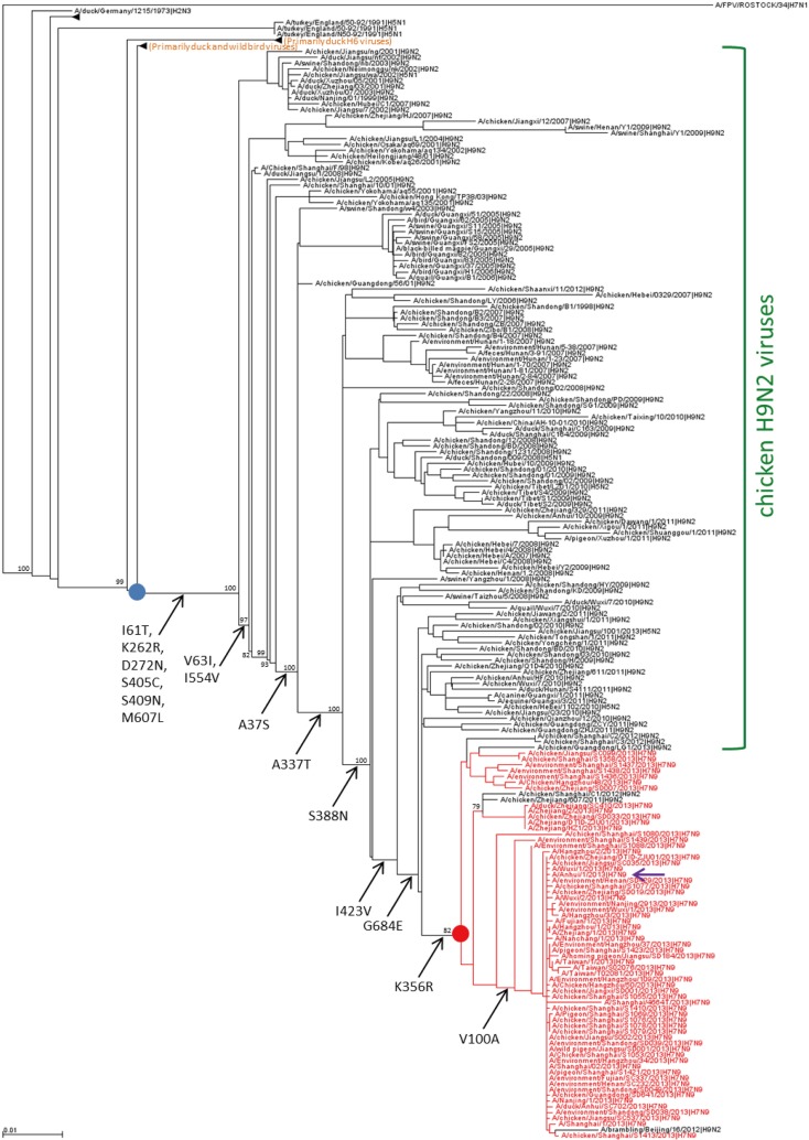
Using sequences downloaded from GenBank (http://www.ncbi.nlm.nih.gov/genomes/FLU/FLU.html) or GISAID (http://gisaid.org), we generated a phylogenetic tree for the PA segment of the Eurasian influenza A viruses from all host species, with the exception of human H1, H2, and H3 viruses. Sequences were aligned using CLUSTAL W (26), with subsequent manual editing. We also removed duplicate and short gene sequences (i.e., those missing >5% of the nucleotides at either end). For the resulting 4,160 PA sequences, we inferred the maximum-likelihood phylogeny under the GTR+Γ model of evolution using RAxML 7.6.3 on the CIPRES Science Gateway (27). From this tree, we selected the A(H7N9) sublineage and all neighboring and ancestral sublineages that potentially contained pertinent information on the emergence of the A(H7N9) viruses. We then inferred the maximum-likelihood phylogeny for all selected sequences using PhyML 3.0 (28) under the GTR+Γ model of evolution. Support for the maximum-likelihood tree topology was calculated from 100 bootstrap replicates using PhyML under the HKY+Γ model of evolution. Branches in the topology with at least 70% bootstrap support are indicated in Fig. 1. Finally, the ancestral amino acid sequence at each internal node between the selected ancestral node (blue circle in Fig. 1) and the most recent common ancestor of A(H7N9) viruses (MRCA, red circle in Fig. 1) was inferred using the codeml routine of PAML 4.4 (29) holding the maximum-likelihood tree fixed. When the inferred ancestral amino acid at a site differed between the two ends of a branch, and the posterior probability of both inferred ancestral amino acids exceeded 90%, then we assumed that a substitution occurred on this branch. Inferred amino acid changes are shown in Fig. 1.
FIG 1.
Phylogenetic tree and inferred ancestral changes of influenza A virus PA. A phylogenetic tree of the PA gene, which includes the A(H7N9) viruses and neighboring and ancestral sequences that may have played a role in the genesis of A(H7N9) PA, is shown. A(H7N9) PA genes are shown in red; the group of chicken H9N2 viruses is indicated in green, while the groups of duck and wild-bird viruses are indicated in orange. The Anhui/1 virus used in the present study is indicated by a purple arrow. The MRCA of A(H7N9) PA genes is indicated by a red circle. Ancestral inference was carried out for branches on the path to A(H7N9) PA located between an ancestral node (blue circle) and the MRCA; all significant bootstrap values (i.e., those >70) are shown. Also shown is the potentially mammalian-adapting PA-V100A substitution, which occurred within the group of A(H7N9) PA genes.
RESULTS AND DISCUSSION
Identification of amino acid changes in A(H7N9) PA that are associated with the genesis of A(H7N9) viruses.
To identify amino acid changes that are associated with the genesis of A(H7N9) viruses, we first inspected the amino acid sequences of A(H7N9) virus proteins (obtained from GISAID, www.gisaid.org, and NCBI, http://www.ncbi.nlm.nih.gov/genomes/FLU/FLU.html). The polymerase PA protein of A(H7N9) viruses possesses several amino acid changes that were predicted by computational analysis (20, 30–32) to be host species specific. Therefore, we focused on the PA protein for further analysis and experimental studies. The predicted host species-specific amino acid substitutions in PA include alanine/valine at position 100, arginine/lysine at position 356, and asparagine/serine at position 409, which are characteristic of human/avian influenza viruses, respectively (5, 20, 30–33) (Table 1). At all three of these positions, A(H7N9) PA proteins encode the human virus-type amino acid. To test the biological significance of these potentially mammalian-adapting amino acids, we converted them individually and in combination to their avian virus-type counterparts. The respective mutations were introduced into A/Anhui/1/2013 [Anhui/1; a prototype A(H7N9) virus recently characterized by us (5)] PA protein and virus; the resulting PA protein expression plasmids and viruses are referred to as PA-A100V, PA-R356K, PA-N409S, and PA-A100V/R356K/N409S.
TABLE 1.
PA amino acid substitutions with a potential role in A(H7N9) genesis
| Source | No. of virusesa | Representative isolate | PA amino acid at positionb: |
||||||||||||||
|---|---|---|---|---|---|---|---|---|---|---|---|---|---|---|---|---|---|
| 37 | 61 | 63 | 100 | 262 | 272 | 337 | 356 | 388 | 405 | 409 | 423 | 554 | 607 | 684 | |||
| Human | 16 | A/Anhui/1/2013 | S | T | I | A | R | N | T | R | N | C | N | V | V | L | E |
| 3 | A/Zhejiang/2/2013 | S | T | I | V | R | D | T | R | N | C | S | V | V | L | E | |
| Avian or environmental | 31 | A/chicken/Shanghai/S1053/2013 | S | T | I | A | R | N | T | R | N | C | N | V | V | L | E |
| 4 | A/chicken/Hangzhou/48/2013 | S | T | I | V | K | N | T | R | N | C | S | V | V | L | E | |
| 3 | A/pigeon/Shanghai/S1421/2013 | S | I | I | A | R | N | T | R | N | C | N | V | V | L | E | |
| 3 | A/chicken/Shanghai/S1358/2013 | S | T | I | V | K | N | T | R | N | C | N | V | V | L | E | |
| 2 | A/chicken/Zhejiang/SD033/2013 | S | T | I | V | R | D | T | R | N | C | S | V | V | L | E | |
| 1 | A/chicken/Shanghai/S1080/2013 | S | T | I | V | R | N | T | R | N | C | N | V | V | L | E | |
| 1 | A/environment/Shanghai/S1439/2013 | S | T | I | A | K | N | T | R | N | C | N | V | V | L | E | |
| Avian | A | I | V | V (96) | K | D | A | K (99) | S | S | S (92) | I | I | M | G | ||
| H9N2 | A | I | V | V (92) | K | D | A | K (96) | S | S | S (72) | I | I | M | G | ||
| Human H1N1 pdm2009 | A | I | V | V (99) | R | D | A | R (100) | G | S | N (100) | I | I | M | G | ||
| Human seasonal H1N1 | A | I | V | A (97) | K | N | S | R (91) | S | S | N (99) | I | I | M | G | ||
| Human H3N2 | A | I | V | A (99) | K | N | S | R (98) | S | S | N (92) | I | I | M | G | ||
That is, the number of viruses with the indicated amino acids at the respective positions; note that PA proteins may differ at amino acid positions not shown here.
Predicted human virus-type amino acids at PA-100, -356, and -409 are indicated in boldface. Amino acids found in the majority of viruses are listed. For the predicted host species-specific amino acids at positions 100, 356, and 409, the percentages for the respective amino acids are shown in parentheses; numbers are rounded to the nearest whole number. The reference sequences are based on the inspection of 8,224, 377, 4,238, 1,457, and 3,692 full-length avian, avian H9N2, human H1N1 pdm2009, human seasonal H1N1, and human H3N2 PA sequences, respectively.
In addition to these predicted host species substitutions, our inspection of A(H7N9) PA sequences also identified several amino acids that are not commonly found among avian or human PA proteins (Table 1). To investigate the evolutionary history of these amino acid changes, we performed phylogenetic and ancestral inference analyses. First, we generated a phylogenetic tree of all of the Eurasian influenza A virus PA genes (excluding human H1, H2, and H3 viruses). From this tree, we selected all A(H7N9) PA sequences, as well as viruses of neighboring lineages and ancestral sequences located on the evolutionary path to A(H7N9); for these selected PA sequences, we inferred a phylogenetic tree (Fig. 1). This tree shows that the PA genes of A(H7N9) viruses have closest sequence similarity with the PA genes of chicken H9N2 viruses, as previously described by us (12) and others (6, 9, 13, 15). To identify amino acid changes that may have been critical to the evolution of A(H7N9) viruses, we inferred ancestral sequences of all nodes between a selected ancestral node (blue circle in Fig. 1) and the most recent common ancestor (MRCA; red circle in Fig. 1) of the A(H7N9) PA genes. The particular ancestral node chosen for the starting point for our ancestral inference analysis is the point of divergence of a subtree containing A(H7N9) and chicken H9N2 viruses (indicated in green in Fig. 1) from a subtree of primarily duck and wild-bird viruses (the latter subtree is collapsed into a triangle in Fig. 1 and labeled in orange). Ancestral inference was not performed for descendants of the MRCA; these are very closely related, resulting in low bootstrap support for the topology of this subtree and therefore low-confidence ancestral inferences.
Our ancestral inference analysis identified several amino acid substitutions on the branches to A(H7N9) PA (Fig. 1). The PA changes identified by ancestral inference analysis included the potentially mammalian-adapting PA-K356R and -S409N mutations (see above); the potentially mammalian-adapting PA-V100A mutation that occurred within the group of A(H7N9) viruses is also indicated in Fig. 1.
We focused on amino acid changes located near the base of the subtree from which the A(H7N9) PA genes evolved, since some subsets of these ancestral amino acid changes may have defined the path for A(H7N9) PA evolution. A relatively long branch separates the PA genes of A(H7N9) and chicken H9N2 viruses from the PA genes of contemporary ducks and wild birds. A pilot ancestral inference analysis based on a small number of PA sequences identified eight amino acid changes on this branch, prompting us to introduce the respective “back-mutations” (PA-T61I/I63V/R262K/N272D/C405S/N409S/V554I/L607M) into the background of Anhui/1 (PA-8Mut). A subsequent, comprehensive ancestral inference analysis based on >4,100 PA sequences (see Materials and Methods) revealed that these changes occurred on two consecutive branches with six and two amino acid substitutions, respectively (Fig. 1). These amino acids are not commonly found among avian and human influenza viruses (see Table 1) with the exception of PA-S409N, which is predicted to be host species-specific (see above and Table 1). Next toward A(H7N9) PA genesis is a PA-A37S substitution (Fig. 1 and Table 1); we tested this mutation alone since PA-37A is highly conserved among human influenza viruses, and the amino acid at this position may affect the PA endonuclease activity (21). Since our focus was on “early” and potentially mammalian-adapting mutations, we did not test the PA-A337T, -S388N, -I423V, and -G684E substitutions, for which no functions have been described to date. Since the mutations selected here for experimental testing may have cooperative effects on PA function, we also created a mutant PA protein expression plasmid and virus possessing all 11 of these amino acid changes in combination (PA-11Mut; PA-S37A/T61I/I63V/A100V/R262K/N272D/R356K/C405S/N409S/V554I/L607M).
Polymerase activity of mutant PA proteins in A549 and DF-1 cells.
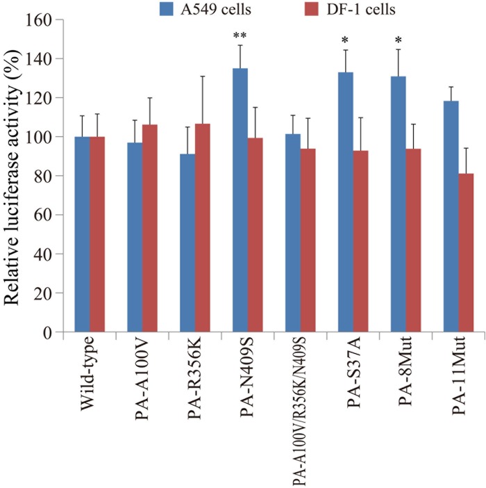
Since PA is one of the components of the viral replication complex, we evaluated viral polymerase activity in human A549 and avian DF-1 cells by using a luciferase-based mini-replicon assay, as previously described (24, 25). A549 or DF-1 cells were transfected with viral protein expression plasmids for NP, PB1, PB2, and PA or its mutants (i.e., PA-A100V, PA-R356K, PA-N409S, PA-A100V/R356K/N409S, PA-S37A, PA-8Mut, or PA-11Mut), a plasmid expressing viral RNA encoding firefly luciferase, and pRL-null, which expresses Renilla luciferase, as a transfection control. At 24 h posttransfection, viral polymerase activities were measured by using the dual-luciferase assay (Fig. 2). In human A549 cells, PA-N409S, PA-S37A, and PA-8Mut moderately increased the viral polymerase activity (P < 0.01, P < 0.05, and P < 0.05, respectively). The viral polymerase activity was not, however, affected by the other mutations tested. In avian DF-1 cells, the viral polymerase activity was not affected by any of the mutations tested. These results indicate that some of the PA amino acid changes identified through sequence alignments and ancestral inference analysis affect Anhui/1 polymerase activity in a host-specific manner.
FIG 2.
Viral polymerase activity in vitro. A549 or DF-1 cells were transfected with plasmids encoding the PB2, PB1, NP, and wild-type or mutant PA proteins, with a plasmid for the expression of a virus-like RNA encoding the firefly luciferase gene, and with a control plasmid encoding Renilla luciferase. At 24 h posttransfection, firefly and Renilla luciferase activities were measured by means of a dual-luciferase assay. Polymerase activity was calculated by normalization of the firefly luciferase activity to the Renilla luciferase activity. The data are shown as the relative polymerase activity with the standard deviation (SD; n = 3). The polymerase activity of the wild-type PA was set to 100%. * and **, P < 0.05 and P < 0.01, respectively, according to a one-way analysis of variance (ANOVA), followed by a Dunnett's test.
Growth kinetics of PA mutant viruses in A549 and DF-1 cells.
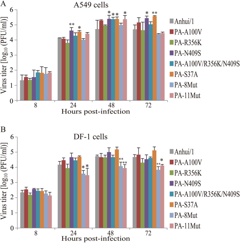
Next, we compared the viral growth kinetics of the PA mutant viruses with those of Anhui/1 in human A549 and avian DF-1 cells. Cells were infected with Anhui/1 or Anhui/1-PA mutant viruses at an MOI of 0.001 and then incubated at 37 or 39°C, respectively. At 8, 24, 48, and 72 h postinfection, virus titers in the cell culture media were determined in MDCK cells (Fig. 3). In A549 cells, the PA-N409S and PA-S37A viruses grew better than did Anhui/1, except at 8 h postinfection. At 48 h postinfection, the PA-A100V/R356K/N409S and PA-11Mut viruses also displayed increased replicative ability compared to Anhui/1. In DF-1 cells, the PA-8Mut and PA-11Mut viruses grew less efficiently than did Anhui/1 at 24, 48, and 72 h postinfection. The other mutants replicated with similar kinetics to Anhui/1.
FIG 3.
Growth kinetics of mutant viruses in vitro. A549 (A) or DF-1 (B) cells were infected with the indicated viruses at an MOI of 0.001. The supernatants of the infected cells were collected at the indicated time points. Virus titers were determined by use of a plaque assay in MDCK cells. The virus titers are means ± the SD (n = 3). * and **, P < 0.05 and P < 0.01, respectively, according to a one-way ANOVA, followed by a Dunnett's test.
Replicative ability and pathogenicity of PA mutant viruses in a mouse model.
To assess the importance of the selected PA mutations in vivo, we compared the virulence of the PA mutant viruses in a mouse model. First, to evaluate replicative ability in vivo, we compared virus titers in nose tissue, lungs, brains, spleens, kidneys, livers, and colons of infected mice. At 3 days postinoculation, the virus titers in the nose tissue and lungs of mice infected with the PA-8Mut virus were significantly higher than those in the nose tissue and lungs of mice infected with Anhui/1 (P < 0.01) (Table 2). The PA-8Mut virus was also recovered from the brain of a mouse at 101.71 PFU/g (data not shown). At 6 days postinoculation, the virus titers in the lungs of mice infected with the PA-S37A virus were significantly lower than those in the lungs of mice infected with Anhui/1 (P < 0.05). In addition, the PA-11Mut virus was recovered from the brain of a mouse at 101.87 PFU/g (data not shown). The titers of all other viruses tested here were not statistically different from those obtained for the wild-type Anhui/1 virus. With the exceptions listed above, the viruses tested here did not spread beyond the respiratory organs.
TABLE 2.
Average virus titers in the organs of infected micea
| Virus | Avg titer (mean log10 PFU ± SD/g) |
|||
|---|---|---|---|---|
| Day 3 |
Day 6 |
|||
| Nose tissue | Lung | Nose tissue | Lung | |
| Anhui/1 | 6.05 ± 0.42 | 6.89 ± 0.23 | 6.20 ± 0.74 | 5.95 ± 0.09 |
| PA-A100V | 6.49 ± 0.23 | 6.98 ± 0.07 | 5.76 ± 0.39 | 6.55 ± 0.51 |
| PA-R356K | 6.21 ± 0.17 | 6.62 ± 0.26 | 5.93 ± 0.17 | 5.69 ± 0.14 |
| PA-N409S | 6.29 ± 0.15 | 7.05 ± 0.04 | 6.04 ± 0.40 | 5.75 ± 0.16 |
| PA-A100V/R356K/N409S | 6.33 ± 0.09 | 7.27 ± 0.19 | 5.80 ± 0.66 | 6.57 ± 0.26 |
| PA-S37A | 5.97 ± 0.53 | 7.15 ± 0.21 | 5.63 ± 0.34 | 5.17 ± 0.24* |
| PA-8Mut | 7.23 ± 0.22** | 7.81 ± 0.45** | 6.61 ± 0.58 | 6.40 ± 0.21 |
| PA-11Mut | 6.43 ± 0.33 | 7.36 ± 0.22 | 6.78 ± 0.67 | 6.28 ± 0.44 |
Mice were inoculated with 106 PFU of each virus intranasally. Three animals per group were euthanized on days 3 and 6 postinfection. Statistically significant differences in the virus titers of Anhui/1-infected mice and of mice infected with the other viruses were assessed by use of a one-way ANOVA, followed by a Dunnett's test (*, P < 0.05; **, P < 0.01). No virus was recovered from the brains, spleens, kidneys, livers, or colons of infected animals, except on days 3 and 6 postinfection from the brain of one mouse each infected with PA-8Mut (1.71 log10 PFU/g) or PA-11Mut (1.87 log10 PFU/g), respectively.
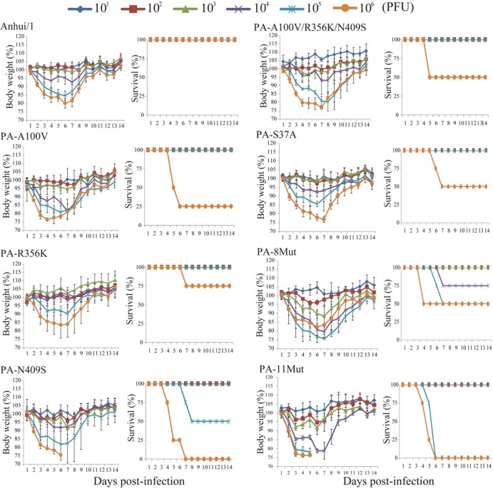
Second, we evaluated the pathogenicity of the mutants in mice. Four mice per group were intranasally infected with the indicated virus at 101, 102, 103, 104, 105, and 106 PFU. Body weight and mortality of each infected mouse were observed daily for 14 days (Fig. 4). Although the original Anhui/1 virus (8), which contains a mixture of at least five variants of HA, was lethal to mice, the reverse genetics Anhui/1 virus used here (which encodes an HA consensus sequence) did not kill mice, even at the highest dose tested. Infection of mice with PA-A100V, -R356K, -A100V/R356K/N409S, or -S37A killed some of the animals infected with 106 PFU, resulting in slightly higher mouse 50% lethal dose values (MLD50; Table 3); however, these differences were not statistically significant. Three viruses, PA-N409S, -8Mut, and -11Mut, caused the deaths of a subset of animals infected with 105 PFU of viruses (or 104 PFU for PA-8Mut), leading to statistically lower MLD50 values (Table 3); hence, these three viruses were more virulent in mice than was the control Anhui/1 virus generated by using reverse genetics. However, none of the mutant viruses tested here possessed an MLD50 value higher than that of the original Anhui/1 isolate (MLD50, 103.5 PFU/ml [8]).
FIG 4.
Virulence of mutant viruses in mice. Four mice per group were intranasally inoculated with 101, 102, 103, 104, 105, or 106 PFU (each in 50 μl) of the indicated viruses. Body weight and survival were monitored daily. Mice that lost >25% of their baseline weight were euthanized. The values represent the average of body weight compared to the baseline weight ± the SD from four mice.
TABLE 3.
MLD50 values of mutant virusesa
| Virus | MLD50b (log10 PFU) |
|---|---|
| Anhui/1 | >6.5 |
| PA-A100V | 5.7 |
| PA-R356K | 6.3 |
| PA-N409S | 5.0 |
| PA-A100V/R356K/N409S | 6.0 |
| PA-S37A | 6.0 |
| PA-8Mut | 5.3 |
| PA-11Mut | 4.5 |
Mice were infected with 101, 102, 103, 104, 105, or 106 PFU.
MLD50, 50% mouse lethal dose.
Here, we have identified several amino acid changes in A(H7N9) PA proteins that may have provided functional benefits that allow A(H7N9) viruses to replicate in mammals. Recent studies suggest that increases in replicative ability may be critical to allow avian influenza virus to replicate in mammals (16–19, 34). Back-mutations to avian virus-type or commonly found amino acids at a particular position may thus be expected to reduce the replicative ability of these viruses in mammals. Interestingly, we observed the opposite trend: for example, back-mutation of the human virus-characteristic PA-409N to the avian virus-characteristic PA-409S residue resulted in slightly increased replication and/or virulence in human cells and/or mice in the background of Anhui/1. On the other hand, back-mutation to amino acids commonly found among avian H9N2 viruses reduced the replicative ability of those viruses in avian cells (see PA-8Mut and PA-11Mut in DF-1 cells). These findings suggest that an ancestor of the A(H7N9) PA genes accumulated several amino acid changes that increased its replicative ability in avian cells but decreased its replicative capacity in mammalian systems; nonetheless, A(H7N9) viruses possessing such a mammalian-adapted PA replicate efficiently in mammals. We recently found the same phenomenon for the NS gene of H5N1 highly pathogenic avian influenza viruses (35), which possesses two mutations that synergistically attenuate the H5N1 virulence in mammalian hosts. Collectively, these data indicate that avian influenza virus replication in mammals is associated with mutations that increase (for example, PB2-627K) or decrease (for example, some of the PA mutations tested here) virus replicative ability in mammals; in the latter case, we speculate that the ancestral virus may have been “too aggressive” in mammals.
The most significant increases in virulence in mice were detected for the PA-11Mut, PA-8Mut, and PA-N409S viruses (Table 3). PA-8Mut and PA-11Mut showed slightly increased replicative ability in minireplicon assays, cultured cells, and/or mice, although not all values reached statistical significance. Among the eight amino acid changes shared by these mutants, PA-N409S may be the most important since this mutation, when tested individually, increased the polymerase activity as measured by minireplicon assays and viral growth in human cells. In mice, the PA-N409S virus replicated to slightly higher titers on day 3 but slightly lower titers on day 6 compared to Anhui/1; however, these differences were not statistically significant. Several computational studies have described PA-409S as avian-, and PA-409N as human-virus specific (5, 20, 30–32); as discussed earlier, we found that the avian virus-type amino acid (PA-409S) increased virus replicative ability in mammalian systems, in contrast to what may have been expected. Inspection of the X-ray crystallographic structure of PA amino acids 239 to 716 in complex with the 15 N-terminal amino acids of PB1 (PDB 2ZNL) (36) revealed a likely interaction between PA-409 and the PB1 N-terminal region. Mutations at PA-409 may therefore alter this interaction with PB1 and through this mechanism affect virus replication and virulence. Two other amino acid changes that were predicted to be host species-specific (valine or alanine at position 100 and arginine or lysine at position 356) did not show statistically significant effects on replication or virulence in the background of Anhui/1.
In summary, we identified several amino acids in A(H7N9) PA proteins that affect virus replicative ability and virulence in mice in the background of a prototype A(H7N9) virus. Some of these amino acid changes attenuated an A(H7N9) virus in human cells and in mice.
ACKNOWLEDGMENTS
We thank Yuelong Shu (WHO Collaborating Center for Reference and Research on Influenza, Chinese National Influenza Center, and National Institute for Viral Disease Control and Prevention China CDC, Beijing, People's Republic of China) for providing A/Anhui/1/2013 (H7N9) viruses. We also thank Hiroaki Katsura and Reina Yamaji for assistance with experiments, Hrishikesh Lokhande of the Los Alamos National Laboratory for valuable computer programming support, and Susan Watson for editing the manuscript.
This study was supported by the Japan Initiative for Global Research Network on Infectious Diseases from the Ministry of Education, Culture, Sports, Science, and Technology of Japan, by grants-in-aid from the Ministry of Health, Labor, and Welfare, Japan, by ERATO (Japan Science and Technology Agency), and by a NIAID-funded Center for Research on Influenza Pathogenesis grant (HHSN266200700010C).
This study was approved by the local Institutional Biosafety Committed (IBC); in addition, the IBC and NIAID evaluated this study and concluded that it does not involve Dual Use Research or Concern (DURC).
Footnotes
Published ahead of print 26 December 2013
REFERENCES
- 1.Gao R, Cao B, Hu Y, Feng Z, Wang D, Hu W, Chen J, Jie Z, Qiu H, Xu K, Xu X, Lu H, Zhu W, Gao Z, Xiang N, Shen Y, He Z, Gu Y, Zhang Z, Yang Y, Zhao X, Zhou L, Li X, Zou S, Zhang Y, Li X, Yang L, Guo J, Dong J, Li Q, Dong L, Zhu Y, Bai T, Wang S, Hao P, Yang W, Zhang Y, Han J, Yu H, Li D, Gao GF, Wu G, Wang Y, Yuan Z, Shu Y. 2013. Human infection with a novel avian-origin influenza A (H7N9) virus. N. Engl. J. Med. 368:1888–1897. 10.1056/NEJMoa1304459 [DOI] [PubMed] [Google Scholar]
- 2.Li Q, Zhou L, Zhou M, Chen Z, Li F, Wu H, Xiang N, Chen E, Tang F, Wang D, Meng L, Hong Z, Tu W, Cao Y, Li L, Ding F, Liu B, Wang M, Xie R, Gao R, Li X, Bai T, Zou S, He J, Hu J, Xu Y, Chai C, Wang S, Gao Y, Jin L, Zhang Y, Luo H, Yu H, Gao L, Pang X, Liu G, Shu Y, Yang W, Uyeki TM, Wang Y, Wu F, Feng Z. 24 April 2013. Preliminary report: epidemiology of the avian influenza A (H7N9) outbreak in China. N. Engl. J. Med. 10.1056/NEJMoa1304617. [DOI] [Google Scholar]
- 3.Kageyama T, Fujisaki S, Takashita E, Xu H, Yamada S, Uchida Y, Neumann G, Saito T, Kawaoka Y, Tashiro M. 2013. Genetic analysis of novel avian A(H7N9) influenza viruses isolated from patients in China, February to April 2013. Euro Surveill. 18(15):pii=20453 http://www.eurosurveillance.org/ViewArticle.aspx?ArticleId=20453 [PMC free article] [PubMed] [Google Scholar]
- 4.Shi J, Deng G, Liu P, Zhou J, Guan L, Li W, Li X, Guo J, Wang G, Fan J, Wang J, Li Y, Jiang Y, Liu L, Tian G, Li C, Chen H. 2013. Isolation and characterization of H7N9 viruses from live poultry markets: implication of the source of current H7N9 infection in humans. Chin. Sci. Bull. 58:1857–1863. 10.1007/s11434-013-5873-4 [DOI] [Google Scholar]
- 5.Liu Q, Lu L, Sun Z, Chen GW, Wen Y, Jiang S. 2013. Genomic signature and protein sequence analysis of a novel influenza A (H7N9) virus that causes an outbreak in humans in China. Microbes Infect. 15:432–439. 10.1016/j.micinf.2013.04.004 [DOI] [PubMed] [Google Scholar]
- 6.Lam TT, Wang J, Shen Y, Zhou B, Duan L, Cheung CL, Ma C, Lycett SJ, Leung CY, Chen X, Li L, Hong W, Chai Y, Zhou L, Liang H, Ou Z, Liu Y, Farooqui A, Kelvin DJ, Poon LL, Smith DK, Pybus OG, Leung GM, Shu Y, Webster RG, Webby RJ, Peiris JS, Rambaut A, Zhu H, Guan Y. 2013. The genesis and source of the H7N9 influenza viruses causing human infections in China. Nature 502:241–244. 10.1038/nature12515 [DOI] [PMC free article] [PubMed] [Google Scholar]
- 7.Chen Y, Liang W, Yang S, Wu N, Gao H, Sheng J, Yao H, Wo J, Fang Q, Cui D, Li Y, Yao X, Zhang Y, Wu H, Zheng S, Diao H, Xia S, Zhang Y, Chan KH, Tsoi HW, Teng JL, Song W, Wang P, Lau SY, Zheng M, Chan JF, To KK, Chen H, Li L, Yuen KY. 2013. Human infections with the emerging avian influenza A H7N9 virus from wet market poultry: clinical analysis and characterisation of viral genome. Lancet 381:1916–1925. 10.1016/S0140-6736(13)60903-4 [DOI] [PMC free article] [PubMed] [Google Scholar]
- 8.Watanabe T, Kiso M, Fukuyama S, Nakajima N, Imai M, Yamada S, Murakami S, Yamayoshi S, Iwatsuki-Horimoto K, Sakoda Y, Takashita E, McBride R, Noda T, Hatta M, Imai H, Zhao D, Kishida N, Shirakura M, de Vries RP, Shichinohe S, Okamatsu M, Tamura T, Tomita Y, Fujimoto N, Goto K, Katsura H, Kawakami E, Ishikawa I, Watanabe S, Ito M, Sakai-Tagawa Y, Sugita Y, Uraki R, Yamaji R, Eisfeld AJ, Zhong G, Fan S, Ping J, Maher EA, Hanson A, Uchida Y, Saito T, Ozawa M, Neumann G, Kida H, Odagiri T, Paulson JC, Hasegawa H, Tashiro M, Kawaoka Y. 2013. Characterization of H7N9 influenza A viruses isolated from humans. Nature 501:551–555. 10.1038/nature12392 [DOI] [PMC free article] [PubMed] [Google Scholar]
- 9.Zhu H, Wang D, Kelvin DJ, Li L, Zheng Z, Yoon SW, Wong SS, Farooqui A, Wang J, Banner D, Chen R, Zheng R, Zhou J, Zhang Y, Hong W, Dong W, Cai Q, Roehrl MH, Huang SS, Kelvin AA, Yao T, Zhou B, Chen X, Leung GM, Poon LL, Webster RG, Webby RJ, Peiris JS, Guan Y, Shu Y. 2013. Infectivity, transmission, and pathology of human-isolated H7N9 influenza virus in ferrets and pigs. Science 341:183–186. 10.1126/science.1239844 [DOI] [PubMed] [Google Scholar]
- 10.Mok CK, Lee HH, Chan MC, Sia SF, Lestra M, Nicholls JM, Zhu H, Guan Y, Peiris JM. 2013. Pathogenicity of the novel A/H7N9 influenza virus in mice. mBio 4:e00362-13. 10.1128/mBio.00362-13 [DOI] [PMC free article] [PubMed] [Google Scholar]
- 11.Zhou J, Wang D, Gao R, Zhao B, Song J, Qi X, Zhang Y, Shi Y, Yang L, Zhu W, Bai T, Qin K, Lan Y, Zou S, Guo J, Dong J, Dong L, Zhang Y, Wei H, Li X, Lu J, Liu L, Zhao X, Li X, Huang W, Wen L, Bo H, Xin L, Chen Y, Xu C, Pei Y, Yang Y, Zhang X, Wang S, Feng Z, Han J, Yang W, Gao GF, Wu G, Li D, Wang Y, Shu Y. 2013. Biological features of novel avian influenza A (H7N9) virus. Nature 499:500–503. 10.1038/nature12379 [DOI] [PubMed] [Google Scholar]
- 12.Belser JA, Gustin KM, Pearce MB, Maines TR, Zeng H, Pappas C, Sun X, Carney PJ, Villanueva JM, Stevens J, Katz JM, Tumpey TM. 2013. Pathogenesis and transmission of avian influenza A (H7N9) virus in ferrets and mice. Nature 501:556–559. 10.1038/nature12391 [DOI] [PMC free article] [PubMed] [Google Scholar]
- 13.Zhang Q, Shi J, Deng G, Guo J, Zeng X, He X, Kong H, Gu C, Li X, Liu J, Wang G, Chen Y, Liu L, Liang L, Li Y, Fan J, Wang J, Li W, Guan L, Li Q, Yang H, Chen P, Jiang L, Guan Y, Xin X, Jiang Y, Tian G, Wang X, Qiao C, Li C, Bu Z, Chen H. 2013. H7N9 influenza viruses are transmissible in ferrets by respiratory droplet. Science 341:410–414. 10.1126/science.1240532 [DOI] [PubMed] [Google Scholar]
- 14.Richard M, Schrauwen EJ, de Graaf M, Bestebroer TM, Spronken MI, van Boheemen S, de Meulder D, Lexmond P, Linster M, Herfst S, Smith DJ, van den Brand JM, Burke DF, Kuiken T, Rimmelzwaan GF, Osterhaus AD, Fouchier RA. 2013. Limited airborne transmission of H7N9 influenza A virus between ferrets. Nature 501:560–563. 10.1038/nature12476 [DOI] [PMC free article] [PubMed] [Google Scholar]
- 15.Rogers GN, Paulson JC, Daniels RS, Skehel JJ, Wilson IA, Wiley DC. 1983. Single amino acid substitutions in influenza haemagglutinin change receptor binding specificity. Nature 304:76–78. 10.1038/304076a0 [DOI] [PubMed] [Google Scholar]
- 16.Hatta M, Gao P, Halfmann P, Kawaoka Y. 2001. Molecular basis for high virulence of Hong Kong H5N1 influenza A viruses. Science 293:1840–1842. 10.1126/science.1062882 [DOI] [PubMed] [Google Scholar]
- 17.Subbarao EK, Kawaoka Y, Murphy BR. 1993. Rescue of an influenza A virus wild-type PB2 gene and a mutant derivative bearing a site-specific temperature-sensitive and attenuating mutation. J. Virol. 67:7223–7228 [DOI] [PMC free article] [PubMed] [Google Scholar]
- 18.Gabriel G, Dauber B, Wolff T, Planz O, Klenk HD, Stech J. 2005. The viral polymerase mediates adaptation of an avian influenza virus to a mammalian host. Proc. Natl. Acad. Sci. U. S. A. 102:18590–18595. 10.1073/pnas.0507415102 [DOI] [PMC free article] [PubMed] [Google Scholar]
- 19.Li Z, Chen H, Jiao P, Deng G, Tian G, Li Y, Hoffmann E, Webster RG, Matsuoka Y, Yu K. 2005. Molecular basis of replication of duck H5N1 influenza viruses in a mammalian mouse model. J. Virol. 79:12058–12064. 10.1128/JVI.79.18.12058-12064.2005 [DOI] [PMC free article] [PubMed] [Google Scholar]
- 20.Chen GW, Chang SC, Mok CK, Lo YL, Kung YN, Huang JH, Shih YH, Wang JY, Chiang C, Chen CJ, Shih SR. 2006. Genomic signatures of human versus avian influenza A viruses. Emerg. Infect. Dis. 12:1353–1360. 10.3201/eid1209.060276 [DOI] [PMC free article] [PubMed] [Google Scholar]
- 21.Dias A, Bouvier D, Crepin T, McCarthy AA, Hart DJ, Baudin F, Cusack S, Ruigrok RW. 2009. The cap-snatching endonuclease of influenza virus polymerase resides in the PA subunit. Nature 458:914–918. 10.1038/nature07745 [DOI] [PubMed] [Google Scholar]
- 22.Neumann G, Watanabe T, Ito H, Watanabe S, Goto H, Gao P, Hughes M, Perez DR, Donis R, Hoffmann E, Hobom G, Kawaoka Y. 1999. Generation of influenza A viruses entirely from cloned cDNAs. Proc. Natl. Acad. Sci. U. S. A. 96:9345–9350. 10.1073/pnas.96.16.9345 [DOI] [PMC free article] [PubMed] [Google Scholar]
- 23.Niwa H, Yamamura K, Miyazaki J. 1991. Efficient selection for high-expression transfectants with a novel eukaryotic vector. Gene 108:193–199. 10.1016/0378-1119(91)90434-D [DOI] [PubMed] [Google Scholar]
- 24.Ozawa M, Fujii K, Muramoto Y, Yamada S, Yamayoshi S, Takada A, Goto H, Horimoto T, Kawaoka Y. 2007. Contributions of two nuclear localization signals of influenza A virus nucleoprotein to viral replication. J. Virol. 81:30–41. 10.1128/JVI.01434-06 [DOI] [PMC free article] [PubMed] [Google Scholar]
- 25.Murakami S, Horimoto T, Yamada S, Kakugawa S, Goto H, Kawaoka Y. 2008. Establishment of canine RNA polymerase I-driven reverse genetics for influenza A virus: its application for H5N1 vaccine production. J. Virol. 82:1605–1609. 10.1128/JVI.01876-07 [DOI] [PMC free article] [PubMed] [Google Scholar]
- 26.Larkin MA, Blackshields G, Brown NP, Chenna R, McGettigan PA, McWilliam H, Valentin F, Wallace IM, Wilm A, Lopez R, Thompson JD, Gibson TJ, Higgins DG. 2007. CLUSTAL W and CLUSTAL X version 2.0. Bioinformatics 23:2947–2948. 10.1093/bioinformatics/btm404 [DOI] [PubMed] [Google Scholar]
- 27.Miller MA, Pfeiffer W, Schwartz T. 2010. Creating the CIPRES Science Gateway for inference of large phylogenetic trees, p 1–8 Proceedings of the Gateway Computing Environments Workshop, New Orleans, LA [Google Scholar]
- 28.Guindon S, Dufayard JF, Lefort V, Anisimova M, Hordijk W, Gascuel O. 2010. New algorithms and methods to estimate maximum-likelihood phylogenies: assessing the performance of PhyML 3.0. Syst. Biol. 59:307–321. 10.1093/sysbio/syq010 [DOI] [PubMed] [Google Scholar]
- 29.Yang Z. 2007. PAML 4: phylogenetic analysis by maximum likelihood. Mol. Biol. Evol. 24:1586–1591. 10.1093/molbev/msm088 [DOI] [PubMed] [Google Scholar]
- 30.Finkelstein DB, Mukatira S, Mehta PK, Obenauer JC, Su X, Webster RG, Naeve CW. 2007. Persistent host markers in pandemic and H5N1 influenza viruses. J. Virol. 81:10292–10299. 10.1128/JVI.00921-07 [DOI] [PMC free article] [PubMed] [Google Scholar]
- 31.Miotto O, Heiny AT, Albrecht R, Garcia-Sastre A, Tan TW, August JT, Brusic V. 2010. Complete-proteome mapping of human influenza A adaptive mutations: implications for human transmissibility of zoonotic strains. PLoS One 5:e9025. 10.1371/journal.pone.0009025 [DOI] [PMC free article] [PubMed] [Google Scholar]
- 32.Shaw M, Cooper L, Xu X, Thompson W, Krauss S, Guan Y, Zhou N, Klimov A, Cox N, Webster R, Lim W, Shortridge K, Subbarao K. 2002. Molecular changes associated with the transmission of avian influenza a H5N1 and H9N2 viruses to humans. J. Med. Virol. 66:107–114. 10.1002/jmv.2118 [DOI] [PubMed] [Google Scholar]
- 33.Xu W, Sun Z, Liu Q, Xu J, Jiang S, Lu L. 2013. PA-356R is a unique signature of the avian influenza A (H7N9) viruses with bird-to-human transmissibility: potential implication for animal surveillances. J. Infect. 67:490–494. 10.1016/j.jinf.2013.08.001 [DOI] [PubMed] [Google Scholar]
- 34.Manz B, Schwemmle M, Brunotte L. 2013. Adaptation of avian influenza A virus polymerase in mammals to overcome the host species barrier. J. Virol. 87:7200–7209. 10.1128/JVI.00980-13 [DOI] [PMC free article] [PubMed] [Google Scholar]
- 35.Fan S, Macken CA, Li C, Ozawa M, Goto H, Iswahyudi NF, Nidom CA, Chen H, Neumann G, Kawaoka Y. 2013. Synergistic effect of the PDZ and p85β-binding domains of the NS1 protein on virulence of an avian H5N1 influenza A virus. J. Virol. 87:4861–4871. 10.1128/JVI.02608-12 [DOI] [PMC free article] [PubMed] [Google Scholar]
- 36.Obayashi E, Yoshida H, Kawai F, Shibayama N, Kawaguchi A, Nagata K, Tame JR, Park SY. 2008. The structural basis for an essential subunit interaction in influenza virus RNA polymerase. Nature 454:1127–1131. 10.1038/nature07225 [DOI] [PubMed] [Google Scholar]


