Abstract
Purpose
To assess the reproducibility of circumpapillary retinal nerve fiber layer (cpRNFL) thickness measurement (measurement agreement) and its color-coded classification (classification agreement) by Cirrus spectral domain optical coherence tomography (OCT) in pseudophakic eyes.
Methods
Two-hundred five participants having glaucoma or glaucoma suspected eyes underwent two repeated Cirrus OCT scans to measure cpRNFL thickness (optic disc cube 200 × 200). After classifying participants into three different groups according to their lens status (clear media, cataract, and pseudophakic), values of intra-class coefficient (ICC), coefficient of variance, and test-retest variability were compared between groups for average retinal nerve fiber layer (RNFL) thicknesses and that corresponding to four quadrant maps. Linear weighted kappa coefficients were calculated as indicators of agreement of color code classification in each group.
Results
ICC values were all excellent (generally defined as 0.75 to 1.00) for the average and quadrant RNFL thicknesses in all three groups. ICC values of the clear media group tended to be higher than those in the cataract and pseudophakic groups for all quadrants and average thickness. Especially in the superior and nasal quadrants, the ICC value of the cataract group was significantly lower than that of the clear media and pseudophakic groups. For average RNFL thickness, classification agreement (kappa) in three groups did not show a statistically significant difference. For quadrant maps, classification agreement (kappa) in the clear media group was higher than those in the other two groups.
Conclusions
Agreement of cpRNFL measurement and its color code classification between two repeated Cirrus OCT scans in pseudophakic eyes was as good as that in eyes with clear crystalline lens. More studies are required to ascertain the effect of lens status on the reproducibility of Cirrus OCT according to different stages of glaucoma patients.
Keywords: Optical coherence tomography, Pseudophakia, Reproducibility of results
Optical coherence tomography (OCT) has become a popular means of diagnosing and determining progression of glaucoma as it allows noninvasive imaging of the retinal nerve fiber layer (RNFL). Cirrus spectral domain (SD) OCT (software version 4.0.1; Carl Zeiss Meditec, Dublin, CA, USA) utilizes SD OCT technology to acquire OCT data with better resolution (5 µm compared with approximately 10 µm axial resolution in tissue) about 70 times faster (27,000 vs. 400 A scans/sec) than time-domain (TD) OCT technology. Previous studies confirmed that peripapillary RNFL thickness measurements by Cirrus SD OCT were very reproducible both in healthy and glaucomatous eyes [1,2].
When analyzing circumpapillary RNFL (cpRNFL) thickness in glaucoma patients, reproducibility is an important reliability index, regardless of the imaging technique used. There have been many potential explanations for variability in cpRNFL thickness measurements using OCT. Factors such as cataract, signal strength (SS), pupil size, type of scan, the quadrant measured, and the thickness of RNFL all may influence the overall scan quality and calculated RNFL thickness [3,4,5,6,7]. Youm et al. [8] reported that the average SS and relative SS change were correlated with the reproducibility in RNFL thickness measurement using Stratus OCT, and glaucomatous eyes tend to be more variable than normal eyes.
Glaucoma and cataract are frequent causes of vision loss among the elderly, and these conditions often coexist [9]. Cataract is a common cause of media opacity that can affect the quality of diagnostic imaging for glaucoma in the elderly. Although prior studies reported that the presence of cataract and cataract surgery might significantly affect the RNFL thickness measurement by OCT [10], no prior study has simultaneously compared the reproducibility of RNFL thickness measurement by OCT in pseudophakic eyes with those in clear media and cataract eyes.
Thus, the current study aims to assess the reproducibility of RNFL thickness measurement and its color code classification using Cirrus SD OCT in pseudophakic eyes, and then compare the obtained results with those in clear media and cataract eyes simultaneously.
Materials and Methods
Glaucoma and glaucoma suspect patients were consecutively enrolled from the Glaucoma-Cataract Clinic of Siloam Eye Hospital from March 2012 to May 2012. The study was approved by the institutional review board and ethics committee of Siloam Eye Hospital, and complied using the tenets of the Declaration of Helsinki. Informed consent was obtained from all participants prior to their participation in the study.
Upon initial evaluation, all participants underwent a complete ophthalmologic examination including best-corrected visual acuity (BCVA), intraocular pressure measurement using a Goldmann applanation tonometer, and slit lamp and fundus examination. Perimetry was performed by experienced technicians using the Humphrey Visual Field Analyzer (Cirrus OCT, Carl Zeiss Meditec) with the 30-2 SITA Standard algorithm. All cataracts were staged according to the slit lamp-based Lens Opacities Classification System (LOCS) III.
Participants included those with a diagnosis of any form of glaucoma or glaucoma suspect with an age greater than 60 years. Glaucomatous eyes were defined as those having glaucomatous visual field (VF) defects confirmed by at least two reliable VF examinations and the presence of a compatible glaucomatous optic disc that showed increased cupping (a vertical cup-disc ratio >0.7), a difference in vertical cup-disc ratio of >0.2 between eyes, diffuse or focal neural rim thinning, disc hemorrhage, or RNFL defects. A glaucomatous VF defect was defined as having three or more significant (p < 0.05) contiguous points with at least one at the p < 0.01 level on the same side of the horizontal meridian in the pattern deviation plot, classified as outside normal limits using the glaucoma hemifield test. Glaucoma-suspect eyes were defined as those with glaucomatous optic disc, but showing normal VF test data. Individuals with 1) any other ocular pathology except glaucoma and cataract, 2) refractive error >5.0 or <-5.0 diopters, 3) history of ocular trauma or surgery (except for uncomplicated cataract surgery), 4) recent history of cataract surgery (<6 months prior to the examination), or 5) presence of posterior capsular opacity or continuous curvilinear capsulorhexis contracture were excluded from this study.
All participants were categorized into three groups according to their lens status. Patients with a degree of cataract more than grade II by LOCS classification were grouped as the cataract group, and patients who had received uncomplicated cataract surgery with "in the bag" implantation of a Biovue (Ophthalmic Innovations International, Ontario, CA, USA) intraocular lens were grouped as the pseudophakic group. Participants with clear media were classified as clear media group.
All individuals were evaluated with the Cirrus SD OCT (software version 4.0.1, Carl Zeiss Meditec) to measure cpRNFL thickness (optic disc cube 200 × 200 scan) twice during the same day, with short breaks between each measurement. The OCT scan was performed by a single well-trained technician through a dilated pupil. Between each scan, the patient was asked to sit back and rest for a few minutes before proceeding to the next scan. The technician applied artificial tears before the subsequent scan, and then repositioned the participants.
Optic disc cube protocol generates a cube of data through a 6 mm square grid by acquiring a series of 200 horizontal scan lines each composed of 200 A-scans. For analysis, Cirrus algorithms identify the center of the optic disc and automatically place a calculation circle of 3.46 mm diameter around it. The anterior and posterior margins of the RNFL are delineated, and after extracting from the data cube 256 A-scan samples along the path of the calculation circle, the system calculates the cpRNFL thickness at each point on the circle. Scans with misalignment, segmentation failure, or decentration of the measurement circle were excluded from analysis. From the OCT database, the following data were collected: two repeated measures of cpRNFL thickness and their stoplight color code from quadrant maps, two repeated measures of average RNFL thickness with stoplight color classification, and SS. Scans with high SS values were defined as those with scores greater than or equal to 5 in two consecutive scans, while other scans were defined as scans with low SS (scans showing a SS of <5).
Statistical methods
When data from both eyes were eligible for analysis, only one eye from each patient was randomly selected and used for data analysis. No participant was excluded based on SS score.
The method employed to determine the reproducibility of RNFL thickness measurement between two repeated OCT scans (measurement agreement) was the intra-class correlation coefficient (ICC). Linear weighted kappa coefficients were calculated as indicators of agreement for RNFL thickness classification between two repeated OCT scans (classification agreement). When the number of categories was less than two by two, the result was marked as 'not available' due to insufficient number of categories to perform the classification agreement test. Coefficient of variation (CV) and test-retest variability were also computed to determine the reproducibility of the Cirrus OCT scan used in this study. The CV, expressed as a percentage (%), was calculated as the square root of the variance divided by the mean RNFL thickness of two repeated measures. Test-retest variability, measured in micrometers, was calculated as twice the square root of the variance among two repeated measures.
Inter-scan measurement difference, calculated by the RNFL thickness difference obtained between two repeated measures, was compared among groups using ANOVA and post-hoc multiple comparisons test.
To determine the effect of signal strength on Cirrus OCT reproducibility, we performed Pearson's correlation analysis. After we categorized participants into two different SS groups (high SS and low SS), we calculated ICC and kappa values for each group.
Data were analyzed using statistical software SPSS ver. 15.0 (SPSS Inc., Chicago, IL, USA) and R software ver. 2.10.1 (GNU General Public License). A p-value less than 0.05 was considered statistically significant.
Results
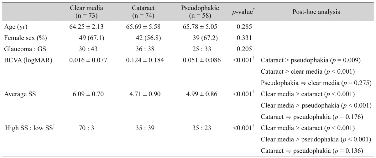
Of the 300 individuals initially enrolled in the study, 95 eyes were excluded due to segmentation error, or scan circle decentration, in any of the repeated Cirrus OCT scans. In total, 205 eyes of 205 individuals were included for the final analysis. Participants' demographic and ophthalmic characteristics are shown in Table 1.
Table 1.
Participant characteristics
GS = glaucoma suspect; BCVA = best-corrected visual acuity; logMAR = logarithm of the minimum angle of resolution; SS = signal strength.
'≒' indicates similar values without statistically significant difference between two groups.
*Value from ANOVA or chi-square test; †Pairwise comparison after ANOVA with Bonferroni correction; ‡High SS (SS ≥5 in two consecutive scans) and low SS (SS <5 in either scan).
After categorizing all participants into three groups (clear media, cataract, and pseudophakia), there were 73 eyes in the clear media group, 74 eyes in the cataract group, and 58 eyes in the pseudophakic group, respectively. Among those three groups, there was no statistically significant difference in relation to age, gender, and proportion of glaucoma or glaucoma suspect. Logarithm of the minimum angle of resolution BCVA in cataract group was significantly worse than that in the clear media group (p < 0.001) and pseudophakic group (p = 0.009). The average SS in the clear media group was significantly greater than that in the cataract (p < 0.001) and pseudophakic groups (p < 0.001). Although the average SS score in pseudophakic eyes (4.99 ± 0.86) was greater than that in cataract eyes (4.71 ± 0.90), the difference did not reach statistical significance (p = 0.176).
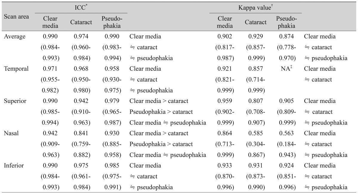
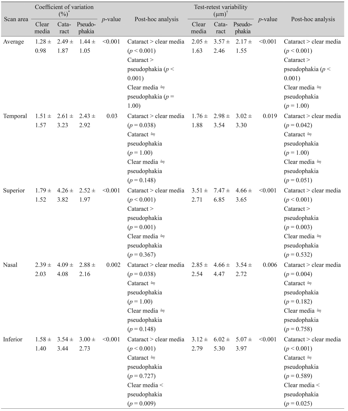
To compare the measurement reproducibility of RNFL thickness between the three groups, ICC, CV, and test-retest variability values were calculated. Also, linear weighted kappa coefficients were calculated as agreement indicators of the probability code results for RNFL thickness analysis. Those values are shown in Tables 2 and 3. ICC values were all excellent (generally defined as 0.75 to 1.00) in average and quadrant RNFL thicknesses for all three groups. Especially in the superior and nasal quadrants, the ICC value of the cataract group was significantly lower than that of the clear media and pseudopahkic groups. However, the ICC values of clear media and pseudophakic groups were similar. In addition, in terms of inferior, temporal quadrant, and average RNFL thickness, differences of ICC values among the three groups did not reach statistical significance. In terms of the classification agreement (kappa) also shown in Table 2, the differences among the three groups did not reach statistical significance in the superior, nasal, and inferior quadrants and average RNFL thickness, though kappa value from the pseudophakic group in the temporal quadrant was not available due to an insufficient number of categories to perform test. As shown in Table 3, the test/retest variability values of cataract group were significantly higher than those of the clear media group (in all quadrants including average RNFL thickness) as well as those of pseudophakic group (superior quadrant and average RNFL thickness). CV and test retest variability values of the pseudophakic group were similar to those of the clear media group in all scan areas, except the inferior quadrant.
Table 2.
ICC and Fleiss' generalized kappa of probability codes of circumpapillary retinal nerve fiber layer thickness measurements obtained using Cirrus optical coherence tomography
ICC = intra-class correlation coefficient; NA = not available.
'≒' indicates similar values without statistically significant difference between two groups.
*ICC values with 95% confidence interval in parenthesis, calculated with the two-way random effects model using the absolute agreement definition; †Mean on top with 95% confidence interval in parentheses; ‡Due to insufficient number of categories to perform test.
Table 3.
Coefficient of variation and test-retest variability of circumpapillary retinal nerve fiber layer thickness measurements with the Cirrus optical coherence tomography
'≒' indicates similar values without statistically significant difference between two groups.
*Calculated as the square root of the variance divided by the mean thickness of two repeated measures; †Calculated as two times the square root of the variance of two repeated measures.
Table 4 provides the comparison of the inter-scan measurement differences in RNFL thickness between three groups. Just like the CV and test retest variability, the inter-scan measurement difference value of the cataract group was significantly higher than those of the clear media group (temporal and nasal quadrant) or even than those of the pseudophakic group (superior quadrant and average RNFL thickness). Also, the inter-scan measurement difference value of the pseudophakic group was similar to those of the clear media group in all scan areas except the inferior quadrant.
Table 4.
Comparison of inter-scan measurement difference between two repeated Cirrus optical coherence tomography scans among three groups
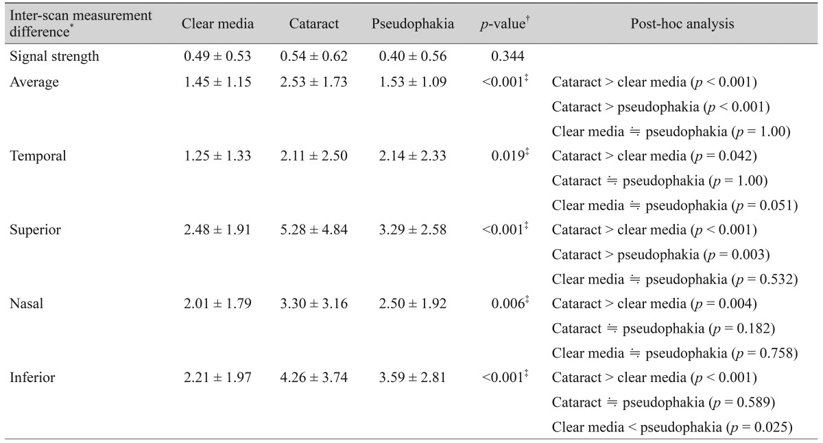
'≒' indicates similar values without statistically significant difference between two groups.
*Obtained by subtracting value of optical coherence tomography scan with lower one from that of scan with the greater value; †Value from ANOVA; ‡Pairwise comparison after ANOVA with Bonferroni correction.
The relationship between SS and inter-scan measurement difference with respect to RNFL thickness is summarized in Table 5. There was a significant negative correlation between mean SS of two repeated OCT scans and inter-scan measurement difference for superior (r = -0.184, p = 0.008), nasal (r = -0.174, p = 0.013), inferior (r = -0.152, p = 0.029), and average RNFL thickness (r = -0.242, p < 0.001), which means that the scans with lower signal strength tend to show increased measurement variability. In addition, the difference in SS, had a significant positive correlation with inter-scan measurement variation for all quadrants (p < 0.05) and average RNFL thickness (r = 0.426, p < 0.001). In other words, as the difference in SS between two repeated Cirrus OCT scans increases, the inter-scan measurement difference also increases.
Table 5.
Relationship between signal strength and inter-scan retinal nerve fiber layer thickness measurement difference of repeated optical coherence tomography scans

SS = signal strength.
*Obtained by obtaining the difference between optical coherence tomography scan values; †Correlation coefficient from Pearson correlation with p-value in parentheses.
As OCT manufacturers recommended that SS score of an acceptable image quality is 5 or higher, we categorized all participants into two groups according to their SS values, participants with high SS (SS ≥5 in two consecutive scans, n = 140) and those with low SS (SS <5 in either scan, n = 65), then calculated ICC and kappa values for each group (Table 6). In comparison with Table 2, the scan results with all SS showed some differences. Considering the scan results of only the high SS, the ICC value of clear media was significantly higher than that of the pseudophakic group in the temporal quadrant. In addition, the ICC value of the pseudophakic group showed no statistical difference from that of the cataract group in superior and nasal quadrants. Considering the low SS scan results, the reproducibility parameters (ICC, kappa value) showed no significant difference between cataract and pseudophakic groups and those in the clear group were not able to be calculated due to insufficient number of categories to perform test.
Table 6.
ICC and kappa values in each group classified by signal strength
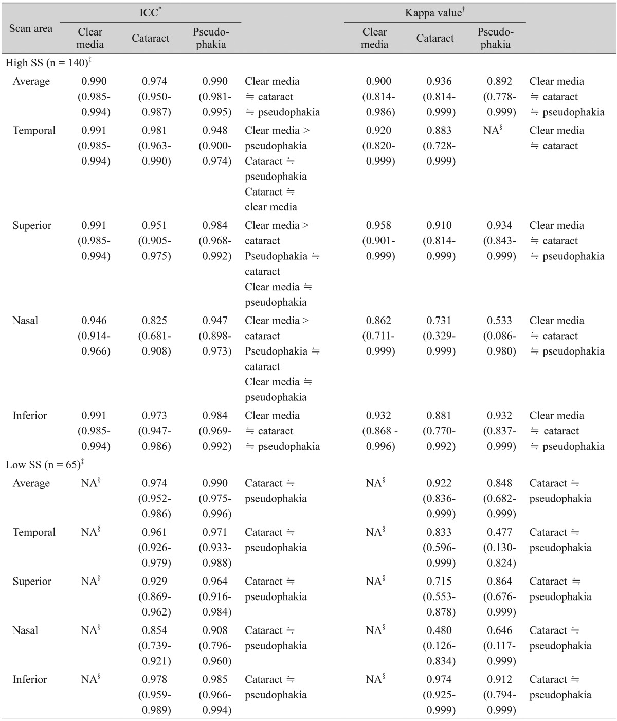
ICC = intra-class correlation coefficient; NA = not available.
'≒' indicates similar values without statistically significant difference between two groups.
*ICC values with 95% confidence interval in parenthesis, calculated with the two-way random effects model using the absolute agreement definition; †Mean on top with 95% confidence interval in parentheses; ‡High SS (SS ≥5 in two consecutive scans) and low SS (SS <5 in either scan); §Due to insufficient number of categories to perform test.
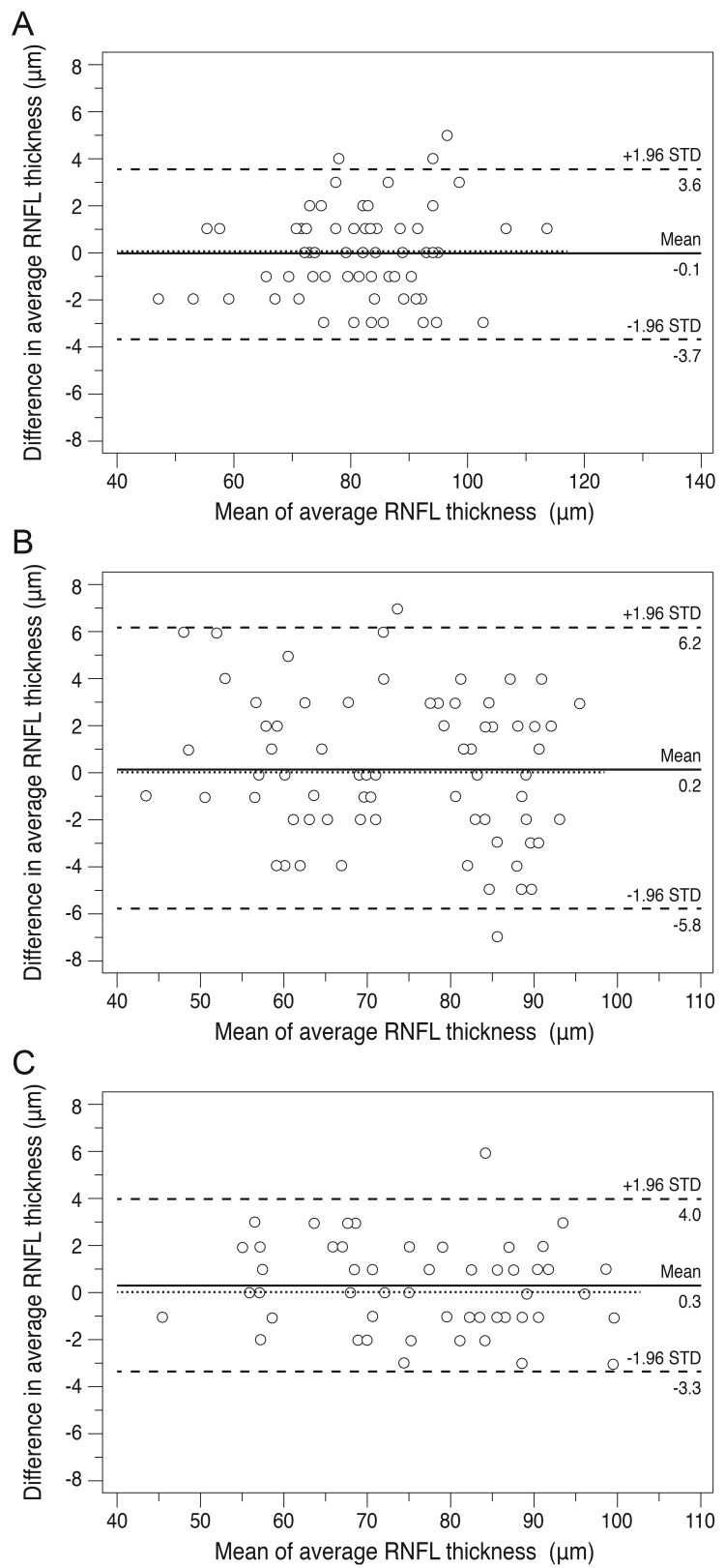
Fig. 1 shows a Bland-Altman plot analysis of average RNFL thickness measured by two repeated Cirrus OCT scans in three groups respectively (A, B, and C). There was greater variation between two repeated average RNFL thickness measurements in the cataract group than in other groups.
Fig. 1.
Bland-Altman plot analysis of the average retinal nerve fiber layer (RNFL) thickness measured by two repeated Cirrus optical coherence tomography scans. Note the greater variation between two repeated average RNFL thickness measurements in the cataract group than in the other groups. (A) Clear media. (B) Cataract. (C) Pseudophakic. STD = standard deviation.
Discussion
In the present study, we evaluated the reproducibility of cpRNFL thickness measurement and its color code classification by Cirrus SD OCT in pseudophakic eyes, and then compared the results with those in clear media and cataract eyes. As a result, ICC and kappa values of pseudophakic eyes were as good as that of clear media eyes despite a slightly lower average signal strength. CV and test retest variability of the pseudophakic group were also similar to those of clear media group except in the inferior quadrant. To our best knowledge, there has been no prior study that simultaneously compares reproducibility of Cirrus SD OCT in three different groups (clear media, cataract, and pseudophakic eyes).
OCT is an important method used in diagnosing and determining the progression of glaucoma [11,12]. The newer SD OCT provides much faster and more detailed structural information than the previous TD OCT method [13,14], and has potentially improved its ability to diagnose and observe glaucoma progression [15]. Hong et al. [16] reported that the reproducibility of Cirrus HD OCT to analyze peripapillary RNFL thickness in healthy eyes was excellent compared with time domain Stratus OCT. In the current study, the agreement for average RNFL thickness measurement was excellent in all three groups (ICC ranged 0.974 to 0.990), thereby confirming that RNFL thickness measurements by Cirrus SD OCT are highly repeatable.
Cataract is a common cause of media opacity that can affect the quality of diagnostic imaging devices for glaucoma like OCT in the elderly. Clear optical media will optimize both forward and back light scattering, whereas media opacity caused by cataracts will increase light scattering and absorption. Previous studies using Stratus TD OCT reported that lens opacities may affect the RNFL thickness measurements [4,6,17], and the RNFL thickness measurement can be increased after uncomplicated cataract surgery [6]. The increase in signal strength and RNFL thickness measurement after cataract surgery may be the result of improved transmittance and reflectivity of the RNFL boundary after removal of opacified media, rather than actual RNFL thickening after cataract surgery. In terms of measurement agreement expressed as ICC, the cataract group showed the smallest ICC values compared to clear media and pseudophakic groups. Especially in some quadrants (superior and nasal), this difference reached statistical significance. There was no significant difference in ICC value between clear media and pseudophakic groups in all scan areas, implying that measurement agreement in pseudophakic eyes is as good as that in clear media eyes. High ICC value in pseudophakic eyes as in clear media eyes in results of all scan areas was similar with results of scans only with high SS. When we calculated the ICC of scans only with high SS, ICC in pseudophakic group also showed no difference with that of cataract group in the superior and nasal quadrants, which was different from the scan results considering all SS.
One possible explanation for this is that the effect of low SS caused by lens opacity in the cataract group on RNFL measurement variability was minimized by exclusion of scans with low SS.
Results regarding the RNFL color code classification from Cirrus OCT were quite interesting. For quadrant maps, the color code classification of the nasal quadrant appeared to be the most variable among all three groups as previously investigated [16]. Although the mean kappa values for the cataract group in temporal, superior, and nasal quadrants were slightly low, the mean kappa value of average RNFL thickness did not show a similar pattern. Moreover, these observations were consistent with a previous study, which reported poorer reproducibility in a smaller scan area using Stratus OCT [18]. No statistically significant difference in color code agreement was found among the three groups in all scan areas, except the temporal quadrant, in which test data was not available due to insufficient number of categories. Similar results were observed for scans with only a high SS score. A relatively comparable agreement of color code was observed among the three groups in all scan areas despite the significantly lower ICC value of the superior and nasal quadrants in the cataract group. This observation might be due to rough classification of color code, which categorizes RNFL thickness into only four codes (white, green, yellow, and red) through comparison with an age-matched normative database. Therefore, small gaps in RNFL thickness measurements between two consecutive scans would be masked via the color code classification.
The result of inter-scan measurement difference, CV, and test-retest variability were found to be higher in the cataract group, while values of the remaining two groups (pseudophakic and clear media group) were similar to each other in temporal, superior, nasal quadrants, and average RNFL thickness. However, only in the inferior quadrant, the inter-scan measurement difference, CV, and test-retest variability in the pseudophakic group were higher than those of the clear media group. There are some potential explanations for this observation. Of several statistical methods for assessing reliability, CV and test-retest variability can be caused by both intra-individual variability and inter-individual variability, in contrast to ICC values. In this study population, the ratio of glaucoma patients to glaucoma suspects was higher in the pseudophakic group (25 : 33, 75.8%) than in the clear media group (30 : 43, 69.8%), as shown in Table 1. In eyes with optic nerve damage, localized RNFL defects are most often found in the temporal inferior sector, followed by the temporal superior sector [19]. Therefore, a relatively higher ratio of glaucoma patients to glaucoma suspects in the pseudophakic group and their higher inter-individual variability from different degrees of glaucomatous RNFL damage might contribute to higher inter-scan measurement differences, CV, and test-retest variability in the pseudo-phakic group than in the clear media group only in the inferior quadrant despite of similar ICC values.
A previous study performed by the authors demonstrated that the presence of cataract significantly affects the RNFL thickness measurements by Cirrus SD OCT and Stratus TD OCT. The changes in the RNFL thickness measurements following cataract surgery were more prominent for Cirrus SD OCT than Stratus OCT [20]. The fact that Cirrus OCT showed more apparent changes of RNFL thickness measurements after cataract surgery does not necessarily mean a lower performance in terms of RNFL thickness measurements in eyes with media opacity. As Stratus OCT and Cirrus OCT use a different data acquisition process with a different speed, we could speculate that more reliable measurements in pseudophakic eyes based on Cirrus OCT might have affected the results of the previous study [20]. Cirrus OCT uses a line scanning ophthalmoscope with a 750 nm superluminescent diode as a light source and this provides clear pupil visualization and fundus image focusing. Thus, the Cirrus OCT scan quality may be better than those obtained with Stratus OCT. Moreno-Montanes et al. [21] also revealed that Cirrus OCT has better SS than Stratus OCT with similar conditions of pupillary dilatation or lens transparency. From all these considerations, similar reproducibility in RNFL thickness measurement using Cirrus OCT between pseudophakic and clear media eyes in the current study offers some support to our hypothesis that RNFL thickness measurement obtained by Cirrus OCT in pseudophakic eyes might be less variable than that obtained by Stratus OCT.
As we expected, the average SS in the pseudophakic group was somewhat lower than that in the clear media group. Na et al. [22] demonstrated that age, visual acuity, and extent of cataracts were associated with SS obtained by Cirrus OCT. One possible explanation for lower SS in the pseudophakic group is that surface light scattering or reflection on the intraocular lens implanted after cataract surgery might induce optical disturbance and impair light transmission. Alternatively, the presence of slight posterior capsular opacity might decrease the amount of light that passes through optical media, which SS strongly depends on. However, despite of lower SS values, the reproducibility of RNFL thickness and color code values in the pseudophakic group was comparable with that in the clear media group. It is likely that the SS of the pseudophakic group was not poor enough to reduce the reproducibility of RNFL thickness or color code in this study using Cirrus OCT. Wu et al. [23] reported that lower SS is associated with thinner RNFL thickness obtained by Stratus OCT in scans with SS values less than 7. On the other hand, Ha et al. [24] suggested a SS score of 4 was the lowest acceptable limit of SS for reproducible Stratus OCT scanning. Thus, special attention should be given to changes of SS when assessing RNFL thickness, particularly in glaucomatous patients with combined cataract or in those who have undergone cataract surgery recently.
Cheng et al. [10] also investigated the influence of cataract on RNFL thickness measurements with Cirrus OCT and Stratus OCT. After removal of cataract, the observed SS value was significantly increased in both Cirrus and Stratus OCT scans, while ICC value appeared to be significantly increased only via Stratus OCT. However, in our study using Cirrus OCT, the observed SS value was slightly higher in the pseudophakic group than in cataract group without statistical significance. Furthermore, the ICC value was significantly higher in the pseudophakic group than in the cataract group, especially in the superior and nasal quadrants. The discrepancy of results between the two studies might come from different study designs. The study by Cheng et al. [10] enrolled only small number of cataract patients without any other ocular pathology and compared reproducibility of preoperative and postoperative OCT evaluation in the same population. On the other hand, our study enrolled a large number of glaucomatous patients and compared reproducibility of Cirrus OCT evaluation among different groups classified according to patients' lens status.
There are several strengths of our study. First, a large population of 205 patients were enrolled indicating that our study has sufficient statistical power. Second, all participants were classified into three different groups according to lens status by a single experienced ophthalmologist (JHK). Third, as a single experienced technician operated all OCT scans, inter-operator variability was minimized.
The current study has several limitations. First, some patients with minimal lens opacity might be classified into clear media group considering the mean age of study population was over 60 years old. These ambiguities in group classification may have adverse effects on our results. Second, a direct comparison of reproducibility with Cirrus OCT and Stratus OCT RNFL thickness measurements in pseudophakic eyes was not allowed since we did not have both devices in our clinic. Third, due to limited participation of patients, we were not able to compare the reproducibility observed with preoperative and postoperative scans in the same patients. Further studies are required to ascertain the effect of lens status on reproducibility of Cirrus OCT according to different stages of glaucoma.
The present study demonstrated that the Cirrus OCT measurements of cpRNFL thickness were very reproducible in pseudophakic eyes, as well as in clear media eyes. However, even after successful cataract surgery, there is a possibility of SS attenuation in comparison with clear media eyes, probably due to the interference of light transmittance by implanted intraocular lens. Thus, when interpreting the results of Cirrus OCT scans in pseudophakic eyes, factors that can compromise the test results should be carefully considered.
Footnotes
No potential conflict of interest relevant to this article was reported.
References
- 1.Carpineto P, Nubile M, Agnifili L, et al. Reproducibility and repeatability of Cirrus HD-OCT peripapillary retinal nerve fibre layer thickness measurements in young normal subjects. Ophthalmologica. 2012;227:139–145. doi: 10.1159/000334967. [DOI] [PubMed] [Google Scholar]
- 2.Cremasco F, Massa G, Goncalves Vidotti V, et al. Intrasession, intersession, and interexaminer variabilities of retinal nerve fiber layer measurements with spectral-domain OCT. Eur J Ophthalmol. 2011;21:264–270. doi: 10.5301/EJO.2010.5469. [DOI] [PubMed] [Google Scholar]
- 3.Pareja-Esteban J, Teus-Guezala MA, Drake-Casanova P, Dapena-Sevilla I. Retinal nerve fiber layer changes after cataract surgery measured by OCT: a pilot study. Arch Soc Esp Oftalmol. 2009;84:305–309. doi: 10.4321/s0365-66912009000600006. [DOI] [PubMed] [Google Scholar]
- 4.El-Ashry M, Appaswamy S, Deokule S, Pagliarini S. The effect of phacoemulsification cataract surgery on the measurement of retinal nerve fiber layer thickness using optical coherence tomography. Curr Eye Res. 2006;31:409–413. doi: 10.1080/02713680600646882. [DOI] [PubMed] [Google Scholar]
- 5.Mwanza JC, Bhorade AM, Sekhon N, et al. Effect of cataract and its removal on signal strength and peripapillary retinal nerve fiber layer optical coherence tomography measurements. J Glaucoma. 2011;20:37–43. doi: 10.1097/IJG.0b013e3181ccb93b. [DOI] [PubMed] [Google Scholar]
- 6.Savini G, Zanini M, Barboni P. Influence of pupil size and cataract on retinal nerve fiber layer thickness measurements by Stratus OCT. J Glaucoma. 2006;15:336–340. doi: 10.1097/01.ijg.0000212244.64584.c2. [DOI] [PubMed] [Google Scholar]
- 7.Lee ES, Kim H, Kim JM. Effect of signal strength on reproducibility of peripapillary retinal nerve fiber layer thickness measurement and its classification by time-domain optical coherence tomography. Jpn J Ophthalmol. 2010;54:414–422. doi: 10.1007/s10384-010-0850-9. [DOI] [PubMed] [Google Scholar]
- 8.Youm DJ, Kim H, Shim SH, et al. The effect of various factors on variability of retinal nerve fiber layer thickness measurements using optical coherence tomography. Korean J Ophthalmol. 2012;26:104–110. doi: 10.3341/kjo.2012.26.2.104. [DOI] [PMC free article] [PubMed] [Google Scholar]
- 9.Quillen DA. Common causes of vision loss in elderly patients. Am Fam Physician. 1999;60:99–108. [PubMed] [Google Scholar]
- 10.Cheng CS, Natividad MG, Earnest A, et al. Comparison of the influence of cataract and pupil size on retinal nerve fibre layer thickness measurements with time-domain and spectral-domain optical coherence tomography. Clin Experiment Ophthalmol. 2011;39:215–221. doi: 10.1111/j.1442-9071.2010.02460.x. [DOI] [PubMed] [Google Scholar]
- 11.Huang ML, Chen HY. Development and comparison of automated classifiers for glaucoma diagnosis using Stratus optical coherence tomography. Invest Ophthalmol Vis Sci. 2005;46:4121–4129. doi: 10.1167/iovs.05-0069. [DOI] [PubMed] [Google Scholar]
- 12.Parikh RS, Parikh S, Sekhar GC, et al. Diagnostic capability of optical coherence tomography (Stratus OCT 3) in early glaucoma. Ophthalmology. 2007;114:2238–2243. doi: 10.1016/j.ophtha.2007.03.005. [DOI] [PubMed] [Google Scholar]
- 13.Wojtkowski M, Bajraszewski T, Gorczynska I, et al. Ophthalmic imaging by spectral optical coherence tomography. Am J Ophthalmol. 2004;138:412–419. doi: 10.1016/j.ajo.2004.04.049. [DOI] [PubMed] [Google Scholar]
- 14.Chen TC, Cense B, Pierce MC, et al. Spectral domain optical coherence tomography: ultra-high speed, ultra-high resolution ophthalmic imaging. Arch Ophthalmol. 2005;123:1715–1720. doi: 10.1001/archopht.123.12.1715. [DOI] [PubMed] [Google Scholar]
- 15.Leung CK, Liu S, Weinreb RN, et al. Evaluation of retinal nerve fiber layer progression in glaucoma a prospective analysis with neuroretinal rim and visual field progression. Ophthalmology. 2011;118:1551–1557. doi: 10.1016/j.ophtha.2010.12.035. [DOI] [PubMed] [Google Scholar]
- 16.Hong S, Kim CY, Lee WS, Seong GJ. Reproducibility of peripapillary retinal nerve fiber layer thickness with spectral domain cirrus high-definition optical coherence tomography in normal eyes. Jpn J Ophthalmol. 2010;54:43–47. doi: 10.1007/s10384-009-0762-8. [DOI] [PubMed] [Google Scholar]
- 17.Van Velthoven ME, van der Linden MH, de Smet MD, et al. Influence of cataract on optical coherence tomography image quality and retinal thickness. Br J Ophthalmol. 2006;90:1259–1262. doi: 10.1136/bjo.2004.097022. [DOI] [PMC free article] [PubMed] [Google Scholar]
- 18.Budenz DL, Chang RT, Huang X, et al. Reproducibility of retinal nerve fiber thickness measurements using the stratus OCT in normal and glaucomatous eyes. Invest Ophthalmol Vis Sci. 2005;46:2440–2443. doi: 10.1167/iovs.04-1174. [DOI] [PubMed] [Google Scholar]
- 19.Jonas JB, Dichtl A. Evaluation of the retinal nerve fiber layer. Surv Ophthalmol. 1996;40:369–378. doi: 10.1016/s0039-6257(96)80065-8. [DOI] [PubMed] [Google Scholar]
- 20.Kim NR, Lee H, Lee ES, et al. Influence of cataract on time domain and spectral domain optical coherence tomography retinal nerve fiber layer measurements. J Glaucoma. 2012;21:116–122. doi: 10.1097/IJG.0b013e31820277da. [DOI] [PubMed] [Google Scholar]
- 21.Moreno-Montañés J, Olmo N, Alvarez A, et al. Cirrus high-definition optical coherence tomography compared with Stratus optical coherence tomography in glaucoma diagnosis. Invest Ophthalmol Vis Sci. 2010;51:335–343. doi: 10.1167/iovs.08-2988. [DOI] [PubMed] [Google Scholar]
- 22.Na JH, Sung KR, Lee Y. Factors associated with the signal strengths obtained by spectral domain optical coherence tomography. Korean J Ophthalmol. 2012;26:169–173. doi: 10.3341/kjo.2012.26.3.169. [DOI] [PMC free article] [PubMed] [Google Scholar]
- 23.Wu Z, Huang J, Dustin L, Sadda SR. Signal strength is an important determinant of accuracy of nerve fiber layer thickness measurement by optical coherence tomography. J Glaucoma. 2009;18:213–216. doi: 10.1097/IJG.0b013e31817eee20. [DOI] [PMC free article] [PubMed] [Google Scholar]
- 24.Ha MM, Kim JM, Kim HJ, et al. Low limit for effective signal strength in the Stratus OCT in imperative low signal strength cases. Korean J Ophthalmol. 2012;26:182–188. doi: 10.3341/kjo.2012.26.3.182. [DOI] [PMC free article] [PubMed] [Google Scholar]


