Hepatic pulmonary fusion is a rare association of right-sided congenital diaphragmatic hernia. The repair and reduction in this case depends on the extent of fusion to the lungs and the associated mediastinal structures [1]. This case has thumb and index finger syndactyly and multiple clefts in vertebrae besides hepatic pulmonary fusion which makes it unique, the first of its kind.
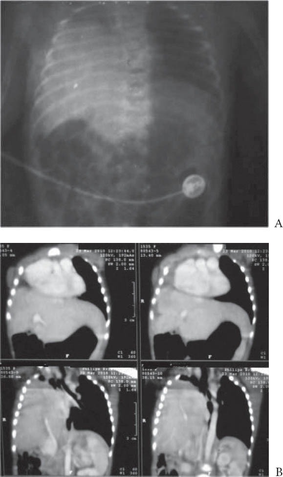
This is a case of a full-term neonate who presented with signs of respiratory distress at 13 h of life. On examination, trachea was central in position with decreased air entry on right side of chest and right thumb and index finger syndactyly. Chest x-ray showed homogenous opacity involving right hemithorax mainly in mid and lower zone, with well-defined superior margin with no mediastinal shift and clefts in vertebrae (Fig. 1 A). Contrast-enhanced computed tomography of thorax revealed migration of liver into the thoracic cavity in the posterior aspect with normal appearing anterior diaphragm with right lung hypoplasia or collapse and multiple cleft vertebrae (Fig. 1 B). Peroperatively, the entire liver was found to be displaced into the right thoracic cavity with pneumatization of right lobe of liver and the diaphragm was stuck to remnant pulmonary tissue and adjacent mediastinal tissue. It was difficult to achieve a clear-cut line of cleavage. The diaphragmatic defect was partially approximated and sutured. Postoperatively, the patient died at day 11 of life due to respiratory insufficiency.
Figure 1.

(A) X-ray film of chest and abdomen showing homogenous opacity involving right hemithorax without mediastinal shift and presence of clefts in vertebrae. (B) Contrast-enhanced computed tomography of chest and abdomen showing migration of liver into the thoracic cavity in the posterior aspect with right lung hypoplasia/collapse
The association of hepatic pulmonary fusion to congenital diaphragmatic hernia is still incompletely understood. There may be primary fusion between liver and lungs followed by lack of development of embryonic structures later on, however the most logical explanation is failure to fuse rather than failure to form. The failure to fuse the diaphragmatic membranes may allow the fusion of adjacent future pulmonary and hepatic tissue [2]. The association of limb defects like syndactyly and polydactyly with congenital diaphragmatic hernia suggests disturbed migration of neural crest cells and mutation of FGF10 [3]. These gene mutations might explain the occurrence of congenital diaphragmatic hernia with vertebral and digital anomalies in our case.
It is important to consider hepatic pulmonary fusion in the differential diagnosis when congenital diaphragmatic hernia is suspected because the presence of this entity makes the reduction of hernia and preservation of the lungs difficult [4]. Magnetic resonance imaging (MRI) is very important in this regard as it may show some unique findings suggestive of hepatic pulmonary fusion [4]. Most of the case reports favor that non-shifting of mediastinal structures away from the affected side in a right-sided congenital diaphragmatic hernia is an indicator of hepatic pulmonary fusion [4-6]. This is also true in our case as there was no evidence of shifting of mediastinum in x-ray film.
In summary, in a suspected case of right-sided congenital diaphragmatic hernia, hepatic pulmonary fusion should be suspected whenever imaging findings show intrathoracic liver and lack of mediastinal shift. Proper radiological imaging especially MRI should be considered for a better surgical outcome.
Biography
Vardhman Mahavir Medical College and Safdarjung Hospital, New Delhi, India
Footnotes
Conflict of Interest: None
References
- 1.Katz S, Kidron D, Litmanovitz I, Erez I, Dolfin Z, Saba K. Fibrous fusion between liver and lungs: An unusual complication of right congenital diaphragmatic hernia. J Paediatr Surg. 1998;33:766–767. doi: 10.1016/s0022-3468(98)90214-7. [DOI] [PubMed] [Google Scholar]
- 2.Slovis TL, Farmer DL, Berdon WE, Rabah R, Campbell JB, Philippart AI. Hepatic pulmonary fusion in neonates. Am J Roentgenol. 2000;174:229–233. doi: 10.2214/ajr.174.1.1740229. [DOI] [PubMed] [Google Scholar]
- 3.Pober BR, Russell MK, Ackerman KG. Congenital Diaphragmatic Hernia Overview 2006 Feb 1 [Updated 2010 Mar 16] In: Pagon RA, Bird TD, Dolan CR, et al., editors. GeneReviews™ [Internet] Seattle (WA): University of Washington, Seattle; 1993. Available from: http://www.ncbi.nlm.nih.gov/books/NBK1359 . [PubMed] [Google Scholar]
- 4.Keller RL, Aaroz PA, Hawgood S, Higgins CB. MR imaging of hepatic pulmonary fusion in neonates. Am J Roentgenol. 2003;180:438–440. doi: 10.2214/ajr.180.2.1800438. [DOI] [PubMed] [Google Scholar]
- 5.Gander JW, Kandenhe-Chiweshe A, Fisher JC, et al. Hepatic pulmonary fusion in an infant with right-sided congenital diaphragmatic hernia and contralateral mediastinal shift. J Paediatr Surg. 2010;45:265–268. doi: 10.1016/j.jpedsurg.2009.10.090. [DOI] [PMC free article] [PubMed] [Google Scholar]
- 6.Taide DV, Bendre P, Kirtane JM, Mukunda R. Hepatic pulmonary fusion: a rare case. Afr J Pediatr Surg. 2010;7:28–29. doi: 10.4103/0189-6725.59357. [DOI] [PubMed] [Google Scholar]


