Abstract
Mating induces changes in female insects, including in egg production, ovulation and laying, sperm storage, and behavior. Several molecules and effects that induce these changes have been identified, but their proximate effects on females remain unexplored. We examined whether vesicle release occurs as a consequence of mating; we used transgenic Drosophila that allow monitoring of secretory granule release at nerve termini. Changes in release occur at specific times postmating in different regions of the female reproductive tract: soon after mating in the lower reproductive tract, and later in the upper reproductive tract. Some changes are triggered by receipt of sperm, others by male seminal proteins, and still others by the act of mating itself (or other unidentified effectors). Our findings indicate that the female reproductive tract is a multi-organ system whose regions are modulated separately by mating and mating components. This modulation could create an environment conducive to increased reproductive capacity.
Keywords: accessory gland proteins, seminal proteins, ovulation, sperm storage, neuromodulators
For fertilization and subsequent zygotic development in animals with internal fertilization, gametes must meet in the female reproductive tract when the female, eggs, and sperm are reproductively competent. Sperm and oocytes form separately in specialized organs and then enter a common region of the female's reproductive tract. It is essential that they encounter an environment that supports their final maturation and promotes their union. A paradigm for this situation comes from mammals, whose oviducts undergo hormonally mediated cyclic modifications climaxing during the preovulatory period. Oviducts respond to hormone levels with morphological and secretory changes that promote a suitable environment for gametes and fertilization (1). Less is known about this phenomenon in insects, although female insects require a period of sexual maturation to become reproductively competent. During this period, changes in secretory activity of the oviduct epithelium are correlated with both juvenile hormone and 20-hydroxyecdysone levels in the circulatory system as shown in locusts (Schistocerca gregaria; ref. 2), but the specific triggers for the changes are unknown.
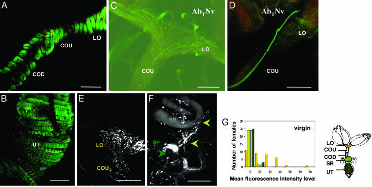
The Drosophila melanogaster female reproductive tract is primarily an epithelium surrounded by circular muscles (Fig. 1 A and B). Unique characteristics and thickness of the epithelium in specific regions of the reproductive tract (4) suggest that secretion patterns may differ between regions. Terminal branches of the abdominal nerves (AbTNv) innervate the female reproductive tract (ref. 4 and this study), providing the potential to modulate the responsive capacity of the musculature and epithelium.
Fig. 1.
Drosophila female reproductive tract. (A and B) Musculature of oviduct (A) and uterus (B) stained with anti-serotonin (red) and with Oregon green 488 phalloidin to label F-Actin (green). Lateral oviducts (LO) and common oviduct (CO) are surrounded by a single layer of circular muscle fibers. Several layers of circular muscle fibers surround the uterus (UT). Schematic of the female reproductive tract, modified from Mahowald and Kambysellis (3), shows reproductive tract regions: LO (yellow), CO (dark gray), and UT (brown). (Scale bars = 50 μm.) (C) Upper reproductive tract stained with anti-Fascilin II and viewed with confocal microscopy to visualize the terminal branches of the abdominal nerve center (AbTNv) that innervate the LO and the upper common oviduct (COU, pale yellow in the diagram). (Scale bar = 50 μM.) (D) Terminal branch of AbTNv along the common oviduct, visualized by anti-Fascilin II staining. (Scale bar = 50 μm.) (E and F) Distribution of proANF-EMD-containing vesicles in regions of virgin Drosophila female reproductive tracts. (E) Upper reproductive tract. Note the high intensity of proANF-EMD fluorescence in the LO at the base of the ovary (adjacent to “LO” label in the figure), as compared with that at the LO junction with the COU. (F) Seminal receptacle (SR; light green in the diagram). (Scale bars = 50 μm.) (G) The sum of proANF-EMD fluorescence intensity level (OMFILF and LMFILF) at each region of the female reproductive tract: LO, COU, COD, SR, and UT of all 36 virgin females examined.
During and after mating, sperm transferred to the female must be stored, and egg production must increase, for high fertility (5–8). To accomplish this transition at high efficiency, the female's reproductive tract must become “aware” that insemination has occurred. In most insects, mating stimulates the female's egg production, sperm storage, and behavioral changes, by means of neural and chemical cues (e.g., substances in the seminal fluid; refs. 9–16). This finding and the notion that in vertebrates the oviduct actively responds to endocrine stimuli (1) led us to test whether seminal fluid components could induce changes in the female D. melanogaster reproductive tract conducive to secretory and muscular activity.
Mating Drosophila males transfer sperm and associated seminal fluid, which includes accessory gland proteins (Acps), to females (12–16). Acps decrease sexual receptivity, stimulate a rapid increase in egg production, ovulation, and deposition, mediate sperm storage, and decrease females' longevity; sperm also contribute to some of these changes. In addition to, and as a result of, their proximate action on females, Acps have been suggested to be important players in evolution, reflecting roles in sperm competition and/or sexual conflict (15, 17–21). To evaluate these models, it is important to know how Acps and other mating components affect females at both physiological and molecular levels, and the timing and positions of their effects.
Because virgin female Drosophila make, ovulate, and deposit eggs at a low rate, we hypothesize that the female reproductive tract can perceive and respond to hormonal stimuli, but that mating, including receipt of seminal proteins, induces optimal physiological conditions required for rapid and successful fertilization. Mating might stimulate secretion of molecules from reproductive tract tissues into the lumen of the reproductive tract to act as organic osmolytes, membrane stabilizers, or facilitators of sperm motility or maturation. Alternatively, mating and/or seminal fluid components may modulate release of neurotransmitters to change the membrane potential of postsynaptic neurons, of neurohormones to change the function or direct the activity of organ(s) or tissue(s), or of neuromodulators to influence neurons' responses to neurotransmitters. Any of these effectors could potentially modulate the contractile activity of reproductive tract musculature. A paradigm for what could be modulated is offered by locusts (Locusta migratoria), whose oviducts are innervated by neurons whose neuroactive substances (including the neuropeptides SchistoFLRFamide and proctolin, the neuromodulator octopamine, and probably the neurotransmitter glutamate) mediate muscle contraction (22–27). Two recent reports indicate that octopamine also regulates ovulation in D. melanogaster (28, 29).
Here, we test whether mating and/or seminal fluid could modulate the release of neuroactive substances into and/or onto the Drosophila female reproductive tract. Specifically, we examined effects of mating and seminal fluid components on vesicle release in transgenic D. melanogaster that express throughout their nervous system a fusion of the neuropeptide rat atrial natriuretic factor (ANF) with the emerald variant of GFP (EMD) (28). Rao et al. (30) showed that this fusion protein is proteolytically processed and stored in secretory granules that are transported down axons and become concentrated in nerve termini, preferentially in peptidergic nerve termini. This transgenic system allowed us to visualize nerve termini and test whether their vesicle release was modulated; we could measure this release as changes in fluorescence intensity level as a consequence of mating. Staining with antibodies against SNAP-25 (synaptosome-associated protein of 25 kDa) separately confirmed the presence of synaptic contacts in this region of the reproductive tract.
We compared vesicle release in the reproductive tracts of virgin females with that in females mated to normal males or to males deficient in sperm and/or Acps. We found that, immediately postmating, there is vesicle release in the lower reproductive tract (lower common oviduct, seminal receptacle, and uterus). Later (3 h postmating), when females are ovulating at high rates and egg production has reached maximal levels, net vesicle release is inhibited in the upper reproductive tract (upper common oviduct and lateral oviducts). By comparing vesicle release in females mated to WT, spermless, or seminal fluid-deficient Drosophila males, we teased apart the contribution of mating, sperm, and seminal proteins to the regulation of vesicle release.
Materials and Methods
Flies. Flies were maintained, collected, and aged (as virgins) for 3 days before testing, as described (31). All females were homozygous for P[UAS proANF-EMD], P[elav-GAL4] (30); this UAS-proANF-EMD stock was kindly provided by D. Deitcher (Cornell University). WT males were Canton S. Transgenic “DTA-E” males deficient in Acps and sperm and were described in Kalb et al. (32). DTA-E males make no detectable Acps in the main cells of their accessory glands (96% of the gland) and no sperm. They make secondary cell, ejaculatory bulb, and ejaculatory duct proteins (32). Spermless males (TUD) were sons of Canton S males and bw sp tud1 females (33).
Reproductive Tract Bioassay. We generated a map of vesicle release patterns by examining the fluorescence intensity level of proANF-EMD in different regions of the female reproductive tract and the distribution of the fluorescence signal within a given region, at different times after mating. Reproductive tracts from virgin and mated females were analyzed. Single virgin females were placed with virgin males (WT, DTA-E, TUD) and timed as soon as mating began. At the end of copulation (≈20 min after the start of mating), females were either placed on ice or were aspirated into fresh vials with yeasted food and held singly at 25 ± 2°C for 90 min [start of ovulation (31); time of maximal sperm storage (34)], or 180 min [start of egg deposition; fertilization also is occurring frequently by this time (31, 35)] before being placed on ice. Female reproductive tracts were dissected on ice in PBS containing 0.1% Triton X-100 (PBST; pH 7.4). Dissected reproductive tracts were washed additionally in PBST and then mounted in Antifade (Molecular Probes). Each slide held reproductive tracts from virgin females and from females mated to different males to normalize imaging (see also Fig. 5, which is published as supporting information on the PNAS web site).
To examine whether exposure to males affected vesicle release, we watched virgin females as they interacted with males. Females were removed for analysis after males had begun courting them, but before copulation occurred.
Immunocytochemistry. Reproductive tracts were fixed in 4% paraformaldehyde and immunostained as in Heifetz et al. (31). Anti-SNAP-25 polyclonal rabbit antibody [kind gift of D. Deitcher, Cornell University (36)], Alexa Fluor (488) secondary antibody (Molecular Probes), mouse anti-Fascilin II [kind gift of C. Goodman, University of California, Berkeley (37)], rhodamine conjugated goat anti-horseradish peroxidase (Jackson ImmunoResearch), and rabbit anti-serotonin antibodies (kind gift of R. Hoy, Cornell University) were used at 1:50, 1:200, 1:4, 1:200, and 1:100 dilutions, respectively.
Microscopy. Reproductive tracts were imaged within 30 min of dissection by using a laser scanning confocal microscope (Bio-Rad MRC 600 system on a Zeiss inverted microscope); samples were kept on ice in the dark until imaged. A ×40-water objective and comos software (Bio-Rad) were used to collect images. Optical sections of 1 μm from different focal planes were collected and processed with confocal assistant 4.02 (T. C. Brelje, University of Minnesota) to reconstruct a three-dimensional image.
Evaluation of Fluorescence Intensity Level of Dissected Reproductive Tracts. Fluorescence was quantitated in imagej 1.25s (National Institutes of Health) by two approaches that cross-validate; their joint use increases reliability. Details are provided in Supporting Materials and Methods, which is published as supporting information on the PNAS web site. (i) Overall mean fluorescence intensity level (OMFIL) quantifies the average gray value within the contours of a selected region. It is the sum of the gray values of all pixels within the region divided by the number of pixels within the region. (ii) Local mean fluorescence intensity level (LMFIL) quantifies mean fluorescence intensity level at five different locations within the selected reproductive tract region.
To supplement these quantitative measures, we determined the distribution of the fluorescence signal (DFS) to determine the uniformity of signal within the selected region. These quantities were evaluated in the lateral oviducts (LO), upper common oviduct (COU), lower common oviduct (COD), seminal receptacle (SR), spermathecae (SP), and uterus (UT) (Fig. 1). Signals were not evaluated in ovaries.
Statistics. One-way ANOVA (spss 8.0 for Windows) were used to measure the differences in intensity level and signal distribution between each pair of treatments. To define mating-induced changes, we compared signals in virgins with those in genetically identical females that had mated to WT males. To define sperm-dependent changes, we compared mates of WT or TUD males. To define Acp-dependent changes, we compared mates of TUD or DTA-E males (see also Supporting Materials and Methods).
Results and Discussion
Region-Specific Innervation Pattern in the Drosophila Female Reproductive Tract. Using anti-Fascilin II to stain neural cell membranes, we observed terminal branches of the abdominal nerves (AbTNv) innervating the female reproductive tract (Fig. 1C). The abdominal ganglionic center branches at its terminus (4). Nerves from either or both branches pass above the lateral oviducts and then run posteriorly along the common oviduct to the sperm storage organs and the uterus (Fig. 1D). Each nerve fiber then splits further into several fine branches, which spread over the surface of the reproductive tract muscle fibers. To examine the behavior of secretory granules in the female reproductive tract, we used the proANF-EMD system, which has previously documented accumulation of secretory granules at nerve termini in the larval neuromuscular junction (30). proANF-EMD fluorescence was observed at nerve termini in all regions of the female reproductive tract [LO and COU, Fig. 1E; sperm storage organs (SP and SR), Fig. 1F; and UT, data not shown], indicating that nerve termini containing proANF-EMD vesicles branch on the surface of the reproductive tract musculature. Staining of proANF-EMD females' reproductive tracts with rhodamine-coupled anti-horseradish peroxidase antibody, which stains all neural cell membranes (30, 38), revealed an innervation pattern similar to that of proANF-EMD fluorescence (data not shown).
Regions of the female reproductive tract [lateral oviducts, common oviduct, sperm storage organs (SR and SP), and UT] differ in innervation patterns. Some are more intensely innervated (e.g., LO, Fig. 1E) than others (e.g., COU, Fig. 1E; SR, Fig. 1F). To see whether the innervation patterns in the different reproductive tract regions correlated with their quantified fluorescence intensity levels (OMFILF and LMFILF; see Fig. 1 legend), we calculated the total fluorescence intensity level at each region of all examined females. proANF-EMD fluorescence intensity level (OMFILF and LMFILF) differed between the regions of female reproductive tract and was highest in lateral oviducts (Fig. 1G). This region is also the most heavily innervated of the reproductive tract. proANF-EMD fluorescence was lower in less innervated regions. Presence of vesicle-bound molecules in all reproductive tract regions gives the potential to modulate release of neuromodulators to mediate postmating responses in the reproductive tract.
Mating Induces Changes in proANF-EMD Staining Levels in Nerve Termini of the Oviducts and the Seminal Receptacle. During exocytosis, when vesicles at nerve termini release their contents, proANF-EMD fluorescence becomes diffuse and decreases in intensity (30, 39). Because actual measured fluorescence intensity level (OMFILF and LMFILF) reflects the balance between (i) accumulation of proANF-EMD in vesicles and (ii) release of proANF-EMD from vesicles, an increase in fluorescence intensity level indicates net accumulation of vesicles at nerve terminals whereas a decrease in fluorescence intensity level indicates net release.
To test whether mating induces a change in neuropeptide distribution or release in nerve termini innervating female reproductive tracts, we compared the proANF-EMD fluorescence intensity levels in virgin females with those in females after mating to WT males. If mating promotes changes in nerve termini in a region of the female reproductive tract, proANF-EMD fluorescence intensity levels should change after mating and/or there should be a significant change in the distribution of proANF-EMD within that region. Mated females were examined at different times after the start of mating (20 min, the end of copulation; 90 min, the start of ovulation and maximal sperm storage; 180 min, the start of egg deposition). Quantitative analyses of the fluorescence images showed that mating induces changes in proANF-EMD fluorescence at nerve termini in all regions of the female reproductive tract (Fig. 2). These changes were due to mating because, if females were exposed to courting males but were not allowed to mate with them, fluorescence intensity levels (OMFILF and LMFILF) in the reproductive tract regions did not differ significantly from those in virgin females (data not shown). Thus, mating affects the nerve termini innervating the female reproductive tract.
Fig. 2.
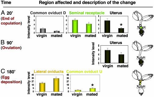
Significant postmating changes in intensity of proANF-EMD fluorescence at nerve termini innervating the female reproductive tract. All regions of the reproductive tract were examined (see Table 1). Graphs are shown only for regions in which substantive changes were observed. (A) Reproductive tract regions with differences in proANF-EMD fluorescence intensity level (OMFILF and LMFILF) between virgin females and mates of WT males at 20 min after the start of mating: lower part of the common oviduct (COD; gray), SR and UT at 20 min postmating; only UT shows a statistically significant difference. (B) UT at 90 min postmating. (C) LO and COU at 180 min postmating. Plotted are means ± SEs of virgins and females mated to WT males. Thirty-five to 40 female reproductive tracts were examined for each treatment at each time point (see also Table 1).
The pattern of mating-induced changes in fluorescence differed between reproductive tract regions, and at different times after mating (Fig. 2). Shortly after the start of mating, changes are seen mainly in the lower reproductive tract: in the uterus, lower part of the common oviduct, and seminal receptacle. Their fluorescence intensity levels tended to decrease soon after mating although the changes were significant only in the uterus at this time. Later, changes occur mainly in the upper part of the common oviduct and the lateral oviducts. By 180 min after mating, there is a relatively high intensity of fluorescence in the upper reproductive tract, significant in the lateral oviducts.
At 20 min after mating (end of copulation), there was a significant decrease in fluorescence intensity level in the uterus (2.6-fold; OMFILF: T = 2.61, P < 0.0013; LMFILF: T = 3.11, P < 0.004; Fig. 2 A). This decreased intensity level suggests a net release of the contents of the proANF-EMD secretory granules at nerve termini in the uterus as a result of mating. A less dramatic decrease in intensity level was also observed in the lower part of the common oviduct (2.3-fold) and in the seminal receptacles (1.4-fold); neither was statistically significant (Fig. 2B). Other regions showed no significant change in intensity level (see Table 1, which is published as supporting information on the PNAS web site).
At 90 min after mating, when ovulation begins and active sperm storage is maximal, the fluorescence intensity in the uteri of mated females still differed from that in virgin females although the intensity difference was smaller than at 20 min (1.5-fold) and no longer statistically significant (Fig. 2B). Other regions were unchanged (see Table 1).
By 180 min after the start of mating (start of egg deposition), a significant increase (2.2-fold; OMFILF: T = -2.81, P < 0.008; LMFILF: T = -2.91, P < 0.006) in fluorescence intensity level was observed in the upper part of the common oviduct (Fig. 2C), suggesting net accumulation of proANF-EMD. An increase in intensity also occurred in the lateral oviducts (1.6-fold), but it was not statistically significant (Fig. 2C), nor were changes elsewhere (see Table 1). Thus, whereas in the lower part of the reproductive tract mating induces net vesicle release shortly after mating, in the upper reproductive tract, mating inhibits net release or increases accumulation of proANF-EMD vesicles in nerve termini at later times postmating. In addition to changes in intensity level, we also observed significant changes in distribution of proANF-EMD fluorescence signal (DFS, see Materials and Methods) in some regions (DFS(virgin); DFS(mated): 20 min UT [29.27 ± 2.66; 18.50 ± 2.76, T = 2.75; P < 0.009], SR [34.48 ± 2.47; 25.43 ± 3.93, T = 2.11; P < 0.041]; 180 min LO [53.25 ± 3.77; 68.09 ± 4.25; T = -2.7; P < 0.0012], COU [23.29 ± 2.06; 35.96 ± 4.2, T = -2.93; P < 0.006]). Changes in distribution of proANF-EMD suggest that mating affects specific nerve termini within a region. Because not all termini respond in the same way to mating, it is possible that stimuli other than mating are involved in mediating release or accumulation of proANF-EMD in the above regions.
We also examined the intensity of staining for SNAP-25, a protein that localizes to the presynaptic plasma membrane and is part of the t-SNARE complex that mediates docking of vesicles to the presynaptic terminal membrane (36, 40–42). SNAP-25 staining was seen in the regions of the reproductive tract in which we saw vesicle release by the proANF-EMD assay, indicating the presence of synapses in this region. Although our original intent was simply to confirm the presence of synapses in this region, we were intrigued to observe changes in the intensity of anti-SNAP-25 label in some regions after mating. Specifically, by the end of mating and when females start to ovulate (20 min and 90 min, respectively), most changes in SNAP-25 fluorescence intensity level were at nerve termini innervating the lower part of the female reproductive tract (SR, increase of 1.5-fold; LMFILF: T = 2.17, P < 0.0022; and SP, decrease of 2.4-fold; Fig. 6 A and B, respectively, which is published as supporting information on the PNAS web site), the regions that showed changes in proANF-EMD fluorescence. Later, when females are actively ovulating and depositing eggs (Fig. 6C, 180 min postmating), changes are mainly seen in the upper part of the oviduct [LO, increase of 1.6-fold (LMFILF: T = 2.07, P < 0.016]; COU, increase of 1.4-fold (LMFILF: T = 2.13, P < 0.033); SP decrease of 1.7-fold (LMFILF: T = 2.1, P < 0.029)], again, the regions in which changes in proANF-EMD fluorescence occur at this time. One explanation for these observations is that they reflect changes in vesicle fusion or in the state of nerve termini. In this model, the temporary increase in plasma membrane due to vesicle fusion (43) would distribute the existing amount of SNAP-25 over a larger area, diluting its effective concentration and thus decreasing anti-SNAP-25 staining intensity. Increased staining intensity could reflect net accumulation or redistribution of vesicles, analogous to the increased staining due to aggregation of a constant amount of acetylcholine receptors on agrin addition (44) or to increased accumulation of SNAP-25 due to its rapid transport into nerve termini (45). The observations likely monitor activity of a broad group of vesicles, including the peptidergic vesicles monitored with proANF-EMD, but also vesicles containing other neuromodulators, such as octopamine and/or neurotransmitters.
Effects of mating can be caused by seminal proteins (Acps), by sperm, or by the physical act of mating and/or other seminal fluid components. To determine which components mediate the postmating changes in the female reproductive tract, we compared the intensity and distribution of proANF-EMD in reproductive tracts of females mated with normal males (WT) with those of females mated to males lacking specific seminal components. Comparison of proANF-EMD fluorescence in mates of WT males vs. spermless males (TUD) defined roles of sperm. Further comparison between mates of TUD males vs. DTA-E males (which lack sperm and Acps) identified Acp effects (see Materials and Methods and ref. 35). Changes in nerve termini in regions of the female reproductive tract are mediated by different male contributions. Examples are presented below (see also Table 1).
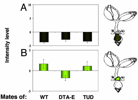
Immediate Postmating Effects on the Lower Reproductive Tract Are Mediated by the Act of Mating and/or Seminal Fluid Components Other than Acps and Sperm. As reported above, at 20 min after the start of mating, we detected in the uterus a significant decrease in fluorescence intensity level and in the distribution of the fluorescence (Figs. 2 A and 3A). These changes occurred in mates of WT, spermless, and DTA-E males (Fig. 3A). Because the change occurs whether or not Acps or sperm are present, the act of mating and/or seminal fluid components other than Acps and sperm must mediate this change. Not all nerve termini innervating the uterus responded to the mating signal because the lower DFS values that we see when females mate to normal, spermless, or DTA-E males indicate a local response (see Table 1). This finding suggests that mating and/or seminal fluid components other than Acps and sperm mediate changes in specific nerve termini innervating the uterus, rather than in all nerve termini in this region.
Fig. 3.
Examples of modulation of vesicle release by specific aspects of mating. (A) The physical act of mating and/or other seminal fluid components other than Acps and sperm induce immediate (20 min) postmating changes in nerve termini innervating the UT. (B) Later (180 min) changes in nerve termini innervating the seminal receptacles are induced by Acps. Values of proANF-EMD fluorescence are plotted as means (OMFILF and LMFILF) ± SEs of mates of WT, DTA-E, and spermless males (TUD). The plotted means are minus virgin female mean values. Thirty-five to 40 female reproductive tracts were examined for each treatment at each time point (see also Table 1).
Changes in activity of nerve termini innervating the uterus immediately after mating could affect contractile activity of uterine muscles and/or induce changes in uterine secretion. This change could modify the uterine environment to promote efficient movement of sperm and seminal proteins toward the sperm storage organs and the upper part of the oviduct after mating, as well as providing optimal conditions for sperm motility and seminal fluid activity.
Later, Acps Induce Changes in Nerve Termini Innervating the Seminal Receptacle. Comparison between virgin females and mates of WT or spermless males at 180 min postmating reveals no change in the fluorescence intensity level of nerve termini innervating the seminal receptacle. Thus, the levels of vesicle release did not seem to change on mating. However, when females were mated to DTA-E males, who do not provide Acps (or sperm), proANF-EMD fluorescence intensity level in nerve termini innervating the seminal receptacles decreased significantly relative to mates of WT (or spermless) males (Fig. 3B; 2-fold decrease relative to mates of WT males, LMFILF: T = -2.59, P < 0.0182; a significant decrease relative to spermless males, LMFILF: T = 2.37, P < 0.029). That these changes are due to Acps is consistent with time that Acps are detectable in females [<6 h; (46–48)] and act on the female reproductive tract (31). Our data suggest that Acps are essential to keep a constant level of neuroactive substance in nerve termini innervating the seminal receptacles, either by mediating the balance between release and accumulation or by inhibiting the release from nerve termini.
Sperm Mediate Changes in Nerve Termini Innervating the Upper Reproductive Tract at Time of Egg Deposition. At 180 min after mating, we also observed a significant change in distribution of proANF-EMD fluorescence in the upper part of the common oviduct. We observed a significant difference in distribution of proANF-EMD between mates of WT and spermless males (DFS(WT); DFS(spermless): 35.96 ± 4.2; 21.77 ± 3.82, T = -2.65, P < 0.017), but no difference between mates of DTA-E males (which are also spermless) and mates of spermless males. Thus, this change correlates with the transfer of sperm during the mating, suggesting that sperm mediate this change. Lower DFS values observed when females mate to spermless males indicate a local response, which means that not all nerve termini innervating the upper part of the common oviduct responded to the signal from sperm. Given that not all nerve termini respond to mating when females mate with spermless males, sperm are needed to mediate changes in most but not all nerve termini innervating the upper common oviduct.
The presence of sperm in the female is essential for rapid onset and high levels of egg deposition. Females that receive no sperm begin to deposit eggs later than females mated to WT males (35), and, if sperm are not stored, egg deposition is low (6). Our finding here, that vesicle release in the COU depends on the presence of sperm from the mating, suggests that this aspect of the sperm effect may be mediated by release of neuromodulators at specific nerve termini innervating the upper common oviduct that could subsequently regulate the oviduct muscles' contractile activity.
Conclusion
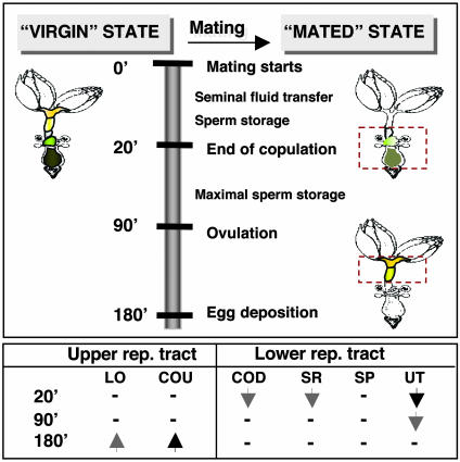
Producing large numbers of progeny shortly after mating ultimately depends on a female's ability to produce mature oocytes ready for fertilization, to manage sperm, and to provide optimal conditions for fertilization. We have shown that mating induces immediate changes in vesicle release/accumulation, at least at peptidergic nerve termini, initially in those innervating the lower part of the Drosophila female reproductive tract. Later, at 3 h postmating, changes are also seen in the upper part of the reproductive tract. We suggest that the immediate postmating changes are the first step in a multiphasic switch in the “state” of the female reproductive tract (Fig. 4). We propose that, before mating, the Drosophila female reproductive tract is poised for future production of large numbers of fertilized eggs but is arrested in a “virgin” state. An example of this arrest occurs in egg production. Oogenesis in unmated females arrests with a maximum of 1–2 mature oocytes per ovariole, and these oocytes are ovulated at a very low rate, ≈2 per day in our strains. Mating causes a drastic change in the physiology of the reproductive tract, to a “mated” state, in part by modulating the activity at nerve termini in the female's reproductive tract. Egg production (49) and ovulation increase (to as much as 50 per day in our strains), sperm are stored, and fertilization occurs very efficiently. We propose that the initial events after mating begin the transition that leads to a reproductive tract environment conducive to efficient production of fertilized eggs. They may also sensitize the female, or some of her reproductive tract tissues, to Acps whose subsequent actions facilitate egg release, sperm storage, etc. to allow the reproductive tract to operate at highest efficiency and coordination. It will be important in the future to identify the effectors whose release is being modulated by mating and Acps to “trip” this switch in state.
Fig. 4.
Postmating changes in vesicle release at nerve termini innervating the female reproductive tract. Net vesicle release, reflected in decreased proANF-EMD fluorescence, is indicated by down arrows (filled = significant, shaded = non-statistically significant trend). Up arrows show net vesicle accumulation, reflected in increased proANF-EMD fluorescence. —, No change.
By elucidating neural effects of Acps and/or sperm in the female reproductive tract, our results begin to address mechanisms by which these effectors cause changes in mated females. These changes are compartmentalized in time and space. In addition, their understanding can assist in elucidating the basis for the interesting evolutionary dynamics of Acps. An unusually high proportion of Acps show characteristics of rapid evolution (19, 50–53). This rapid evolution has been suggested to stem from aspects of sexual conflict and/or sperm competition that drive male effector molecules to change (20, 21). It is of interest to determine whether the female partners of Acps show similar evolutionary dynamics, but those partners remain unidentified. Elucidation of the physiological events triggered by Acps is a first step toward identifying the signaling pathways headed by their partners.
Supplementary Material
Acknowledgments
We thank D. Deitcher, C. Goodman, and R. Hoy for antibodies; D. Deitcher, S. Rao, and P. Rivlin for proANF-EMD flies and advice; and D. Deitcher, J. Ewer, R. Hoy, M. Bloch Qazi, P. Rivlin, Y. Gottlieb, and an anonymous reviewer for comments on the manuscript. We appreciate support from National Science Foundation Grant IAB99-04824 and National Institutes of Health Grant HD38921 (to M.F.W.).
Abbreviations: Acp, accessory gland protein; ANF, atrial natriuretic factor; EMD, emerald variant of GFP; SNAP-25, synaptosome-associated protein of 25 kDa; OMFIL, overall mean fluorescence intensity level; LMFIL, local mean fluorescence intensity level; DFS, distribution of the fluorescence signal; LO, lateral oviduct; COU, upper common oviduct; COD, lower common oviduct; SR, seminal receptacle; SP, spermathecae; UT, uterus; TUD, spermless male; DTA-E, sperm-less, Acp-less male.
References
- 1.Salamonsen, L. A. & Nancarrow, C. D. (1994) in Molecular Biology of the Female Reproductive System., ed. Findlay, J. K. (Academic, San Diego), pp. 289-328.
- 2.Szopa, T. M. (1979) J. Insect Physiol. 28, 475-483. [Google Scholar]
- 3.Mahowald, A. P. & Kambysellis, M. P. (1980) in The Genetics and Biology of Drosophila, eds. Ashburner, M. & Wright, T. R. F. (Academic, New York), Vol. 2D, pp. 141-224. [Google Scholar]
- 4.Miller, A. (1950) in Biology of Drosophila, ed. Demerec, M. (Wiley, New York), pp. 420-534.
- 5.Nachman, R. J., Moyna, G., Williams, H. J., Tobe, S. S. & Scott, A. I. (1998) Bioorg. Med. Chem. 6, 1379-1388. [DOI] [PubMed] [Google Scholar]
- 6.Neubaum, D. M. & Wolfner, M. F. (1999) Genetics 153, 845-857. [DOI] [PMC free article] [PubMed] [Google Scholar]
- 7.Suarez, S. S., Brockman, K. & Lefebvre, R. (1997) Biol. Reprod. 56, 447-453. [DOI] [PubMed] [Google Scholar]
- 8.Bloch Qazi, M. C., Heifetz, Y. & Wolfner, M. F. (2003) Dev. Biol. 256, 195-211. [DOI] [PubMed] [Google Scholar]
- 9.Ottiger, M., Soller, M., Stocker, R. F. & Kubli, E. (2000) J. Neurobiol. 44, 57-71. [PubMed] [Google Scholar]
- 10.Ding, Z., Haussmann, I., Ottiger, M. & Kubli, E. (2003) J. Neurobiol. 55, 372-384. [DOI] [PubMed] [Google Scholar]
- 11.Raabe, M. (1986) in Advances in Insect Physiology, eds. Evans, P. D. & Wigglesworth, V. B. (Academic, London), Vol. 19, pp. 30-154. [Google Scholar]
- 12.Kubli, E. (1992) BioEssays 14, 779-784. [DOI] [PubMed] [Google Scholar]
- 13.Eberhard, W. G. (1996) Female Control: Sexual Selection by Cryptic Female Choice (Princeton Univ. Press, Princeton). [DOI] [PubMed]
- 14.Wolfner, M. F. (1997) Insect Biochem. Mol. Biol. 27, 179-192. [DOI] [PubMed] [Google Scholar]
- 15.Wolfner, M. F. (2002) Heredity 88, 85-93. [DOI] [PubMed] [Google Scholar]
- 16.Gillott, C. (2003) Annu. Rev. Entomol. 48, 163-184. [DOI] [PubMed] [Google Scholar]
- 17.Chapman, T. & Partridge, L. (1996) Nature 381, 189-190. [DOI] [PubMed] [Google Scholar]
- 18.Rice, W. R. (1996) Nature 381, 232-234. [DOI] [PubMed] [Google Scholar]
- 19.Swanson, W. J., Clark, A. G., Waldrip-Dail, H. M., Wolfner, M. F. & Aquadro, C. F. (2001) Proc. Natl. Acad. Sci. USA 98, 7375-7379. [DOI] [PMC free article] [PubMed] [Google Scholar]
- 20.Swanson, W. J. & Vacquier, V. D. (2002) Nat. Rev. Genet. 3, 137-144. [DOI] [PubMed] [Google Scholar]
- 21.Chapman, T. (2001) Heredity 87, 511-521. [DOI] [PubMed] [Google Scholar]
- 22.Kiss, T., Varanka, I. & Benedeczky, I. (1984) Neuroscience 12, 309-322. [DOI] [PubMed] [Google Scholar]
- 23.Lange, A. B., Orchard, I. & Adams, M. E. (1986) J. Comp. Neurol. 254, 279-286. [DOI] [PubMed] [Google Scholar]
- 24.Lange, A. B., Orchard, I. & Brugge, V. A. (1991) Comp. Biochem. Physiol. A 168, 383-391. [Google Scholar]
- 25.Lange, A. B. & Tsang, P. K. C. (1993) J. Insect Physiol. 39, 393-400. [Google Scholar]
- 26.Lange, A. B. & Nykamp, D. A. (1996) Arch. Insect Biochem. Physiol. 33, 183-196. [Google Scholar]
- 27.Lange, A. B. (2002) Peptides 23, 2063-2070. [DOI] [PubMed] [Google Scholar]
- 28.Lee, H. G., Seong, C. S., Kim, Y. C., Davis, R. L. & Han, K. A. (2003) Dev. Biol. 264, 179-190. [DOI] [PubMed] [Google Scholar]
- 29.Monastirioti, M. (2003) Dev. Biol. 264, 38-49. [DOI] [PubMed] [Google Scholar]
- 30.Rao, S., Lang, C., Levitan, E. S. & Deitcher, D. L. (2001) J. Neurobiol. 49, 159-172. [DOI] [PubMed] [Google Scholar]
- 31.Heifetz, Y., Lung, O., Frongillo, E. A., Jr., & Wolfner, M. F. (2000) Curr. Biol. 10, 99-102. [DOI] [PubMed] [Google Scholar]
- 32.Kalb, J. M., DiBenedetto, A. J. & Wolfner, M. F. (1993) Proc. Natl. Acad. Sci. USA 90, 8093-8097. [DOI] [PMC free article] [PubMed] [Google Scholar]
- 33.Boswell, R. E. & Mahowald, A. P. (1985) Cell 43, 97-104. [DOI] [PubMed] [Google Scholar]
- 34.Tram, U. & Wolfner, M. F. (1999) Genetics 153, 837-844. [DOI] [PMC free article] [PubMed] [Google Scholar]
- 35.Heifetz, Y., Tram, U. & Wolfner, M. F. (2001) Proc. R. Soc. London B Biol. Sci. 268, 175-180. [DOI] [PMC free article] [PubMed] [Google Scholar]
- 36.Rao, S. S., Stewart, B. A., Rivlin, P. K., Vilinsky, I., Watson, B. O., Lang, C., Boulianne, G., Salpeter, M. M. & Deitcher, D. L. (2001) EMBO J. 20, 6761-6771. [DOI] [PMC free article] [PubMed] [Google Scholar]
- 37.Schuster, C. M., Davis, G. W., Fetter, R. D. & Goodman, C. S. (1996) Neuron 17, 641-654. [DOI] [PubMed] [Google Scholar]
- 38.Finley, K. D., Taylor, B. J., Milstein, M. & McKeown, M. (1997) Proc. Natl. Acad. Sci. USA 94, 913-918. [DOI] [PMC free article] [PubMed] [Google Scholar]
- 39.Husain, Q. M. & Ewer, J. (2004) J. Neurobiol., in press. [DOI] [PubMed]
- 40.Sollner, T., Bennett, M. K., Whiteheart, S. W., Scheller, R. H. & Rothman, J. E. (1993) Cell 75, 409-418. [DOI] [PubMed] [Google Scholar]
- 41.Schulze, K. L., Broadie, K., Perin, M. S. & Bellen, H. J. (1995) Cell 80, 311-320. [DOI] [PubMed] [Google Scholar]
- 42.Weber, T., Zemelman, B. V., McNew, J. A., Westermann, B., Gmachl, M., Parlati, F., Sollner, T. H. & Rothman, J. E. (1998) Cell 92, 759-772. [DOI] [PubMed] [Google Scholar]
- 43.Klychko, V. A. & Jackson, M. B. (2002) Nature 418, 89-92. [DOI] [PubMed] [Google Scholar]
- 44.Sanes, J. R. & Lichtman, J. W. (1999) Annu. Rev. Neurosci. 22, 389-442. [DOI] [PubMed] [Google Scholar]
- 45.Loewy, A., Liu, W.-S., Baitinger, C. & Willard, M. B. (1991) J. Neurosci. 11, 3412-3421. [DOI] [PMC free article] [PubMed] [Google Scholar]
- 46.Monsma, S. A., Harada, H. A. & Wolfner, M. F. (1990) Dev. Biol. 142, 465-475. [DOI] [PubMed] [Google Scholar]
- 47.Bertram, M. J., Neubaum, D. M. & Wolfner, M. F. (1996) Insect Biochem. Mol. Biol. 26, 971-980. [DOI] [PubMed] [Google Scholar]
- 48.Lung, O. & Wolfner, M. F. (1999) Insect Biochem. Mol. Biol. 29, 1043-1052. [DOI] [PubMed] [Google Scholar]
- 49.Soller, M., Bownes, M. & Kubli, E. (1999) Dev. Biol. 208, 337-351. [DOI] [PubMed] [Google Scholar]
- 50.Aguadé, M., Miyashita, N. & Langley, C. H. (1992) Genetics 132, 755-770. [DOI] [PMC free article] [PubMed] [Google Scholar]
- 51.Tsaur, S.-C., Ting, C.-T. & Wu, C.-I. (1998) Mol. Biol. Evol. 15, 1040-1046. [DOI] [PubMed] [Google Scholar]
- 52.Aguadé, M. (1999) Genetics 152, 543-551. [DOI] [PMC free article] [PubMed] [Google Scholar]
- 53.Begun, D. J., Whitley, P., Todd, B. L., Waldrip-Dail, H. M. & Clark, A. G. (2000) Genetics 156, 1879-1888. [DOI] [PMC free article] [PubMed] [Google Scholar]
Associated Data
This section collects any data citations, data availability statements, or supplementary materials included in this article.


