Summary
Studies have indicated significant pubertal-related differences in hormonal stress reactivity. We report here that prepubertal (30d) male rats display a more protracted stress-induced corticosterone response than adults (70d), despite showing relatively similar levels of adrenocorticotropic hormone (ACTH). Additionally, we show that adrenal expression of the ACTH receptor, melanocortin 2 receptor (Mc2r), is higher in prepubertal compared to adult animals, and that expression of melanocortin receptor accessory protein (Mrap), a molecule that chaperones MC2R to the cell surface, is greater in prepubertal males following stress. Given that these data suggest a pubertal shift in adrenal sensitivity to ACTH, we directly tested this possibility by injecting prepubertal and adult males with 6.25 or 9.375 μg/kg of exogenous rat ACTH and measured their hormone levels 30 and 60 min post-injection. As these doses resulted in different circulating levels of ACTH at these two ages, we performed regression analyses to assess the relationship between circulating ACTH and corticosterone concentrations. We found no difference between the ages in the correlation between ACTH and corticosterone levels at the 30 min time point. However, 60 min following the ACTH injection, we found prepubertal rats had significantly higher corticosterone concentrations at lower levels of ACTH compared to adults. These data suggest that prolonged exposure to ACTH leads to greater corticosterone responsiveness prior to puberty, and indicate that changes in adrenal sensitivity to ACTH may, in part, contribute to the protracted hormonal stress response in prepubertal rats.
Keywords: ACTH, adolescence, adrenal gland, corticosterone, puberty, restraint
1. Introduction
Puberty is marked by many changes in neuroendocrine processes, resulting in significant and extensive influences on an organism’s physiological and neurobehavioral function (Grumbach 2002; Sisk and Foster 2004; Romeo 2005). One such pubertal-related change is the substantial shift in stress reactivity exhibited by the hypothalamic-pituitary-adrenal (HPA) axis, with peripubertal animals showing an extended hormonal stress response compared to adults (McCormick and Mathews 2007; Romeo 2010a, b). For example, following a variety of acute stressors, such as intermittent foot shock, ether inhalation, or restraint, prepubertal male and female rats (i.e., approximately 30 days of age) display adrenal corticosterone responses, both total and free, that last significantly longer (~40 min) than those observed in adults (i.e., greater than 65 days of age; Goldman et al. 1973; Vazquez and Akil 1993; Romeo et al. 2004a; Romeo et al. 2004b; Romeo et al. 2006a; Romeo et al. 2006b; Foilb et al. 2011; Lui et al. 2012). Interestingly, puberty in both human and non-human animal models is also associated with increases in many stress-related physiological and psychological dysfunctions, including depression, anxiety, and drug use and abuse (Andersen 2003; Costello et al. 2003; Dahl 2004; Patton and Viner 2007). Therefore, examining the basic mechanisms that mediate changes in stress reactivity during puberty may contribute to our understanding of the stress-related vulnerabilities often observed during this crucial developmental stage.
The factors that mediate the pubertal change in corticosterone reactivity to stress are presently unclear. As adrenocorticotropic hormone (ACTH) secretion from the anterior pituitary is one of the major signals regulating the release of adrenal corticosterone under stressful conditions (Herman et al. 2003; Bornstein et al. 2008), the slightly higher stress-induced plasma ACTH levels found in prepubertal compared to adult animals may contribute to the age-related difference in corticosterone (Vazquez and Akil 1993; Romeo et al. 2004a; Romeo et al. 2004b). It would appear, however, that a difference in ACTH levels is not the only mediator of the prolonged corticosterone response in prepubertal rats. First, the magnitude of the age-dependent change in stress-evoked ACTH is much smaller than the changes in corticosterone (Vazquez and Akil 1993; Romeo et al. 2004a; Romeo et al. 2004b). Second, and perhaps more importantly, a recent study reported that post-stress ACTH levels are similar across peripubertal animals between 30-50 days, while corticosterone levels were only significantly higher in 30 day old animals (Foilb et al. 2011). As the rate of corticosterone metabolism is similar in peripubertal and adult rats (Schapiro et al. 1971), these data would suggest that pubertal shifts in adrenal responsiveness to ACTH contribute to the prolonged hormonal response observed prior to puberty.
The purpose of the present set of experiments was to test the hypothesis that the greater stress-induced corticosterone response in prepubertal animals is due to increased sensitivity of the prepubertal adrenal glands to ACTH. More specifically, we predict that exogenously administered ACTH will lead to a significantly greater corticosterone response in prepubertal compared to adult rats. We also examined stress-induced changes in the expression of melanocortin 2 receptor (Mc2r) and melanocortin 2 receptor accessory protein (Mrap) mRNA in the prepubertal and adult adrenal gland. MC2R is the receptor for ACTH that when stimulated leads to the production and release of corticosterone from the adrenal gland, while MRAP is an accessory protein to MC2R that helps chaperone the receptor to the membrane surface (Hinkle and Sebag 2009; Webb and Clark 2010; Cooray and Clark 2011). Therefore, based on our prediction of greater ACTH sensitivity prior to puberty, we also hypothesized that prepubertal animals exposed to stress would demonstrate greater expression levels of adrenal Mc2r and/or Mrap compared to adults.
2. Materials and Methods
2.1. Animals and housing
Male Sprague-Dawley rats were obtained from Charles River (Wilmington, MA) at 21 days of age, housed 2 per cage in clear polycarbonate cages (45 × 25 × 20 cm) with wood chip bedding, and were maintained on a 12 h light-dark schedule (lights on at 0900 h). All animals had free access to food and water and the animal room was maintained at 21±2°C. All procedures were carried out in accordance with the guidelines established by the National Institutes of Health Guide for the Care and Use of Laboratory Animals and approved by the Institutional Animal Care and Use Committee (IACUC) of Columbia University.
2.2. Experimental design
Two experiments were conducted. Experiment 1 measured the stress-induced ACTH and corticosterone response, as well as changes in adrenal Mc2r and Mrap expression, in prepubertal (30 days of age) and adult (70 days of age) male rats before, during, and after a 30 min session of restraint stress. Experiment 2 examined the relationship between exogenously administered ACTH and corticosterone secretion in prepubertal and adult male rats, as well as any changes in plasma testosterone levels.
In Experiment 1, prepubertal and adult rats were weighed and rapidly decapitated by a guillotine either before or after a 30 min session of restraint stress. Two time points after the 30 min restraint stress session were examined: immediately after termination of the stressor or 30 min after the stress session (n= 6 per age and time point). These ages and time points were chosen based on previously published studies investigating pubertal-related changes in hormonal stress reactivity (Goldman et al. 1973; Vazquez and Akil 1993; Romeo et al. 2004a). The restraint stress was administered by placing animals in the prone position in wire mesh restrainers, sized so that animals at these different ages were equally restrained. Trunk blood samples were collected in EDTA-coated tubes (Vacutainer K3; Fisher Scientific, Pittsburgh, PA), spun down for 10 min at 2500 rpm in a refrigerated centrifuge, and plasma was removed and stored at −20°C until radioimmunoassays were performed (see below). Adrenal glands were cleaned of fat, weighed, snap frozen on powdered dry ice, and stored at −80°C until they were prepared for determination of Mc2r or Mrap mRNA levels by RT-qPCR (see below). In regards to Mrap, we did not measure the recently characterized Mrap2, as studies in rats indicate that Mrap2 plays little role in mediating ACTH-evoked adrenal corticosterone secretion, particularly in vivo (Gorrigan et al. 2011). Furthermore, we measured mRNA levels of Mc2r and Mrap, rather than protein, as specific, reliable, and quantifiable staining via western blot was not possible with several of the commercially available antibodies we tested (unpublished observation).
For Experiment 2, prepubertal and adult males were weighed and exposed to either 6.25 or 9.375 μg/kg of rat ACTH (Sigma, St. Louis, MO; i.p.; n = 9 and 11, and 7 and 8, respectively) in a 0.9% sterile saline vehicle (1 ml/kg), and decapitated 30 or 60 min after ACTH exposure (n = 4-6 per age and time point). Intraperitoneal injections were chosen as the route of ACTH administration based on a previous study assessing HPA reactivity in adult male rats (Cole et al. 2000). Moreover, these doses of ACTH were chosen based on preliminary studies in our laboratory that indicated that the 6.25 μg/kg dose of ACTH provided physiologically relevant levels of ACTH found in rats exposed to 30 min of restraint stress (~400 pg/ml). We also had initially used a lower dose of ACTH at 3.125 μg/kg, but this dose did not result in any significant increases in ACTH beyond baseline levels at either the 30 or 60 min post-injection time points in prepubertal and adult males (unpublished observation). All animals were injected with the saline vehicle once a day for three days prior to ACTH treatment in order for animals to acclimate to the stress of the handling and injection. Pilot studies indicated that when this pretest procedure is used neither prepubertal nor adult males show significant increases in plasma ACTH, corticosterone, or testosterone to a saline injection on the test day (unpublished observation). Truck blood samples were collected and processed as described above.
2.3. Radioimmunoassays
ACTH, corticosterone, and testosterone assays were conducted using commercially available kits and reagents and were performed as indicated by the supplier. ACTH levels were determined with an ACTH 125I kit (MP Biomedicals; Santa Ana, CA), while corticosterone and testosterone values were determined with Coat-A-Count 125I RIA kits (Siemens Medical Solutions Diagnostics; Malvern, PA). For each assay, all samples were run in duplicate. The lower limits of detectability and intraassay coefficient of variations for each assay were as follows: ACTH, 13.3 pg/ml and 12.5%; corticosterone, 16.52 ng/ml and 8.4%; testosterone, 0.11 ng/ml and 11.6%.
2.4. RT-qPCR
Total RNA was isolated from one adrenal gland using RNeasy Lipid Tissue Mini Kit (Qiagen; Valencia, CA) and included the DNase treatment step with RNase-free DNase (Qiagen) on a QIAcube robot (Qiagen). cDNA was synthesized using above RNA samples as templates and the High-Capacity cDNA Reverse Transcription Kit with RNase Inhibitor (Applied Biosystems; Foster City, CA). RT-qPCR was run on obtained cDNA using TaqMan Fast Advanced Master Mix (Applied Biosystems) and following TaqMan Gene Expression Assays, from Life Technologies: for Mc2r, Assay Id: Rn02082290_s1; for Mrap, Assay Id: Rn01477212_m1; and for housekeeping gene Gapdh, Assay Id: Rn01775763_g1 and run on an Applied Biosystems ViiA7 RT-PCR machine. Raw data were analyzed and CT values were produced using ViiA7 Software v1.1 (Applied Biosystems). Relative gene expression was calculated using the comparative CT method also known as 2−ΔΔCT method (Schmittgen and Livak 2008).
2.5. Statistical Analyses
All data are presented as the mean ± S.E.M. In experiment 1, two-way ANOVAs were used for statistical analyses (age × time point), and significant main effects and interactions were further analyzed with Tukey’s honestly significant difference tests. It should be noted that the ΔCT numbers were used to conduct the statistical analyses for Mc2r and Mrap, while fold change is used to present the data graphically. Linear regression analyses were conducted in Experiment 2 to examine the relationship between plasma corticosterone and ACTH levels. All statistical analyses were performed using GraphPad Prism software (version 5.04) and differences were considered significant when P < 0.05.
3. Results
3.1. Experiment 1
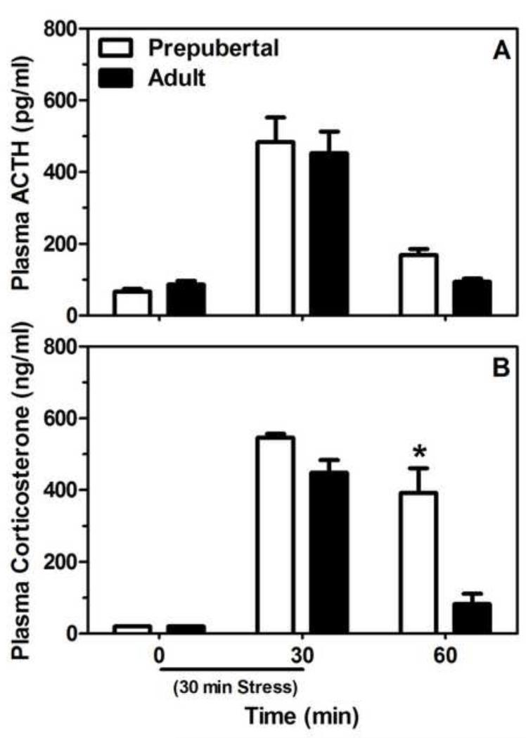
A two-way ANOVA revealed a significant effect of time on ACTH secretion (F (2,26) = 73.74, P < 0.05), such that immediately after termination of the 30 min session of restraint stress prepubertal and adult animals had significantly higher levels of ACTH than animals before or 30 min following restraint (Figure 1A). There was no significant main effect of age or interaction between age and time on ACTH levels. However, there was a significant interaction between age and time on plasma corticosterone levels (F (2,26) = 16.27, P < 0.05) in that restraint led to significant corticosterone responses in both prepubertal and adult males, but prepubertal animals displayed higher levels of corticosterone than adults 30 min following the termination of the stressor (Figure 1B). Together, these data indicate that despite similar stress-induced ACTH levels before and after puberty, prepubertal rats show significantly greater corticosterone responses following stress compared to adults, suggesting a potential pubertal shift in adrenal sensitivity to ACTH.
Figure 1.
Mean (± SEM) plasma ACTH (A) and corticosterone (B) concentrations in prepubertal (30 days of age) and adult (70 days of age) male rats before, during, and after a 30 min session of restraint stress (black bar on the x-axis). The asterisk in panel B indicates a significant difference between the ages at that time point (P < 0.05).
In assessing various parameter of the adrenal gland we found that, although adult body and adrenal weights were significantly higher than those in prepubertal animals (F (1,28) = 1728.0 and 164.5, respectively, P < 0.05; Table 1), the adrenal index (adrenal weight (mg)/ body weight (g)) was significantly greater in prepubertal compared to adult males (F (1, 28) = 73.12, P < 0.05; Table 1). Thus, these data indicate the adrenal gland to body weight ratio is greater prior to pubertal maturation.
Table 1.
Mean (± SEM) body (g) and adrenal (mg) weights and adrenal index (adrenal weight/body weight) of prepubertal and adult male rats. Asterisks indicate a significant difference (P < 0.05) between the ages within that parameter. Note that data are collapsed across time point as only a significant main effect of age was found.
| Age | Body (g) | Adrenal (mg) | Adrenal Index |
|---|---|---|---|
| Prepubertal | 97.7±1.8 | 23.4±1.0 | 0.24±0.01* |
| Adult | 427.4±7.2* | 57.6±2.6* | 0.14±0.01 |
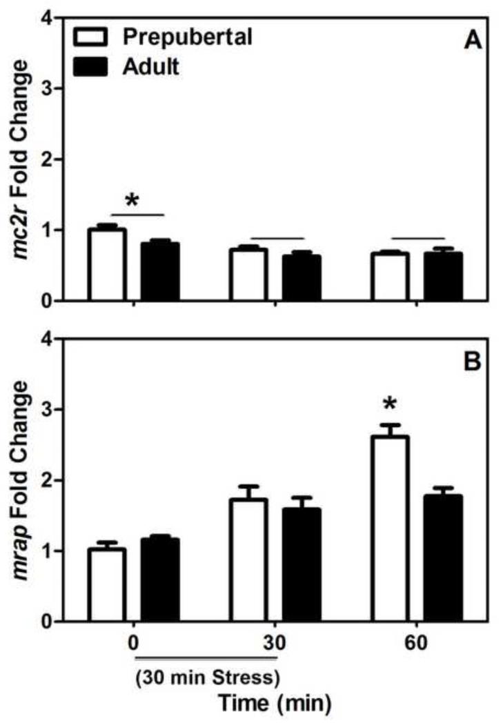
In addition to these gross changes in the adrenal, we found significant main effects of both age and time on Mc2r expression in the adrenal gland such that Mc2r levels are higher in the prepubertal compared to adult animals, and animals at both ages showed lower levels of Mc2r at the 30 and 60 min time points compared to baseline measures (F (1,28) = 4.24 and 9.98, respectively, P < 0.05; Figure 2A). For Mrap expression, we found a significant interaction of age and time (F (2,28) = 5.05, P <0.05; Figure 2B) that indicated that stress resulted in increased adrenal Mrap expression at both ages, but that this increase was significantly higher in prepubertal compared to adult animals at the 60 min time point.
Figure 2.
Mean (± SEM) fold change of adrenal Mc2r (A) and Mrap (B) expression in prepubertal (30 days of age) and adult (70 days of age) male rats before, during, and after a 30 min session of restraint stress (black bar on the x-axis). The asterisk in panel A indicates a significant main effect of time, while the asterisk in panel B indicates a significant difference between the ages at that time point (P < 0.05).
3.2. Experiment 2
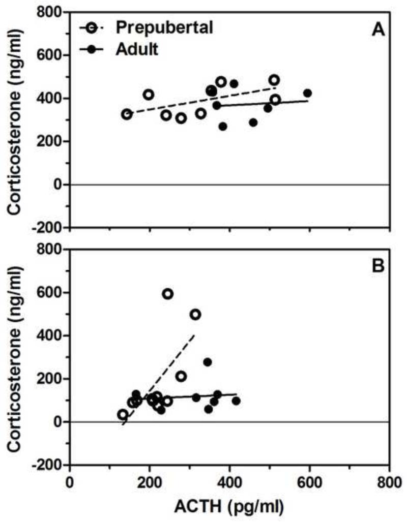
In an effort to further explore this possible age-related change in adrenocortical responsiveness to ACTH, we analyzed the blood samples from ACTH-treated prepubertal and adult males. As the 6.25 and 9.375 mg/kg doses resulted in different plasma levels of ACTH between prepubertal and adult animals after both the post-injection time points, we probed these data with regression analyses comparing the association between ACTH and corticosterone levels at the 30 and 60 min time points. We found that the elevation of the regression lines were not significantly different from each other at the 30 min post-injection time point (P = 0.58, Figure 3A). However, 60 min after the injection, we found that the prepubertal regression line was significantly elevated compared to the adult line (F (1,15) = 5.79, P < 0.05), indicating that lower levels of ACTH are associated with higher levels of corticosterone in animals prior to puberty (Figure 3B).
Figure 3.
Regression analyses of plasma corticosterone and ACTH in prepubertal and adult male rats 30 (A) or 60 (B) min after injection with 6.25 or 9.375 mg/kg ACTH. We found that the prepubertal regression line was significantly elevated (P < 0.05) compared to the adult line 60 min after the injection of ACTH.
In addition to ACTH and corticosterone, we also measured plasma testosterone levels and found no significant association between these ACTH treatments and testosterone levels. However, adult males did show significantly higher plasma testosterone concentrations than prepubertal animals at both post-injection time points (prepubertal = 0.15 ng/ml ± 0.02; adult = 3.23ng/ml ± 0.42). These data indicate that the exogenously administered ACTH did not result in any significant increases in testosterone secretion in either prepubertal or adult male rats, and that our prepubertal animals had not undergone any significant gonadal maturation by 30 days of age.
4. Discussion
These data provide support for the hypothesis that the greater adrenocortical stress response observed in prepubertal compared to adult male rats is, in part, mediated by shifts in adrenal sensitivity to ACTH. Specifically, we found greater stress-induced corticosterone responses in prepubertal compared to adult males in the presence of similar ACTH levels. There also appear to be age- and stress-dependent changes in the expression of signaling molecules in the adrenal gland necessary for ACTH-mediate corticosterone responses, such that prepubertal males have slightly higher levels of Mc2r than adults, and Mrap levels are upregulated by stress to a greater extent in prepubertal compared to adult animals. Moreover, we found that lower levels of ACTH are associated with greater corticosterone responses 60 min after exposure to ACTH in prepubertal compared to adult animals.
Though we attempted to administer similar levels of ACTH to prepubertal and adult animals in our study, the substantial age-dependent changes in body composition and blood volume makes equating hormone levels through exogenous administration at these two developmental stages difficult. Thus, we measured the relationship between ACTH and corticosterone levels in prepubertal and adult animals through regression analyses. It is interesting to note that we found a significant age-related difference in the correlation between ACTH and corticosterone at the 60 min post-injection time point, but not at the 30 min time point. These data are similar to the pubertal differences in the hormonal response we observed after restraint, in that the divergent pattern between these hormones emerges at the later stages of the response (i.e., between 30-60 min). Thus, changes in adrenal sensitivity may only occur after a certain amount or duration of ACTH stimulation has occurred. Future studies will need to further investigate the dynamic nature of this relationship.
Given the observed pubertal-related shift in ACTH responsiveness following restraint stress, the significant age- and stress-dependent changes in adrenal Mc2r and Mrap levels are interesting, as these factors mediate the release of corticosterone in the presence of ACTH (Hinkle and Sebag 2009; Webb and Clark 2010; Cooray and Clark 2011). Though these results lend a mechanistic explanation for the apparent shift in adrenal sensitivity to ACTH, such an interpretation would need to be further strengthened with additional in vitro experiments that could quantify any pubertal differences in MC2R trafficking and/or MC2R and MRAP protein function. Importantly, these data are the first to our knowledge that report the relative expression levels of these transcripts in the adrenals of prepubertal and adult rats before and after exposure to an acute stressor. Independent of any changes in the levels of these signaling molecules, a recent study indicated that the prepubertal adrenal gland appears to be more steroidogenic than the adult adrenal, at least in regards to the synthesis of corticosterone (Foilb et al. 2011). Thus, perhaps the greater capacity of the prepubertal adrenal glands to produce corticosterone also contributes to our observed result of pubertal-related changes in adrenocortical reactivity to stress and ACTH administration.
Though ACTH is a major regulator of adrenal corticosterone secretion during exposure to stressors (Herman et al. 2003; Bornstein et al. 2008), it is not the sole factor that mediate stress-induced corticosterone release. For instance, innervation by the splanchnic nerve can affect corticosterone secretion under stress and non-stress conditions, independent of adrenal stimulation by ACTH (Engeland 1998). Therefore, in addition to pubertal-related shifts in adrenal sensitivity to ACTH, other factors, such as differential splanchnic innervation, may contribute to these different stress-induced corticosterone responses before and after puberty.
It is important to note that while peripheral factors, such as altered adrenal sensitivity to ACTH and sympathetic activation, may contribute to the changes in stress reactivity observed during puberty, central modulation of the HPA axis should not be excluded. A previous study reported reduced negative feedback on the prepubertal compared adult HPA axis, suggesting that the shorter hormone response in adults may be related to greater feedback regulation of the HPA axis through hypothalamic and extrahypothalamic areas (Goldman et al. 1973). A number of experiments have also indicated that prepubertal animals display greater stress-induced activation of the hypothalamus, specifically within the paraventricular nucleus, compared to adult rats (Viau et al. 2005; Romeo et al. 2006a; Lui et al. 2012). Taken together, these studies suggest a role for both the central and peripheral nervous systems in mediating pubertal-related changes in HPA reactivity.
In addition to the differential corticosterone responses observed in prepubertal and adult males, previous studies have found that exposure to acute stress increases plasma testosterone levels in adults, while decreasing it in prepubertal males (Romeo et al. 2004a; Foilb et al. 2011). Our current data indicate that the changes in testosterone secretion following stress are not due to changes in stress-induced ACTH, as we found no effect of exogenously applied ACTH on testosterone secretion in either prepubertal or adult males. Therefore, it would appear the effects of stress on testosterone secretion are through direct activation of elements of hypothalamic-pituitary-gonadal (HPG) axis, such as gonadotropin-releasing hormone and luteinizing hormone (Euker et al. 1975; Lopez-Calderon et al. 1990), instead of through direct modulation by the HPA axis.
In conclusion, these studies provide evidence of age-related changes in adrenal sensitivity to ACTH that may contribute to the significant shifts in hormonal stress reactivity exhibited during pubertal maturation. More specifically, we show that prepubertal male rats display greater stress-induced corticosterone responses compared to adults, despite similar post-stress levels of ACTH. These hormonal responses are paralleled by overall greater expression levels of ACTH signaling molecules in the prepubertal adrenal gland, such as Mc2r and Mrap. Furthermore, we have demonstrated that 60 min after exposure to exogenously administered ACTH prepubertal rats have significantly higher levels of corticosterone in the presence of lower levels of ACTH compared to adults. Though additional work is clearly needed to further parse out the influence of central and peripheral factors on the dramatic change in stress reactivity observed during pubertal development, our data indicate that changes in adrenal sensitivity to ACTH may contribute to these developmental differences in HPA function.
Acknowledgements
We would like to thank Page Buchanan for excellent animal care. This work was supported in part from a grant to Barnard College from the Undergraduate Science Education Program of the Howard Hughes Medical Institute and grants from the National Institute of Mental Health MH-090224 and the National Science Foundation IOS-1022148 (to R.D.R.).
Funding
This work was supported in part from a grant to Barnard College from the Undergraduate Science Education Program of the Howard Hughes Medical Institute and grants from the National Institute of Mental Health MH-090224 and the National Science Foundation IOS-1022148 (to R.D.R.).
Footnotes
Publisher's Disclaimer: This is a PDF file of an unedited manuscript that has been accepted for publication. As a service to our customers we are providing this early version of the manuscript. The manuscript will undergo copyediting, typesetting, and review of the resulting proof before it is published in its final citable form. Please note that during the production process errors may be discovered which could affect the content, and all legal disclaimers that apply to the journal pertain.
Conflict of interest
All authors declare that they have no conflicts of interest.
Contributions
RDR and INK designed the studies, SM, SES, BSH, and MS all collected, analyzed, and managed data files, and all authors have contributed and have approved the final manuscript.
References
- Andersen SL. Trajectories of brain development: point of vulnerability or window of opportunity. Neurosci. Biobehav. Rev. 2003;27:3–18. doi: 10.1016/s0149-7634(03)00005-8. [DOI] [PubMed] [Google Scholar]
- Bornstein SR, Engeland WC, Ehrhart-Bornstein M, Herman JP. Dissociation of ACTH and glucocorticoids. Trend. Endocrinol. Metab. 2008;19:175–180. doi: 10.1016/j.tem.2008.01.009. [DOI] [PubMed] [Google Scholar]
- Cole MA, Kim PJ, Kalman BA, Spencer RL. Dexamethasone suppression of corticosterone secretion: evaluation of the site of action by receptor measures and functional studies. Psychoneuroendocrinology. 2000;25:151–167. doi: 10.1016/s0306-4530(99)00045-1. [DOI] [PubMed] [Google Scholar]
- Cooray SN, Clark AJL. Melanocortin receptors and their accessory proteins. Mol. Cell. Endocrinol. 2011;331:215–221. doi: 10.1016/j.mce.2010.07.015. [DOI] [PubMed] [Google Scholar]
- Costello EJ, Mustillo S, Erkanli A, Keeler G, Angold A. Prevalence and development of psychiatric disorders in childhood and adolescence. Arch. Gen. Psychiatry. 2003;60:837–844. doi: 10.1001/archpsyc.60.8.837. [DOI] [PubMed] [Google Scholar]
- Dahl RE. Adolescent brain development: a period of vulnerabilities and opportunities. Ann. NY Acad. Sci. 2004;1021:1–22. doi: 10.1196/annals.1308.001. [DOI] [PubMed] [Google Scholar]
- Engeland WC. Functional innervation of the adrenal cortex by the splanchnic nerve. Horm. Metab.Res. 1998;30:311–314. doi: 10.1055/s-2007-978890. [DOI] [PubMed] [Google Scholar]
- Euker JS, Meites J, Riegle GD. Effects of acute stress on serum LH and prolactin in intact, castrate and dexamethasone-treated male rats. Endocrinology. 1975;96:85–92. doi: 10.1210/endo-96-1-85. [DOI] [PubMed] [Google Scholar]
- Foilb AR, Lui P, Romeo RD. The transformation of hormonal stress responses throughout puberty and adolescence. J. Endocrinol. 2011;210:391–398. doi: 10.1530/JOE-11-0206. [DOI] [PubMed] [Google Scholar]
- Goldman L, Winget C, Hollingshead GW, Levine S. Postweaning development of negative feedback in the pituitary-adrenal system of the rat. Neuroendocrinology. 1973;12:199–211. doi: 10.1159/000122169. [DOI] [PubMed] [Google Scholar]
- Gorrigan RJ, Guasti L, King P, Clark AJ, Chan LF. Localisation of the melanocortin-2-receptor and its accessory proteins in the developing and adult adrenal gland. J. Mol. Endocrinol. 2011;46:227–232. doi: 10.1530/JME-11-0011. [DOI] [PMC free article] [PubMed] [Google Scholar]
- Grumbach MM. The neuroendocrinology of human puberty revisited. Horm. Res. 2002;57:2–14. doi: 10.1159/000058094. [DOI] [PubMed] [Google Scholar]
- Herman JP, Figueiredo H, Mueller NK, Ulrich-Lai Y, Ostander MM, Choi DC, Cullinan WE. Central mechanisms of stress integration: hierarchical circuitry controlling hypothalamic-pituitary-adrenocortical responsiveness. Front. Neuroendocrinol. 2003;24:151–180. doi: 10.1016/j.yfrne.2003.07.001. [DOI] [PubMed] [Google Scholar]
- Hinkle PM, Sebag JA. Structure and function of the melanocortin2 receptor accessory protein (MRAP) Mol. Cell. Endocrinol. 2009;300:25–31. doi: 10.1016/j.mce.2008.10.041. [DOI] [PMC free article] [PubMed] [Google Scholar]
- Lopez-Calderon A, Gonzalez-Quijano MI, Tresguerres JAF, Ariznavarreta C. Role of LHRH in the gonadotrophin response to restraint stress in intact male rats. J. Endocrinol. 1990;124:241–246. doi: 10.1677/joe.0.1240241. [DOI] [PubMed] [Google Scholar]
- Lui P, Padow VA, Franco D, Hall BS, Park B, Klein ZA, Romeo RD. Divergent stress-induced neuroendocrine and behavioral responses prior to puberty. Physiol. Behav. 2012;107:104–111. doi: 10.1016/j.physbeh.2012.06.011. [DOI] [PMC free article] [PubMed] [Google Scholar]
- McCormick CM, Mathews IZ. HPA function in adolescence: role of sex hormones in its regulation and the enduring consequences of exposure to stressors. Pharmacol. Biochem. Behav. 2007;86:220–233. doi: 10.1016/j.pbb.2006.07.012. [DOI] [PubMed] [Google Scholar]
- Patton GC, Viner R. Pubertal transitions in health. Lancet. 2007;369:1130–1139. doi: 10.1016/S0140-6736(07)60366-3. [DOI] [PubMed] [Google Scholar]
- Romeo RD. Neuroendocrine and behavioral development during puberty: a tale of two axes. Vitam. Horm. 2005;71:1–25. doi: 10.1016/S0083-6729(05)71001-3. [DOI] [PubMed] [Google Scholar]
- Romeo RD. Adolescence: a central event in shaping stress reactivity. Dev. Psychobiol. 2010a;52:244–253. doi: 10.1002/dev.20437. [DOI] [PubMed] [Google Scholar]
- Romeo RD. Pubertal maturation and programming of hypothalamic-pituitary-adrenal reactivity. Front. Neuroendocrinol. 2010b;31:232–240. doi: 10.1016/j.yfrne.2010.02.004. [DOI] [PubMed] [Google Scholar]
- Romeo RD, Bellani R, Karatsoreos IN, Chhua N, Vernov M, Conrad CD, McEwen BS. Stress history and pubertal development interact to shape hypothalamic pituitary adrenal axis plasticity. Endocrinology. 2006a;147:1664–1674. doi: 10.1210/en.2005-1432. [DOI] [PubMed] [Google Scholar]
- Romeo RD, Karatsoreos IN, McEwen BS. Pubertal maturation and time of day differentially affect behavioral and neuroendocrine responses following an acute stressor. Horm. Behav. 2006b;50:463–468. doi: 10.1016/j.yhbeh.2006.06.002. [DOI] [PubMed] [Google Scholar]
- Romeo RD, Lee SJ, Chhua N, McPherson CR, McEwen BS. Testosterone cannot activate an adult-like stress response in prepubertal male rats. Neuroendocrinology. 2004a;79:125–132. doi: 10.1159/000077270. [DOI] [PubMed] [Google Scholar]
- Romeo RD, Lee SJ, McEwen BS. Differential stress reactivity in intact and ovariectomized prepubertal and adult female rats. Neuroendocrinology. 2004b;80:387–393. doi: 10.1159/000084203. [DOI] [PubMed] [Google Scholar]
- Schapiro S, Percin CJ, Kotichas FJ. Half-life of plasma corticosterone during development. Endocrinology. 1971;89:284–286. doi: 10.1210/endo-89-1-284. [DOI] [PubMed] [Google Scholar]
- Schmittgen TD, Livak KJ. Analyzing real-time PCR data by the comparative Ct method. Nat. Protoc. 2008;3:1101–1108. doi: 10.1038/nprot.2008.73. [DOI] [PubMed] [Google Scholar]
- Sisk CL, Foster DL. The neural basis of puberty and adolescence. Nat. Neurosci. 2004;7:1040–1047. doi: 10.1038/nn1326. [DOI] [PubMed] [Google Scholar]
- Vazquez DM, Akil H. Pituitary-adrenal response to ether vapor in the weanling animal: characterization of the inhibitory effect of glucocorticoids on adrenocorticotropin secretion. Pediatr. Res. 1993;34:646–653. doi: 10.1203/00006450-199311000-00017. [DOI] [PubMed] [Google Scholar]
- Viau V, Bingham B, Davis J, Lee P, Wong M. Gender and puberty interact on the stress-induced activation of parvocellular neurosecretory neurons and corticotropin-releasing hormone messenger ribonucleic acid expression in the rat. Endocrinology. 2005;146:137–146. doi: 10.1210/en.2004-0846. [DOI] [PubMed] [Google Scholar]
- Webb TR, Clark AJL. Minireview: the melanocortin 2 receptor accessory proteins. Mol. Endocrinol. 2010;24:475–484. doi: 10.1210/me.2009-0283. [DOI] [PMC free article] [PubMed] [Google Scholar]


