Abstract
Zebrafish are an ideal model organism to research cancer. Zebrafish embryos and larvae are optically translucent, which has made imaging multiple processes in development and disease possible. When coupled with fluorescent imaging techniques, zebrafish are fast becoming a model of choice for following tumor formation. This is highlighted by recent studies using fluorescent proteins to image xenograft transplantation, neovascularization, growth responses to drug treatments, and self-renewal. Fluorescent labeled tumors can be generated in zebrafish by multiple methods including chemical mutagenesis, oncogene expression by mosaic or stable transgenesis, or genetic mutations that are predisposing to cancer. In this chapter, we highlight the studies that have employed fluorescence to follow critical aspects of tumorigenesis, with particular focus on providing methods for labeling, isolating, transplanting, and imaging fluorescently labeled tumors in zebrafish.
I. Introduction
Fluorescent proteins have a long history of being used in experimental model systems to track cell types, monitor molecular pathway regulation, and label intracellular organelles. The importance of fluorescent proteins to biological studies was underscored by the awarding of the noble prize for chemistry in 2008 to Osamu Shimomura, Martin Chalfie, and Roger Y. Tsien for the discovery and development of green fluorescent protein (GFP) as a research tool. Osamu Shimomura first isolated GFP from the jellyfish Aequorea victoria (Shimomura et al., 1962), Martin Chalfie was the first to use GFP to label cells in vivo in Caenorhabditis elegans (Chalfie et al., 1994), and Roger Y Tsien discovered how GFP fluoresces and created stronger fluorescing variants of GFP aswell as many of the color variants (Tsien, 1998, 2010). Because zebrafish are optically translucent during early larval development, the use of fluorescence imaging of cancer has proven to be a major advantage of this system. Moreover, the development of adult zebrafish and Medaka that are translucent as adults further increases the utility of the model to assess cancer in older animals (Wakamatsu et al., 2001; White et al., 2008). In initial studies using transgenic zebrafish that develop Myc-induced T-cell acute lymphoblastic leukemia (T-ALL), we successfully labeled tumor cells by fusing GFP with the Myc oncogene (Langenau et al., 2003). Leukemias could be imaged to assess tumor growth kinetics and introduced into recipient animals to image tumor engraftment. Since this first report was published in 2003, the field has continued to expand the use of imaging to model more complex behaviors of cancer including xenograft transplantation, neovascularization, growth responses to drug treatments, use of unbiased genetic screens to identify mutants associated with cancer, and assessment of self-renewal. Highlighted below are multiple cancer studies that use fluorescent proteins or dyes to track tumor growth in vivo in zebrafish with an emphasis on describing methods that can be used to study these processes.
II. Fluorescent Proteins and Transgenic Models of Cancer
A. Types of Fluorescent Proteins and Practical Considerations
Numerous fluorescent protein variants are currently available and can be used in imaging cancer in zebrafish (Table I). Moreover, several newly described proteins may aid in expanding the colors and combinatorial use of proteins to image tumor cell subpopulations (Table II). For example, E2-crimson is a far shifted red fluorescent protein that can be excited with a 633 nm laser and could be used in combination with multiple fluorescent reporters. Most experimental models have either used GFP or red fluorescent proteins to distinguish tumor cell subpopulations because of the extreme spectral differences between these proteins; however, other fluorescent protein combinations can be used. For example, we have had good success in imaging membrane localized blue fluorescence protein (lyn-amCyan) from GFP and monomeric RFP within the same animal using confocal imaging (unpublished results). In these experiments, GFP and amCyan are expressed in nonoverlapping cell types and amCyan is excited using a 458 nm laser with 475– 525 nm filter, while GFP uses a 488 nm laser and a 505–530 nm filter. Monomeric RFP is visualized with a 543 nm laser with a 560–615 nm filter. Use of cell labeling approaches that target fluorescence to specific organelles will continue to expand the types and numbers of labels that can be delineated within cell populations. For example, use of nuclear localized and membrane localized fluorescent proteins may help in refining fluorescence labeling of discrete cell populations. Another consideration for imaging multiple colors within the same animal are the extent to which each fluorescent protein is expressed. For example, although many proteins have seemingly nonoverlapping spectral properties, if one fluorescent protein is vastly overexpressed in comparison to the others, it will often bleed into the other channels making multispectral imaging difficult. Another consideration is the fluorescent protein half-life and the time required for maturation. For example, fluorescent protein fusions with histones are very long-lived (Foudi et al., 2009) and will likely persist in cells that no longer express the given gene promoter. The time required for proteins to fold correctly and fluoresce also differs greatly between proteins and should be accounted for if one is attempting to capture differentiation within specific cell subpopulations. Finally, cells from fluorescent transgenic tumors can be used in fluorescence activated cell sorting (FACS) to identify populations of cells that may not be able to be imaged using conventional approaches. For example, mCherry and dsRED express are both excited by confocal laser lines in the 543 nm range and cannot be easily delineated. By contrast, FACS using a 488 nm and 561 nm laser line can easily distinguish these fluorophores. In the end, we commonly approach the above problems by assessing various color combinations prior to developing stable transgenic lines, ensuring that our approaches for color combinations will ultimately yield informative results.
Table I.
Selected examples of fluorescent proteins being used in zebrafish cancer models
| Fluorescent protein |
Cancer type | Transgenic/ mosaic expression |
Transplantation Results |
Reference |
|---|---|---|---|---|
| GFP/EGFP | Pancreatic tumors | ptfla-EGFP-kRASG12V, stable line | NA | Park et al., 2008 |
| B-cell acute lymphoblastic leukemia | Xenopus-elongation- factor l -EGFP-TEL-AML1 and zebrafish-beta-actin- EGFP- TEL-AML1. stable lines | IR transplants | Sabaawy et al., 2006 | |
| Melanoma, lymphoma, hepatocarcinoma, gut carcinoma, RMS | Gt(GFP-H-RASV12), stable line | NA | Santoriello et al., 2009 | |
| Liver tumors induced with DMBA and DBP | Ifabp-GFP, stable line | NA | Lam et al., 2006 | |
| T-cell acute lymphoblastic leukemia (T-ALL) | rag2-EGFP-Myc, mosaic expression, rag2-EGFP-Myc. stable line | IR transplants | Langenau et al., 2003 | |
| Genetic screen for T-cell malignancies | lck-EGFP, stable line | IR transplants | Frazer et al., 2009 | |
| T-ALL drug screen | rag2-EGFP-Myc, mosaic | Syngeneic transplants into adults and 5 dpf larvae | Mizgirev and Revskoy, 2010 | |
| ERMS | rag2-GFP+rag-kRASG12D, mosacis | NA | Langenau et al., 2008 | |
| RFP | C8161-RFP labeled melanoma | NA | Xenograft transplants into embryos | Topczewska et al., 2006 |
| Human U251-RFP glioblastoma | NA | Xenograft transplants into embryos | Geiger et al., 2008; Lally et al., 2007 | |
| dsRED/dsREDExpress | T-ALL | rag2-mMyc+rag2-dsRED2. mosaic | Syngeneic transplants | Langenau et al., 2008; Smith et al., 2010 |
| YFP/ zs Ycllow | T-ALL | rag2-mMyc+rag2-zs Yellow, mosaic | Syngeneic transplants | Smith et al., 2010 |
| A mcyan | T-ALL | rag2-mMyc+rag2-Amcyan, mosaic | Syngeneic transplants | Smith et al., 2010 |
Table II.
Fluorescent protein excitation and emission spectra
| Fluorescent protein | Color | Excitation maximum | Emission maximum | Source |
|---|---|---|---|---|
| CFP | Blue | 435 | 485 | Invitrogen |
| Amcyan 1 | Blue | 458 | 489 | Clontech |
| GFP/EGFP | Green | 484 | 510 | Clonetech |
| YFP/zs Yellow | Yellow | 529 | 539 | Clonetech |
| tagRFP | Red | 555 | 584 | Invitrogen |
| dsRED/dsREDExpress | Red | 554–563 | 581–592 | Clonetech |
| mCherry | Red | 587 | 610 | Clonetech |
| m-KATE | Far Red | 588 | 635 | Ervogen |
| Katushka | Far Red | 588 | 635 | Ervogen |
| E2-Crimson | Far Red | 611 | 646 | Clonetech |
B. Creation of Stable Transgenic Cancer Models
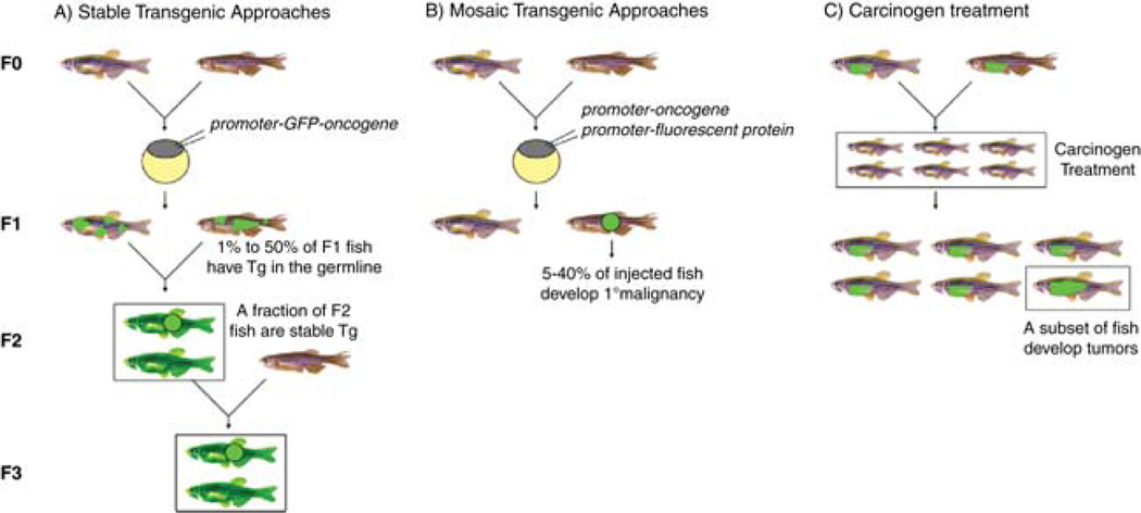
There are several ways to create transgenic zebrafish models of cancer. The most straightforward approach is to create stable transgenic animals that express your oncogene of interest fused with a fluorescent protein driven by a tissue-restricted promoter (Fig. 1A). We have previously used GFP fusions with mouse c-Myc and zebrafish bcl2 to generate T-ALLs and thymic hyperplasia, respectively (Langenau et al., 2003, 2005). Park and colleagues utilized GFP fusions with kRASG12 V to create a model of pancreatic adenocarcinoma, while other groups have utilized GFP and RED fluorescent protein fusions to generate a transgenic model of RAS-induced liver cancer (Park et al., 2008; Santoriello et al., 2009). A major concern with fusion constructs is that the fluorophore does not inhibit the function of the oncogene and that the half-life of the oncogene does not affect expression of the fluorescent protein. For example, the Myc transcription factor has a very short half-life of approximately ~30 min (Sears et al., 2000); and thus, EGFP–Myc fusions cannot be easily detected in normal thymocytes. However, following transformation, GFP expression is elevated and can be easily seen in disseminated leukemias. These experimental observations suggest that Myc may be stabilized during transformation allowing GFP to mature and fluoresce to a greater extent following transformation. To obviate issues surrounding the creation of fusion genes, nonfluorescent stable transgenic lines that express oncogenes can be derived and these lines can be bred to existing fluorescent reporter lines.
Fig. 1.
Methods for creating fluorescent-labeled cancers in zebrafish. Approaches for making stable (A) and mosaic (B) transgenic and chemically induced models (C) of fluorescent-labeled cancer. Stable transgenic animals capable of germline transmission are denoted by boxes inA. (C) F1 stable transgenic zebrafish treated as larva with carcinogen are denoted as smaller fish and contained within a boxed region. These animals are monitored for tumor onset, with a subset of animals developing fluorescent-labeled disease (denoted as an adult sized animalwith an enlarged region of GFP+ cells, single fish boxed). (See color plate.)
Transgenic approaches in zebrafish are not the only option to create robust models of cancer (Fig. 1C). For example, Lam et al. used chemical carcinogens to reproducibly develop lesions in the liver and used bioinformatics approaches to show that these models accurately recapitulate specific features of human disease. By treating lfabp-GFP fish with 7,12-dimethylbenz(a)anthracene (DMBA) and dibenzo(a,l)pyrene (DBP) one could produce liver tumors that are GFP labeled (Lam et al., 2006). Such approaches could easily be extended to germ cell tumors, liver tumors, pancreatic tumors, adenocarcinoma of the intestine, chondrosarcomas, and spindle cell sarcomas where carcinogens reproducibly induce specific subtypes of tumors (Mizgireuv and Revskoy, 2006; Neumann et al., 2009; Spitsbergen et al., 2000a, 2000b). However, stable transgenic lines would be required that drive fluorescent protein expression within targeted cell types. There are also various genetic models that are predisposed to developing cancer, including TILLING mutants that have loss of function mutations in P53, APC, and PTENA and PTENB (Berghmans et al., 2005; Faucherre et al., 2008; Frazer et al., 2009; Neumann et al., 2009; Phelps et al., 2009), viral insertion mutants that disrupt ribosomal protein gene function (Amsterdam et al., 2004), and ENU-induced mutants that have increased susceptibility to developing germ cell tumors when treated with mutagens DMBA and MNNG (Neumann et al., 2009). In these models, transgenic reporters could be bred into mutant lines and the progeny uncrossed to create homozygous mutant animals that contain the transgenes of interest. By contrast, Frazer et al. carried out an ENU-mutagenesis screen to identify genetic mutations that are predisposed to T-cell malignancies (Frazer et al., 2009). This screen was carried out in a lck-EGFP transgenic background allowing for animals to be assessed for expansion of GFP-labeled T-cells outside of the thymus as an indicator of disease. Three recessive mutants and one dominant mutant were obtained from this innovative forward genetic screen. Molecular analysis showed that these leukemias mimic a wide range of human T-cell malignancies (Frazer et al., 2009) and provide exciting opportunities to identify new genetic lesions associated with T-ALL initiation, growth, and dissemination.
C. Mosaic Transgenic Approaches
In addition to well-established models of chemical, genetic, and stable transgenic models of cancer, our laboratory has largely focused on utilizing mosaic transgenic animals to develop models of cancer. In these approaches, animals are microinjected at the one-cell stage of life with multiple linearized transgenes (Fig. 1B). Because transgenes integrate into the genome as concatemeric arrays, tumors coexpress multiple transgenes providing efficient methods to deliver an oncogene and fluorescent reporter to developing tumors (Langenau et al., 2008). Such approaches are particularly advantageous to studying early developing pediatric malignancies like T-ALL and embryonal rhadomyosarcoma (ERMS), because stable transgenic animals would develop tumor prior to reproductive maturity making maintenance of stable transgenic lines difficult (Langenau et al., 2003). Moreover, mosaic transgenic animals likely mimic human disease in that not all cells of a particular lineage are predisposed to cancer. For example, in stable transgenic zebrafish models of Myc-induced T-ALL all thymocytes will express the oncogene; whereas oncogene expression in mosaic animals is confined to a subcompartment of hematopoietic stem cells and thus only a subset of lymphocytes express Myc and become transformed. Coinjection approaches also allow the investigator rapid means of introducing genetic lesions into varied backgrounds that does not require multiple generations of crossing to obtain the desired genotype. Finally, stable transgenic approaches require time to raise injected fish, screen for transgenic founders, and then expand the line, whereas mosaic transgenic animals will develop fluorescent tumors as F0 offspring. The one major confounding issue for mosaic transgenic approaches is that many animals need to be microinjected to create a small cohort of tumor-bearing animals. For example, 100% of stable transgenic rag2-EGFPMyc fish develop leukemia by 80 days of life, while only 5% of surviving mosaic animals injected with rag2-Myc will develop leukemia (Langenau et al., 2003). One investigator can microinject approximately 500 surviving one-cell stage zebrafish in a day; thus, on an average 25 leukemic fish will be produced per day of microinjection. By contrast, 40% one-cell stage animals injected with rag2-kRASG12D develop ERMS, providing larger numbers of animals to be used in a given study (Langenau et al., 2007).
D. Selecting Strains of Zebrafish for Imaging Cancer
Once a strategy is established to create fluorescent-labeled zebrafish cancers, one must consider which strain of animals to use. Recent development of caspar zebrafish provides exciting opportunities to visualize tumor cells in animals using real-time imaging and will likely be useful for assessing cell migration, metastasis, and kinetics of tumor growth (White et al., 2008). These fish lack melanocytes and iridophores and were created by breeding together roy and nacre mutants (White et al., 2008). caspar fish must be maintained as double homozygous mutant animals; thus, for developing stable transgenic models of cancer, multiple rounds of crossing and in crossing will be required to generate a stable transgenic line in the caspar background. Our laboratory has found that for many imaging applications, transparent larval and adult zebrafish are not required. For example, we have had great success in imaging tumor evolution, growth, and dissemination in wild-type AB-strain fish. Fluorescent-labeled T-ALL develops in the thymus and then disseminates widely. T-ALL growth can be easily observed in both primary and transplanted leukemia. Moreover, ERMS develop very early in development during a stage when the larvae are still largely translucent. We find that imaging fish up to 30 days of life by confocal and dual photon imaging is remarkably easy, albeit sometimes one will have to image away from the rare melanocyte.
Use of syngeneic zebrafish is recommended if cell transplantation will be used to image self-renewal or engraftment kinetics (Mizgireuv and Revskoy, 2006, 2010; Smith et al., 2010). These animals are genetically identical and thus, amenable to cell transplantation in the absence of immune ablation. The benefit of using syngeneic strains of zebrafish comes from immune matching rather than optical translucence. For short-term transplant assays, fluorescent tumors can be transplanted into irradiated caspar animals; however, irradiation doses required for immune ablation appear to be lower in these animals (unpublished data) and the immune system will recover by 21 days of life. Recent development of an optically translucent syngeneic zebrafish from the Revskoy laboratory will likely provide robust models for assessing both cell transplantation and imaging (Mizgirev and Revskoy, 2010).
III. Macroscopic Observation of Tumor Growth
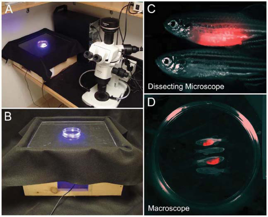
For many applications, imaging fluorescent-labeled cancers in whole zebrafish is advantageous. For example, whole animal imaging can be used to assess growth kinetics and overall dissemination. Cell transplantation can also be used to assess if fluorescent-labeled cell populations are fully transformed – a hallmark of cancer. Finally, limiting dilution cell transplantation into syngeneic zebrafish can be used to quantify self-renewing cell numbers contained within the cancer mass (Smith et al., 2010). We summarize two approaches to image these processes at the macroscopic level using traditional epifluorescence imaging with a dissecting microscope or the LED fluorescence macroscope (Fig. 2).
Fig. 2.
Macroscopic imaging of fluorescent tumor engraftment. (A) The LED fluorescence macroscope (left) juxtaposed with an epifluorescent Olympus SZ16 stereomicroscope (right). (B) Enlarged view of the LED fluorescence macroscope. (C) Images of two adult fish, one with a rag2-dsREDexpress+ T-ALL (top) and one control fish (bottom). (D) Macroscope image of four adult zebrafish two of which have developed rag2-dsREDexpress+ T-ALL. Petri dish is 10 cm in diameter.
A. Stereomicroscopy to Image Tumor Growth
Most zebrafish laboratories are equipped with a fluorescent dissecting microscope capable of imaging embryos, larvae, and adult fish. For macroscopic imaging of embryos and larvae, stereomicroscopy is the preferred method for screening and imaging fish. Our laboratory currently uses an Olympus SZ16 stereomicroscope fitted with a Prior lumen 200 fluorescence emission setup and a DP72 color camera (Fig. 2A). Stereomicroscopy can be used to image single adult animals; however, it is usually not possible to capture the whole fish within the viewing field. Our stereomicroscope can only image a 7 cm2 viewing area, which allows for only approximately 2/3 the length of an adult zebrafish to be imaged (Fig. 2C).
B. LED Fluorescence Macroscope Imaging
To aid in screening transplant animals that have engrafted fluorescent-labeled T-ALL and ERMS, our lab has developed a novel method for imaging a whole plate of adult zebrafish using the LED fluorescence microscope (Fig. 2). This machine consists of LED fluorescent lights that illuminate a 10 by 10 cm viewing area, a series of cameras that interface with a computer, and various filter sets that allow only emitted light of a specific wavelength to be detected. The LED fluorescence macroscope is inexpensive, can image up to 30 animals simultaneously, is capable for real-time imaging, and can capture live, time-lapse dual-fluorescence images using two cameras. We have initially described the use of this machine to image fluorescent-labeled T-ALLs (Smith et al., 2010) and showed that this machine can accurately identify animals that develop fluorescent-labeled T-ALL by 30 days post-transplantation. This machine can easily distinguish Amcyan, GFP, zsYellow, dsREDexpress, and mCherry in both leukemia and muscle transgenic lines; however, fluorescence must be bright within the target cell populations of transgenic animals. Additionally, small focal tumors will be hard to image; however, as tumors continue to grow they can be imaged by the macroscope.Blackburn et al. (2011) provide new experimental protocols to image fluorescently labeled ERMS and to label zebrafish with fluorescent elastomers. This manuscript provides a comprehensive description of how to use, build, and troubleshoot the macroscope (Blackburn et al., 2011).
IV. Microscopic Observation in Tumorigenesis
One of the unique advantages of the zebrafish model is the ability to image individual cells using confocal, spinning disc, and two-photon microscopy. Although elegantly used to image important processes in development including axonal guidance (Kucenas et al., 2008), vascular development (Lawson and Weinstein, 2002a, 2002b; Nicoli et al., 2010), cell movements in embryogenesis (Gong et al., 2004; Keller et al., 2008), and the emergence of hematopoietic stem cells (Bertrand et al., 2010; Kissa and Herbomel, 2010); cellular microscopic observation of the hallmarks of cancer are just beginning to be explored. In this section, we highlight the types of technologies that can be utilized to image at the single-cell level in live zebrafish (Table III).
Table III.
Comparing laser scanning confocal, spinning disc confocal, and two-photon microscopy
| Laser scanning confocal microscopy (LSCM) |
Spinning disc confocal microscopy |
Two-photon microscopy | |
|---|---|---|---|
| Light source | Visible/UV lasers 365–647 nm range | Visible/UV lasers 365–647 nm range – laser excitation not required | Infrared 700–1000 nm range |
| Deep tissue imaging | 100–250 µM | Similar to confocal laser scanning microscope | As deep as 1 mm |
| Tissue damage/bleaching /auto-fluorescence | High | Low | Lowest |
| Time lapse imaging | Not recommended | Medium | Best |
| Fluorophores visible | Most common fluorophores | Most common fluorophores | Far red fluorescent proteins can be difficult to image |
| Imaging rates | Relatively slow | Fast | Fast |
| Image resolution | High quality | Not as good as LSCM | Similar to LSCM |
| Cost | High | Moderate | Considerably higher |
| Comments | Better at imaging ultrathin sections but imaging times are usually higher | Lower resolution but can image specimens at much higher speeds. | Best for time lapse imaging over longer times (hours) |
A. Confocal Microscopy
Confocal microscopy is an optical imaging technique commonly employed to increase the optical resolution and contrast of an image. A routine problem encountered in imaging three-dimensional biological samples with compound light or fluorescence microscopes is that light is captured in multiple focal planes to create a single two-dimensional image. This can result in image distortion and loss of resolution if the sample is sufficiently thick. Image distortion is greatly minimized through the use of confocal microscopy because light is focused on a single point within a defined focal plane, eliminating light noise from other planes. Point-by-point scanning allows a two-dimensional image to be captured. Light, if consecutively captured from multiple planes can then be used to reconstruct a three-dimensional image of the specimen. Laser scanning microscopy is one of the most common confocal techniques and can serially image a sample using various laser excitation lines and emission filters, facilitating detailed cellular fluorescence imaging within single cells.
B. Spinning-Disc Confocal Microscopy
One of the drawbacks of laser scanning confocal microscopy (LSCM) is that the image is captured sequentially one pixel at a time, typically resulting in longer imaging times and higher phototoxicity. To decrease the scan time without compromising the signal to noise ratio, spinning-disc confocal microscopy uses a disc with a series of pinhole apertures to capture multiple pixels simultaneously. Each pinhole aperture illuminates a fixed point in the specimen and the emitted light is captured through another pinhole aperture on the disc. The disc is spun at high speeds, thus pixels from all over the sample are captured simultaneously. The pinholes on a spinning-disc confocal microscope serve both to focus light and also as the pinhole in a standard confocal microscope. Spinning disc imaging can capture samples in near real-time and can substantially limit laser-induced phototoxicity; however, image resolution is usually not as good as LSCM.
C. Two-Photon Microscopy
Two-photon microscopy excitation is based on the principle that two photons of comparably lower energy can excite a fluorophore that normally excites with one high-energy photon. Each photon carries approximately half the energy necessary to excite the molecule. An excitation results in the subsequent emission of a fluorescence photon, typically at a higher energy than either of the two excitatory photons, which can then be captured by the photomultiplier tube. Since the probability of two lower energy photons exiting the same point is extremely small the laser beam can be focused to a small volume resulting in a high degree of rejection of out of focus light in the sample. The excitation spectra used in two-photon imaging is in the 700–1000 nm range. The two-photon microscope can image up to 1 mm into a sample facilitating imaging of deeper structures when compared with traditional LSCM and spinning disc imaging. Given the low amount of energy transferred to the sample using two-photon approaches, this imaging modality is uniquely suited for longterm, time-lapse imaging at high cellular resolution.
V. Confirming Transformation of Fluorescent-Labeled Tumor Cells by Cell Transplantation into Irradiated Recipient Animals
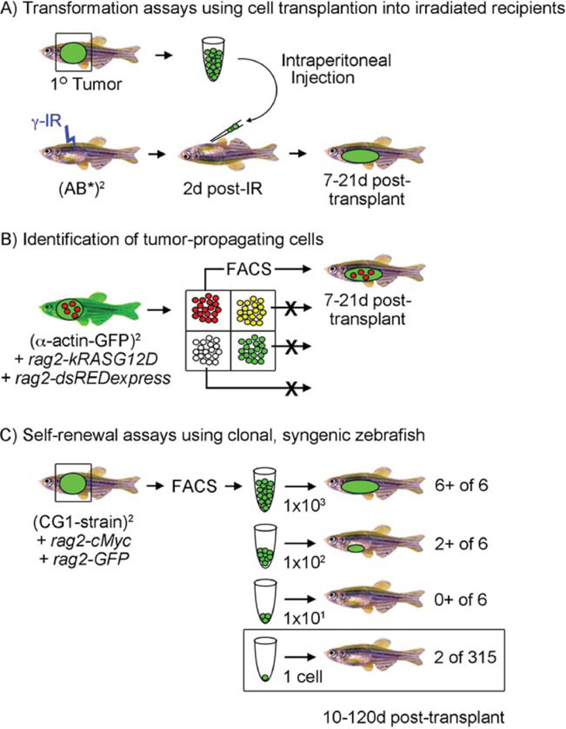
One of the most basic assays used in our laboratory is cell transplantation of fluorescent-labeled tumors into irradiated animals to assess if fluorescent cell populations are fully transformed (Fig. 3A). Sublethal-irradiation is commonly used to transiently ablate the immune system, providing engrafted cancer cells the ability to grow unabated by immune responses (Langenau et al., 2003; Traver et al., 2004). However, irradiation protocols provide only a transient assay for tumor growth, with animals capable of mounting robust immune responses after 21 days post-irradiation (Smith et al., 2010). To obviate these concerns, it is best to assess transplants prior to 20 days post-transplant and/or to transfer large numbers of tumor cells for engraftment where even after recovery of the immune system, the tumor cells overwhelm the immune system and cannot be cleared.
Fig. 3.
Cell transplantation assays in zebrafish. (A) Establishing malignancy by cell transplantation into irradiated recipients. A GFP+ primary tumor can be excised and individual cells isolated by disaggregation and filtering. Cells are induced into irradiated recipient animals (23 Gy, 2 days post-IR) and recipients assessed for engraftment from 7 to 21 days post-transplantation. (B) Identification of tumor-propagating cells. In this published example (Langenau et al., 2007), rag2-kRASG12D was co-injected with rag2-dsREDexpress into one-cell stage alpha-actin-GFP+ animals. Because the rag2 promoter expresses in satellite cells and early myoblasts while alpha-actin is expressed in late myoblasts and fused fibers, four cell populations could be identified following tumor resection, disaggregation, and fluorescent activated cell sorting (FACS). Each cell population is introduced into irradiated recipient animals and assessed for the ability to remake tumor. In this case, only the rag2-dsREDexpress+/alpha-actin-GFP-negative cells were capable of engraftment. (C) Self-renewal and single cell transplant assays using clonal, syngeneic zebrafish. In this example based onSmith et al. (2010), fluorescently labeled T-ALLs were created by microinjecting rag2-mouse cMyc (rag2-cMyc) and rag2-GFP into one-cell stage CG1-strain, syngeneic zebrafish. A subset of animals develop fluorescent-labeled T-ALLs that can be FACS sorted and transplanted at limiting dilution into CG1 recipient animals. Six animals per group were monitored for T-ALL onset up to 120 days post-transplantation. Limiting dilution cell transplantation assays accurately determine the number of leukemia-initiating cells contained within the bulk of the tumor mass and have been reviewed previously (Ignatius and Langenau, 2009). A single cell can also be transplanted into recipient fish, but fewer animals develop disease. (See color plate.)
A. Method
Fluorescent-labeled zebrafish cancer models can be created as outlined above. Tumor-bearing animals are euthanized by tricaine overdose, and then the tumor is excised from the animal using a razor blade. For most applications, the tumor is dissected along with noneffected margins on one side to maximize the amount of tumor cells. On the opposite side of the animal, some of the tumor is left within the body and fixed in 4% paraformaldehyde overnight. Thisway, tumor histology can be completed on transplanted tumors.
Most tumor cells are easily dispersed from resected tumor by repeatedly mincing the tissue with a razor blade in the presence of 250 µL of 0.9 × PBS + 5% FBS. This volume can change based on the size of the resected tumor, but the volume should be limited to increase the efficiency of tumor cell extraction from the tissue. Usually, the tissue is minced 50 to 100 times for 30–60 s at room temperature in a 10 cm Petri dish. Following mincing, 0.9 × PBS+ 5% FBS solution is added to the tumor samples to get a final volume of 10 mL. The sample is aspirated repeatedly 10 times using a 10 mL plastic pipette and a pipette aid. This procedure loosens tumor cells facilitating disassociating from normal tissue. Next, the sample is filtered over a 40-micron filter (BD Biosciences, 352340) into a 50mL conical tube. The filter is then washed with 10mL of 0.9 × PBS+ 5% FBS. Some solid tumors can benefit from treating the samples with collagenase to release tumor from normal unaffected tissue. We have previously used liberase III from Roche to isolate ERMS cells from affected animals; however, this product has recently been replaced by liberase blendzyme III. We have not found an optimal dose for extracting tumor cells using the new liberase blendzyme III reagent. Therefore, we no longer use collagenase or liberase treatment to isolate embryonal rhabdomyosarcoma cells from tumor-bearing fish but still obtain sufficient numbers of tumor cells for most assays.
A hemocytometer is used to count the numbers of cells contained in the tumor cell suspension by (1) vortexing the sample for 5 s at 75% velocity, (2) pipetting 10 mL of sample into 90 mL of trypan blue (Gibco, #15250-061), (3) vortexing this mixture for 5 s at 75% velocity, and (4) transferring 10 mL of the sample to a hemocytometer (Hausser Scientific, #1492). Cell counts are estimated by counting the number of optically clear cells within a 16 square viewing area and then multiplying the number of cells by the dilution factor (10) and then by 10,000 to arrive at the number of cells per mL. For some applications, unsorted cells can be transplanted directly and thus, cell counts can be used to directly make the suspension as concentrated as needed. If the sample is too concentrated, 0.9 × PBS + 5%FBS can be added to the sample. If the sample is too dilute, the sample can be centrifuged at 1000G for 10 min, the supernatant removed, and the sample resuspended to the correct volume required. If pure populations of cells are required, FACS can be used to isolate homogenous fluorescent cells.
Once tumor cell suspensions have been isolated, tumor samples can be introduced into irradiated recipient animals by intraperitoneal injection (5 mL of solution per fish using a #701N Hamilton Syringe, #80366). It is important to irradiate recipient animals 2 day prior to cell transplantation with 23 Gy from a Cesium-137 radiation source to transiently ablate the immune system (Traver et al., 2004). Some strains of fish are much more sensitive to gamma-irradiation, so dose optimization may be required to enhance engraftment of tumors and/or curb death due to irradiation. For example, we find that 23 Gy irradiation doses are lethal to optically clear caspar fish. For cell transplantation of unsorted tumor cells, we commonly utilize 1 × 10⁶ T-ALL cells and 2 × 10⁴ ERMS cells to engraft tumors into irradiated recipients (Langenau et al., 2003, 2007). Cell transplantation of lower numbers of tumor cells can often lead to robust engraftment (Smith et al., 2010).
Imaging fluorescent tumor cell engraftment into irradiated recipients can be completed using an epifluorescence dissecting microscope or the LED fluorescence macroscope (see Section III). Transplant recipients are routinely monitored for engraftment at 7, 14, 21, and 28 days post-transplantation. Photographs can be taken at each time point to visualize fluorescent-labeled tumor growth within the affected animal over time. Engrafted animals are routinely sectioned to confirm tumor histology. Tumor cells can also be isolated from recipient animals and used in serial transplantation experiments to propagate the tumor and to verify long-term tumorigenicity of the target cell population. We caution that tumor growth after 21 days post-irradiation treatment will be greatly impacted by immune system recovery and tumor cells will often begin to be killed by the host. Although this assay is only a short-term assay for transformation, the power of cell transplantation into irradiated recipient fish lies in the ability of tumor-derived cells from any strain of fish to be efficiently engrafted into recipient animals, irrespective of immune matching.
VI. Identifying Tumor-Propagating Cell Subpopulations by Fluorescent Protein Expression and Cell Transplantation into Irradiated Recipient Animals
We have previously used fluorescent transgenic approaches to create zebrafish that develop dual-fluorescent-labeled ERMS (Langenau et al., 2007). Specifically, one-cell stage alpha-GFP transgenic animals were coinjected with rag2-kRASG12D and rag2-dsREDexpress. 20% of injected animals developed ERMS all of which were dual-fluorescent labeled. Because the rag2 promoter drives transgene expression in satellite cells and early stage myoblasts, while alpha-actin-GFP is expressed in late stage myoblasts and fused fibers, four distinct subpopulations of tumor cells could be identified by FACS. ERMS were harvested, minced in the 0.9 × PBS + 5% FBS, and filtered using a 40 micron mesh. Propidium iodide (BD Pharmingen, #51-66211E) was added to the samples to identify dye-excluding, live cells by FACS and samples were double-sorted to increase purity. Purity and viability were assessed during the second sort. Live, fluorescent-sorted ERMS-cell subpopulations were transplanted into irradiated recipient animals at limiting dilution. Animals were assessed for engraftment at intervals up to 21 days post-transplantation to ascertain which cell population was best at remaking tumor (Fig. 3B). Tumor cell subpopulations were subsequently reisolated from engrafted animals and injected into new recipient animals to establish that the dsREDexpress+/alpha-actin-GFP-negative cell subpopulation retained long-term self-renewal ability and could remake tumors. A subset of transplant animals were sectioned to confirm that ERMS cell subpopulations remade tumors with similar histology as the primary malignancy. Although there are numerous caveats for using nonimmune matched animals as recipients, limiting dilution cell transplantation experiments determined that the serially transplantable ERMS-propagating cells was rag2-dsREDexpress+/alpha-actin-GFP-negative and expressed myf5, m-cadherin, and cMet – all markers of activated satellite cells. Taken together, these experiments highlight the use of fluorescent transgenic zebrafish models to label distinct tumor cell populations based on differentiation status and subsequently to use FACS and cell transplantation to assess which tumor cell subpopulations remake tumor.
VII. Use of Syngeneic Zebrafish for Cell Transplantation of Fluorescent-Labeled Tumors and Drug Discovery
Self-renewal is the process by which both normal and malignant cells make more of themselves and other more differentiated cell types. The ability to self-renew indefinitely is one of the hallmarks of cancer (Hanahan and Weinberg, 2000). Several hypotheses can account for how self-renewal occurs in cancer. The cancer stem cell hypothesis proposes that a rare, distinct subpopulation of tumor cells has the capacity to indefinitely self-renew (Reya et al., 2001). These cancer stem cells are thought to escape conventional treatments and lead to tumor relapse in patients. Another hypothesis, the stochastic model suggests that self-renewal is not contained within a specific tumor cell subpopulation (Adams and Strasser, 2008). While examples of tumors whose biology validates each hypothesis can be found in the scientific literature (reviewed in Dalerba et al., 2007; Dick, 2008; Kelly et al., 2007), it is clear that self-renewal occurs in cancer and if this process could be stopped, then tumors would cease to grow. Measuring self-renewal is best accomplished using limiting dilution cell transplantation into immune-compromised or immune-matched animals; this is the gold standard for determining self-renewal potential.
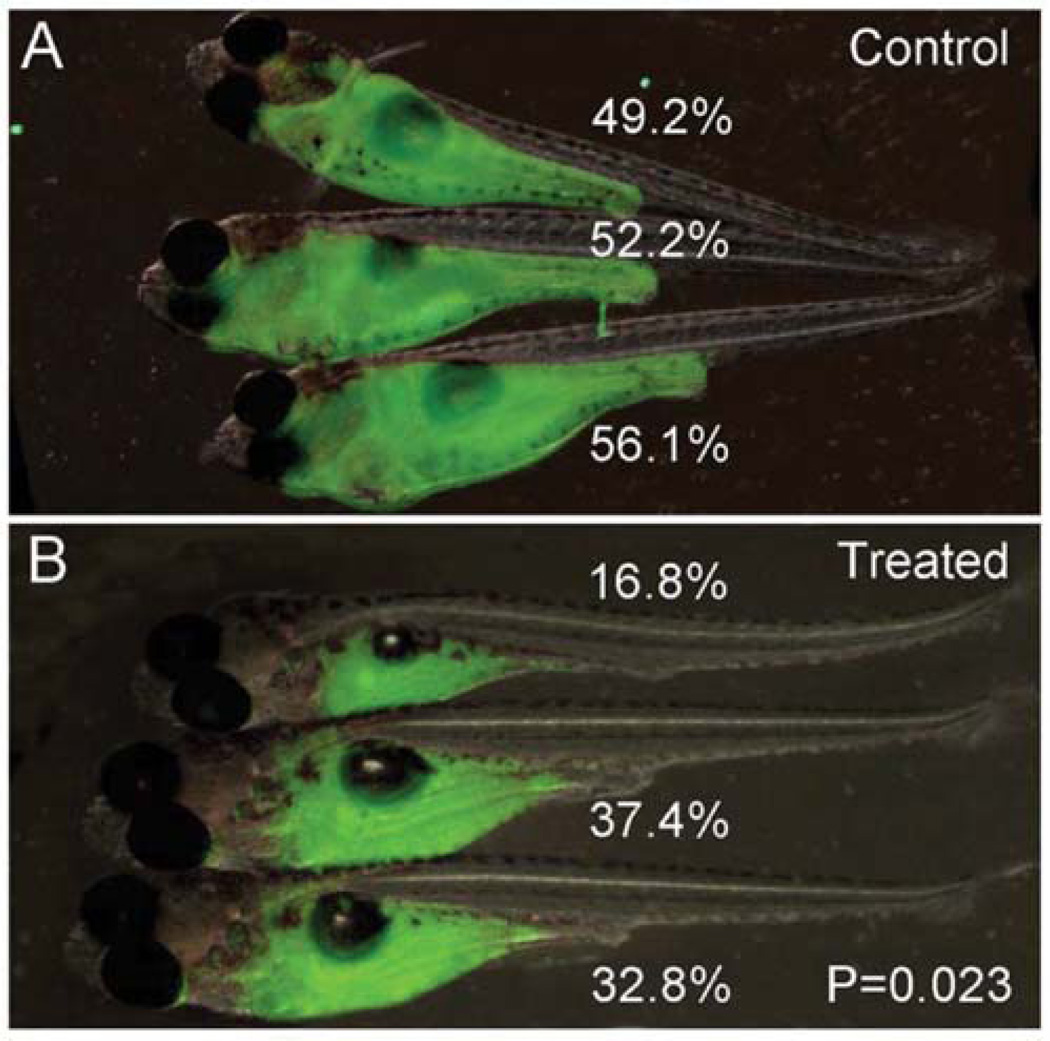
The Revskoy laboratory has been a pioneer in creating clonal, syngeneic zebrafish for use in cell transplantation assays (Mizgireuv and Revskoy, 2006; Mizgirev and Revskoy, 2010). Protocols for making strain specific syngeneic zebrafish have been recently reported (Mizgirev and Revskoy, 2010); however, we provide an abbreviated synopsis of the protocol here. Specifically, their method relies on manually extruding eggs, fertilizing with UV-inactivated sperm, and subjecting eggs to heat-shock to produce gynogenic diploid animals that can be subsequently raised to adulthood. Eggs are again isolated from gynogenic diploid females, fertilized with UV-inactivated sperm, and heat-shocked. The resulting progeny are now genetically similar and can be maintained by incrossing or mating male fish back to the founding mother. Remarkably, a subset of these lines are viable, reproduce efficiently, and have been shown to be genetically similar (Smith et al., 2010). The Revskoy laboratory has used several clonal lines for cell transplantation experiments where liver tumors, pancreatic acinar cell carcinomas, and T-cell leukemias can be passaged into recipient animals (Mizgireuv and Revskoy, 2006; Mizgirev and Revskoy, 2010). In these latter experiments, a serially transplantable fluorescent T-ALL line was developed which could be engrafted into syngeneic larval fish and be used to assess drug effects on leukemia growth (Fig. 4). GFP-labeled T-ALLs could be serially passaged into adult CG2-strain syngeneic animals without immune ablation and engrafted disease when introduced into 5-day-old larvae. Transplant animals developed T-ALL rapidly and became moribund by 5 days post-transplant. This rapid onset of disease could be delayed by treatment with vincristine and cyclophosphamide; however, neither drug could completely ablate tumor (Fig. 4). These experiments established that high-throughput cell transplantation assays can generate large cohorts of animals for drug screens and show that zebrafish T-ALL responds to the same drugs as used to treat patients.
Fig. 4.
Syngeneic zebrafish transplant models of T-ALL are a powerful tool for drug discovery: T-ALL growth is suppressed by cyclophosphamide treatment. Approximately 200 cells/5 nL were engrafted into 5-day-old syngeneic CG2 larvae. Engrafted animals were treated with cyclophosphamide (400 mg/L dissolved in fish water) beginning 5 days post-transplantation. Images of control (A) and treated animals (B) captured 3 days after continuous drug treatment. Tumor growth was assessed based on the percentage of body taken over by GFP+ T-ALL and compared using T-test calculations (Revskoy et al., 2010).
VIII. Cell Transplantation into Syngeneic Zebrafish to Accurately Assess Self-Renewal in Fluorescent-Labeled Cancer
Our laboratory has collaborated with the Revskoy group to develop an in vivo assay for T-ALL in zebrafish to assess self-renewal within the bulk of the tumor mass utilizing clonal-syngeneic zebrafish (Fig. 3C). CG1-strain syngeneic zebrafish were microinjected at the one-cell stage of development with rag2-cMyc and rag2-GFP (other fluorescent reporters can be substituted). Because the CG1-strain fish are severely inbred, they are very sensitive to gamma irradiation and DNA microinjection. We commonly inject only 60 ng/µL of linearized DNA into embryos with 5–10% of surviving animals developing T-ALL by 60 days of life. Myc-induced T-ALLs can be isolated from diseased animals and purified populations isolated by FACS. Because T-ALLs comprise a large portion of moribund zebrafish (70–90% of all cells isolated are fluorescent labeled T-ALLs), double sorting is not required to obtain highly pure and viable fluorescent cells (>95%). We also routinely sort tumor cells directly into 96 well plates supplemented with 100 µL of 0.9 × PBS + 5% FBS containing 2 × 10⁴ red blood cells (RBCs) from unaffected CG1-strain animals. This allows an accurate number of cells to be placed into each well for limiting dilution cell transplantation analysis. A single well that does not contain RBCs is collected, but has 0.9 × PBS + 5% FBS + propidium iodide and then reanalyzed to assess purity and viability following sorting. Once sort plates are obtained, the samples are centrifuged at 1000G for 10 min, the supernatant is removed, the samples are resuspended by repeated pipetting in 5–10 µL of residual 0.9 × PBS + 5% FBS. Next, the volume of each well is transferred to a Hamilton syringe and microinjected into the peritoneal cavity of nonirradiated CG1-strain fish. Animals are monitored for leukemia engraftment at 10, 20, 30, 45, 60, 90, and 120 days post-transplantation. When comparing the results obtained from limiting dilution analysis of the same tumor into irradiated AB-stain animals, it was shown that short-term irradiation transplantation assays lead to more deaths in recipient animals, lower overall engraftment rates, and rejection in a subset of leukemias. In total, the irradiation assay underestimates self-renewal potential by 20–33-fold (Smith et al., 2010). Use of CG1-strain syngeneic zebrafish allows for the accurate assessment of leukemia-propagating frequency contained within the bulk of the tumor mass, and thus, is an unbiased assay for self-renewal.
IX. Use of Syngeneic Zebrafish for Cell Transplantation: Single Cell Transplants
The ability to remake tumor from a single cell will open many new avenues of study including assessing heterogeneity within tumor cells and mimicking relapse. Using syngeneic fluorescent zebrafish models of T-ALL, we have successfully engrafted T-ALL from cell transplantation of a single leukemic cell (Smith et al., 2010). The procedure is similar to what is outlined above, but only one cell is sorted into each 96-well (Fig. 3C). It is imperative, that one assesses how often single cells are allocated to each well. We have used sorting onto slides, followed by visual inspection as a means to assess the accuracy and purity of each one-cell sort. In our hands, 45 of 48 wells contain single cells and all were T-ALL blasts. Moreover, only a subset of leukemia cells can remake tumor. In primary T-ALLs, the reported leukemia initiating frequency is approximately 1%. So even if two cells are contained in each well, it is unlikely that the leukemia is derived from both clones (~1%). Due to the speed of cell transplantation into adult zebrafish and the large numbers of animals available for experimentation, we were able to create two T-ALLs derived from a single primary T-ALL cell (n=315 recipients transplanted), while 7 of 44 animals engrafted tumor from a single cell in a leukemia that had evolved increased self-renewal following serial passaging. The ability to engraft T-ALL from a single fluorescent leukemic clone will facilitate tumor evolution studies to assess heterogeneity of self-renewal and tumor aggression within a tumor.
X. Xenograft Transplantation of Fluorescently Labeled Cells into Zebrafish
Xenograft transplantation experiments using fluorescent proteins in zebrafish have been reported. For example, cell transplantation of human C8161-RFP labeled melanoma cells into blastula stage embryos showed that the nodal pathway was active in melanomas; however, animals never developed robust engraftment of tumors (Lee et al., 2005; Topczewska et al., 2006). By contrast, cell transplantation of the human metastatic melanoma cell line WM-266-4 into the yolk of 2-day-old embryos formed observable tumors by 7 days postinjection (Haldi et al., 2006), suggesting that xenograft engraftment into zebrafish embryos will likely differ between tumors based on growth kinetics and stage of tumor growth. These tumor cells could be easily visualized due to incorporation of Dil, a lipophilic fluorescent tracking dye.
Human U251-RFP marked glioblastoma cells can also engraft into larval fish when transplanted into the yolk sac of blastula-stage embryos. The U251 cell line proliferated, formed tumors, and recruited blood vessels (Geiger et al., 2008). Engrafted glioblastoma cells were also more sensitive to radiation treatment when combined with temozolomide (Geiger et al., 2008). In another study, using the same U251-RFP labeled glioblastoma cells,Lally et al. (2007) identified novel small molecule sensitizers and discovered a compound NS123 (40 -bromo-30 -nitropropiophenone) that enhanced the growth-inhibitory effect of ionizing radiation (Lally et al., 2007).
The ability to recruit vasculature for tumor growth is one of the hallmarks of tumor formation and can be imaged in live zebrafish. Zebrafish provide many advantages over other model organisms in studying neovascularization including ease of manipulating embryos, use of large numbers in a given study, optical clarity during larval stages, and development of blood vessels by 2-days postfertilization. Additionally, several pan endothelial fluorescent transgenic lines are already available that mark blood vessels and vasculature including fli1-EGFP (Lawson and Weinstein, 2002b), tie2-GFP (Motoike et al., 2000), and flk1-EGFP (Cross et al., 2003). Utilizing these stable transgenic reporter lines in conjunction with cell transplantation into larval fish, several groups have imaged interactions between the vasculature and tumors cells including neovascularization, metastasis, migration, and extravasation. For example, Stoletov et al. showed that adenocarcinoma (MDA-435), fibrosarcoma (HT-1080), and melanoma (B16) cell lines that stably express either GFP, dsRED, or CFP can engraft into 30-day-old dexamethasone-treated fli1-EGFP transgenic zebrafish (Stoletov et al., 2007). Because fli1-EGFP transgenic fish have GFP-labeled endothelial cells, vascular reorganization can be easily visualized in engrafted animals (Stoletov et al., 2007). In these experiments, the RAS family homolog member C (RHOC) was required for cell movement and could partially control the early stages of metastasis. More recently, Stoletov et al. were able to visualize the extravasation of human breast adenocarcinoma cells in fli1-EGFP transgenic embryos, taking advantage of the lack of an adaptive immune response in 2-day-old embryos. In this study, stable fluorescent (CFP and dsRED) cell lines for human breast adenocarcinoma (MDA-MB-435, MDA-MB-231), human fibrosarcoma (HT1080), and human colon adenocarcinoma (SW480, SW620) were used for transplantation. Tumor cells in circulation are able to exit the capillaries by local remodeling of the blood vessel that is β-1-integrin dependent. The authors also show that expression of twist can increase intravascular migration and extravasation; however, this process does not require b-1-integrin (Stoletov et al., 2010). Additional studies have visualized tumor angiogenesis in zebrafish embryos. Experiments by Nicoli et al. demonstrated that tumorigenic FGF2-overexpressing mouse aortic endothelial cells transduced with DiL could engraft into 2-day-old transgenic VEGFR2:G-RCFP embryos. These fluorescently labeled FGF2-overexpressing cells stimulated new vascular growth (Nicoli et al., 2007). Lee et al. studied the effects of hypoxia on neovascularization. Murine-T241 fibrosarcoma cells labeled with DiL migrated from the primary site of transplantation only under hypoxic conditions, which was associated with increased vascularization. The hypoxia-induced migratory effected could be mimicked by overexpression of hypoxia-regulated angiogenic factor. Conversely, inhibition of VEGF-receptor signaling via morpholino or pharmacological inhibitor sunitinib abrogated the dissemination of tumor cells (Lee et al., 2009).
Taken together, xenograft cell transplantation experiments have provided new insights into tumor biology and identified potential drugs that impair tumor growth.
XI. Conclusions
The optical clarity of zebrafish embryos and larvae combined with the use of fluorescent proteins and dyes have made zebrafish an ideal model to image complex behaviors of tumor cells including xenotransplant engraftment, neo-vascularization, and growth responses to drug treatments. Moreover, fluorescently labeled proteins can be used in unbiased genetic screens to identify mutants associated with cancer and to directly assess self-renewal by limiting dilution cell transplantation into syngeneic zebrafish. Although much progress has been made in imaging cancer in zebrafish, the ultimate challenge is to follow tumor growth in real-time in live animals. Time-lapse imaging studies have been used to study multiple developmental processes including axonal guidance, vascular development, cell movements during early development, and in hematopoiesis (Bertrand et al., 2010; Gong et al., 2004; Keller et al., 2008; Kissa and Herbomel, 2010; Kucenas et al., 2008; Lawson and Weinstein, 2002a, 2002b; Nicoli et al., 2010). However, the ability to image tumor cell migration, local invasion, metastases to distant sites, and self-renewal in vivo over long-periods of time has yet to be fully described. Uncovering the dynamic processes of tumor growth in vivowill be the next frontier in zebrafish cancer research and will likely provide key insights into our understanding of tumor formation in human disease.
Acknowledgments
The authors thank Sergei Revskoy for sharing his work and high-resolution images that are reproduced here. David Langenau is supported by NIH grants K01AR05562190-01A1 and K01AR05562190-S1, a new investigator grant from Alex Lemonade Stand, the Sarcoma Foundation of America, the Leukemia Research Foundation, and a seed grant from the Harvard Stem Cell Institute.
References
- Adams JM, Strasser A. Is tumor growth sustained by rare cancer stem cells or dominant clones? Cancer Res. 2008;68:4018–4021. doi: 10.1158/0008-5472.CAN-07-6334. [DOI] [PubMed] [Google Scholar]
- Amsterdam A, Sadler KC, Lai K, Farrington S, Bronson RT, Lees JA, Hopkins N. Many ribosomal protein genes are cancer genes in zebrafish. PLoS Biol. 2004;2:E139. doi: 10.1371/journal.pbio.0020139. [DOI] [PMC free article] [PubMed] [Google Scholar]
- Berghmans S, Murphey RD, Wienholds E, Neuberg D, Kutok JL, Fletcher CD, Morris JP, Liu TX, Schulte-Merker S, Kanki JP, Plasterk R, Zon LI, Look AT. tp53 mutant zebrafish develop malignant peripheral nerve sheath tumors. Proc. Natl. Acad. Sci. USA. 2005;102:407–412. doi: 10.1073/pnas.0406252102. [DOI] [PMC free article] [PubMed] [Google Scholar]
- Bertrand JY, Chi NC, Santoso B, Teng S, Stainier DY, Traver D. Haematopoietic stem cells derive directly from aortic endothelium during development. Nature. 2010;464:108–111. doi: 10.1038/nature08738. [DOI] [PMC free article] [PubMed] [Google Scholar]
- Blackburn JS, Liu S, Raimondi AR, Ignatius MS, Salthouse CD, Langenau DM. High-throughput imaging of adult fluorescent zebrafish with an LED fluorescence macroscope. Nat Protoc. 2011;6(2):229–241. doi: 10.1038/nprot.2010.170. [DOI] [PMC free article] [PubMed] [Google Scholar]
- Chalfie M, Tu Y, Euskirchen G, Ward WW, Prasher DC. Green fluorescent protein as a marker for gene expression. Science. 1994;263:802–805. doi: 10.1126/science.8303295. [DOI] [PubMed] [Google Scholar]
- Cross LM, Cook MA, Lin S, Chen JN, Rubinstein AL. Rapid analysis of angiogenesis drugs in a live fluorescent zebrafish assay. Arterioscler. Thromb. Vasc. Biol. 2003;23:911–912. doi: 10.1161/01.ATV.0000068685.72914.7E. [DOI] [PubMed] [Google Scholar]
- Dalerba P, Cho RW, Clarke MF. Cancer stem cells: Models and concepts. Annu. Rev. Med. 2007;58:267–284. doi: 10.1146/annurev.med.58.062105.204854. [DOI] [PubMed] [Google Scholar]
- Dick JE. Stem cell concepts renew cancer research. Blood. 2008;112:4793–4807. doi: 10.1182/blood-2008-08-077941. [DOI] [PubMed] [Google Scholar]
- Faucherre A, Taylor GS, Overvoorde J, Dixon JE, Hertog J. Zebrafish pten genes have overlapping and non-redundant functions in tumorigenesis and embryonic development. Oncogene. 2008;27:1079–1086. doi: 10.1038/sj.onc.1210730. [DOI] [PubMed] [Google Scholar]
- Foudi A, Hochedlinger K, Van Buren D, Schindler JW, Jaenisch R, Carey V, Hock H. Analysis of histone 2B-GFP retention reveals slowly cycling hematopoietic stem cells. Nat. Biotechnol. 2009;27:84–90. doi: 10.1038/nbt.1517. [DOI] [PMC free article] [PubMed] [Google Scholar]
- Frazer JK, Meeker ND, Rudner L, Bradley DF, Smith AC, Demarest B, Joshi D, Locke EE, Hutchinson SA, Tripp S, Perkins SL, Trede NS. Heritable T-cell malignancy models established in a zebrafish phenotypic screen. Leukemia. 2009;23:1825–1835. doi: 10.1038/leu.2009.116. [DOI] [PMC free article] [PubMed] [Google Scholar]
- Geiger GA, Fu W, Kao GD. Temozolomide-mediated radiosensitization of human glioma cells in a zebrafish embryonic system. Cancer Res. 2008;68:3396–3404. doi: 10.1158/0008-5472.CAN-07-6396. [DOI] [PMC free article] [PubMed] [Google Scholar]
- Gong Y, Mo C, Fraser SE. Planar cell polarity signalling controls cell division orientation during zebrafish gastrulation. Nature. 2004;430:689–693. doi: 10.1038/nature02796. [DOI] [PubMed] [Google Scholar]
- Haldi M, Ton C, Seng WL, McGrath P. Human melanoma cells transplanted into zebrafish proliferate, migrate, produce melanin, form masses and stimulate angiogenesis in zebrafish. Angiogenesis. 2006;9:139–151. doi: 10.1007/s10456-006-9040-2. [DOI] [PubMed] [Google Scholar]
- Hanahan D, Weinberg RA. The hallmarks of cancer. Cell. 2000;100:57–70. doi: 10.1016/s0092-8674(00)81683-9. [DOI] [PubMed] [Google Scholar]
- Ignatius MS, Langenau DM. Zebrafish as a model for cancer self-renewal. Zebrafish. 2009;6:377–387. doi: 10.1089/zeb.2009.0610. [DOI] [PMC free article] [PubMed] [Google Scholar]
- Keller PJ, Schmidt AD, Wittbrodt J, Stelzer EH. Reconstruction of zebrafish early embryonic development by scanned light sheet microscopy. Science. 2008;322:1065–1069. doi: 10.1126/science.1162493. [DOI] [PubMed] [Google Scholar]
- Kelly PN, Dakic A, Adams JM, Nutt SL, Strasser A. Tumor growth need not be driven by rare cancer stem cells. Science. 2007;317:337. doi: 10.1126/science.1142596. [DOI] [PubMed] [Google Scholar]
- Kissa K, Herbomel P. Blood stem cells emerge from aortic endothelium by a novel type of cell transition. Nature. 2010;464:112–115. doi: 10.1038/nature08761. [DOI] [PubMed] [Google Scholar]
- Kucenas S, Takada N, Park HC, Woodruff E, Broadie K, Appel B. CNS-derived glia ensheath peripheral nerves and mediate motor root development. Nat. Neurosci. 2008;11:143–151. doi: 10.1038/nn2025. [DOI] [PMC free article] [PubMed] [Google Scholar]
- Lally BE, Geiger GA, Kridel S, Arcury-Quandt AE, Robbins ME, Kock ND, Wheeler K, Peddi P, Georgakilas A, Kao GD, Koumenis C. Identification and biological evaluation of a novel and potent small molecule radiation sensitizer via an unbiased screen of a chemical library. Cancer Res. 2007;67:8791–8799. doi: 10.1158/0008-5472.CAN-07-0477. [DOI] [PMC free article] [PubMed] [Google Scholar]
- Lam SH, Wu YL, Vega VB, Miller LD, Spitsbergen J, Tong Y, Zhan H, Govindarajan KR, Lee S, Mathavan S, Murthy KR, Buhler DR, Liu ET, Gong Z. Conservation of gene expression signatures between zebrafish and human liver tumors and tumor progression. Nat. Biotechnol. 2006;24:73–75. doi: 10.1038/nbt1169. [DOI] [PubMed] [Google Scholar]
- Langenau DM, Jette C, Berghmans S, Palomero T, Kanki JP, Kutok JL, Look AT. Suppression of apoptosis by bcl-2 overexpression in lymphoid cells of transgenic zebrafish. Blood. 2005;105:3278–3285. doi: 10.1182/blood-2004-08-3073. [DOI] [PubMed] [Google Scholar]
- Langenau DM, Keefe MD, Storer NY, Guyon JR, Kutok JL, Le X, Goessling W, Neuberg DS, Kunkel LM, Zon LI. Effects of RAS on the genesis of embryonal rhabdomyosarcoma. Genes Dev. 2007;21:1382–1395. doi: 10.1101/gad.1545007. [DOI] [PMC free article] [PubMed] [Google Scholar]
- Langenau DM, Keefe MD, Storer NY, Jette CA, Smith AC, Ceol CJ, Bourque C, Look AT, Zon LI. Co-injection strategies to modify radiation sensitivity and tumor initiation in transgenic Zebrafish. Oncogene. 2008;27:4242–4248. doi: 10.1038/onc.2008.56. [DOI] [PMC free article] [PubMed] [Google Scholar]
- Langenau DM, Traver D, Ferrando AA, Kutok JL, Aster JC, Kanki JP, Lin S, Prochownik E, Trede NS, Zon LI, Look AT. Myc-induced T cell leukemia in transgenic zebrafish. Science. 2003;299:887–890. doi: 10.1126/science.1080280. [DOI] [PubMed] [Google Scholar]
- Lawson ND, Weinstein BM. Arteries and veins: Making a difference with zebrafish. Nat. Rev. Genet. 2002a;3:674–682. doi: 10.1038/nrg888. [DOI] [PubMed] [Google Scholar]
- Lawson ND, Weinstein BM. In vivo imaging of embryonic vascular development using transgenic zebrafish. Dev. Biol. 2002b;248:307–318. doi: 10.1006/dbio.2002.0711. [DOI] [PubMed] [Google Scholar]
- Lee LM, Seftor EA, Bonde G, Cornell RA, Hendrix MJ. The fate of human malignant melanoma cells transplanted into zebrafish embryos: Assessment of migration and cell division in the absence of tumor formation. Dev. Dyn. 2005;233:1560–1570. doi: 10.1002/dvdy.20471. [DOI] [PubMed] [Google Scholar]
- Lee SL, Rouhi P, Dahl Jensen L, Zhang D, Ji H, Hauptmann G, Ingham P, Cao Y. Hypoxia-induced pathological angiogenesis mediates tumor cell dissemination, invasion, and metastasis in a zebrafish tumor model. Proc. Natl. Acad. Sci. USA. 2009;106:19485–19490. doi: 10.1073/pnas.0909228106. [DOI] [PMC free article] [PubMed] [Google Scholar]
- Mizgireuv IV, Revskoy SY. Transplantable tumor lines generated in clonal zebrafish. Cancer Res. 2006;66:3120–3125. doi: 10.1158/0008-5472.CAN-05-3800. [DOI] [PubMed] [Google Scholar]
- Mizgirev I, Revskoy S. Generation of clonal zebrafish lines and transplantable hepatic tumors. Nat. Protoc. 2010;5:383–394. doi: 10.1038/nprot.2010.8. [DOI] [PubMed] [Google Scholar]
- Motoike T, Loughna S, Perens E, Roman BL, Liao W, Chau TC, Richardson CD, Kawate T, Kuno J, Weinstein BM, Stainier DY, Sato TN. Universal GFP reporter for the study of vascular development. Genesis. 2000;28:75–81. doi: 10.1002/1526-968x(200010)28:2<75::aid-gene50>3.0.co;2-s. [DOI] [PubMed] [Google Scholar]
- Neumann JC, Dovey JS, Chandler GL, Carbajal L, Amatruda JF. Identification of a heritable model of testicular germ cell tumor in the zebrafish. Zebrafish. 2009;6:319–327. doi: 10.1089/zeb.2009.0613. [DOI] [PMC free article] [PubMed] [Google Scholar]
- Nicoli S, Ribatti D, Cotelli F, Presta M. Mammalian tumor xenografts induce neovascularization in zebrafish embryos. Cancer Res. 2007;67:2927–2931. doi: 10.1158/0008-5472.CAN-06-4268. [DOI] [PubMed] [Google Scholar]
- Nicoli S, Standley C, Walker P, Hurlstone A, Fogarty KE, Lawson ND. MicroRNA-mediated integration of haemodynamics and Vegf signalling during angiogenesis. Nature. 2010;464:1196–1200. doi: 10.1038/nature08889. [DOI] [PMC free article] [PubMed] [Google Scholar]
- Park SW, Davison JM, Rhee J, Hruban RH, Maitra A, Leach SD. Oncogenic KRAS induces progenitor cell expansion and malignant transformation in zebrafish exocrine pancreas. Gastroenterology. 2008;134:2080–2090. doi: 10.1053/j.gastro.2008.02.084. [DOI] [PMC free article] [PubMed] [Google Scholar]
- Phelps RA, Chidester S, Dehghanizadeh S, Phelps J, Sandoval IT, Rai K, Broadbent T, Sarkar S, Burt RW, Jones DA. A two-step model for colon adenoma initiation and progression caused by APC loss. Cell. 2009;137:623–634. doi: 10.1016/j.cell.2009.02.037. [DOI] [PMC free article] [PubMed] [Google Scholar]
- Reya T, Morrison SJ, Clarke MF, Weissman IL. Stem cells, cancer, and cancer stem cells. Nature. 2001;414:105–111. doi: 10.1038/35102167. [DOI] [PubMed] [Google Scholar]
- Santoriello C, Deflorian G, Pezzimenti F, Kawakami K, Lanfrancone L, d’Adda di Fagagna F, Mione M. Expression of H-RASV12 in a zebrafish model of Costello syndrome causes cellular senescence in adult proliferating cells. Dis. Model Mech. 2009;2:56–67. doi: 10.1242/dmm.001016. [DOI] [PMC free article] [PubMed] [Google Scholar]
- Sears R, Nuckolls F, Haura E, Taya Y, Tamai K, Nevins JR. Multiple Ras-dependent phosphorylation pathways regulate Myc protein stability. Genes Dev. 2000;14:2501–2514. doi: 10.1101/gad.836800. [DOI] [PMC free article] [PubMed] [Google Scholar]
- Shimomura O, Johnson FH, Saiga Y. Extraction, purification and properties of aequorin, a bioluminescent protein from the luminous hydromedusan, Aequorea. J. Cell. Comp. Physiol. 1962;59:223–239. doi: 10.1002/jcp.1030590302. [DOI] [PubMed] [Google Scholar]
- Smith AC, Raimondi AR, Salthouse CD, Ignatius MS, Blackburn JS, Mizgirev IV, Storer NY, de Jong JL, Chen AT, Zhou Y, Revskoy S, Zon LI, Langenau DM. High-throughput cell transplantation establishes that tumor-initiating cells are abundant in zebrafish T-cell acute lymphoblastic leukemia. Blood. 2010;115:3296–3303. doi: 10.1182/blood-2009-10-246488. [DOI] [PMC free article] [PubMed] [Google Scholar]
- Spitsbergen JM, Tsai HW, Reddy A, Miller T, Arbogast D, Hendricks JD, Bailey GS. Neoplasia in zebrafish (Danio rerio) treated with 7,12-dimethylbenz[a]anthracene by two exposure routes at different developmental stages. Toxicol. Pathol. 2000a;28:705–715. doi: 10.1177/019262330002800511. [DOI] [PubMed] [Google Scholar]
- Spitsbergen JM, Tsai HW, Reddy A, Miller T, Arbogast D, Hendricks JD, Bailey GS. Neoplasia in zebrafish (Danio rerio) treated with N-methyl-N’-nitro-N-nitrosoguanidine by three exposure routes at different developmental stages. Toxicol. Pathol. 2000b;28:716–725. doi: 10.1177/019262330002800512. [DOI] [PubMed] [Google Scholar]
- Stoletov K, Kato H, Zardouzian E, Kelber J, Yang J, Shattil S, Klemke R. Visualizing extravasation dynamics of metastatic tumor cells. J. Cell. Sci. 2010;123:2332–2341. doi: 10.1242/jcs.069443. [DOI] [PMC free article] [PubMed] [Google Scholar]
- Stoletov K, Montel V, Lester RD, Gonias SL, Klemke R. High-resolution imaging of the dynamic tumor cell vascular interface in transparent zebrafish. Proc. Natl. Acad. Sci. USA. 2007;104:17406–17411. doi: 10.1073/pnas.0703446104. [DOI] [PMC free article] [PubMed] [Google Scholar]
- Topczewska JM, Postovit LM, Margaryan NV, Sam A, Hess AR, Wheaton WW, Nickoloff BJ, Topczewski J, Hendrix MJ. Embryonic and tumorigenic pathways converge via nodal signaling: Role in melanoma aggressiveness. Nat. Med. 2006;12:925–932. doi: 10.1038/nm1448. [DOI] [PubMed] [Google Scholar]
- Traver D, Winzeler A, Stern HM, Mayhall EA, Langenau DM, Kutok JL, Look AT, Zon LI. Effects of lethal irradiation in zebrafish and rescue by hematopoietic cell transplantation. Blood. 2004;104:1298–1305. doi: 10.1182/blood-2004-01-0100. [DOI] [PubMed] [Google Scholar]
- Tsien RY. The green fluorescent protein. Annu. Rev. Biochem. 1998;67:509–544. doi: 10.1146/annurev.biochem.67.1.509. [DOI] [PubMed] [Google Scholar]
- Tsien RY. The 2009 Lindau Nobel Laureate Meeting: Roger Y. Tsien, Chemistry, 2008. J. Vis. Exp. 2010;(35) doi: 10.3791/1575. pii: 1575. [DOI] [PMC free article] [PubMed] [Google Scholar]
- Wakamatsu Y, Pristyazhnyuk S, Kinoshita M, Tanaka M, Ozato K. The see-through medaka: A fish model that is transparent throughout life. Proc. Natl. Acad. Sci. USA. 2001;98:10046–10050. doi: 10.1073/pnas.181204298. [DOI] [PMC free article] [PubMed] [Google Scholar]
- White RM, Sessa A, Burke C, Bowman T, LeBlanc J, Ceol C, Bourque C, Dovey M, Goessling W, Burns CE, Zon LI. Transparent adult zebrafish as a tool for in vivo transplantation analysis. Cell Stem Cell. 2008;2:183–189. doi: 10.1016/j.stem.2007.11.002. [DOI] [PMC free article] [PubMed] [Google Scholar]


