Introduction
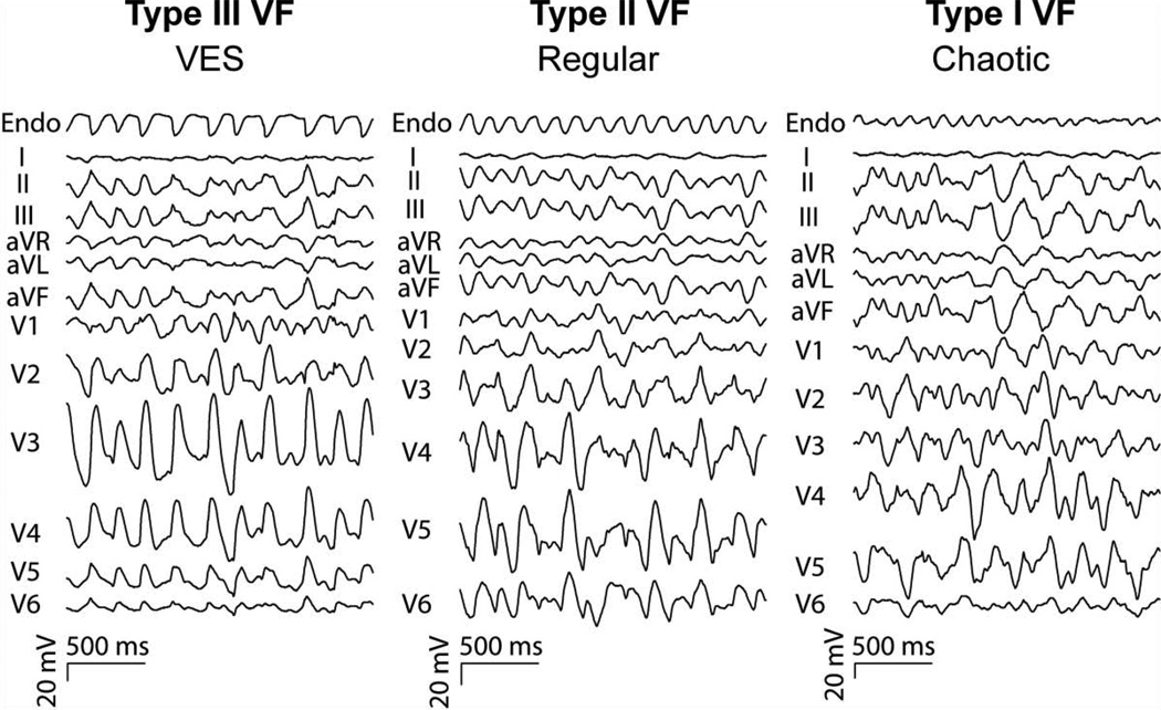
We tested the hypothesis that after 2 min of ventricular fibrillation (VF), in addition to periods with disorganized, chaotic activation (Type I pattern, chaotic), during some periods activation can be highly organized on the endocardium and arise from a mother rotor (Type II pattern, regular) or triggered activity (Type III pattern, ventricular electrical synchrony (VES)) originating in Purkinje fibers (PFs).
Methods
In 6 anesthetized dogs, a 64-electrode basket catheter was inserted into the LV to record from about 2/3rds of the endocardium. Electrically induced VF was recorded from the 64 basket electrodes as well as from a body surface 12 lead ECG for 7 min. The study was repeated in another 12 dogs in which the early afterdepolarization (EAD) blocker pinacidil (loading dosage 0.5 mg/kg over 10 min, maintenance 0.5 mg/kg/hr) was given in 6 animals and the delayed afterdepolarization (DAD) blocker flunarizine (loading dosage 2 mg/kg over 10 min, maintenance 4 mg/kg/hr) was given in the other 6 animals before inducing VF.
Results
Between 3 and 7 min of VF in control animals, in addition to periods of Type I activation, two highly organized endocardial activation patterns were observed in all electrograms from all 64 electrodes simultaneously. In the Type II pattern, activation was highly regular and repeatable, predominantly propagating from the apex towards the base, occasionally from a stable, large rotor centered at the apex. In the Type III pattern, focal endocardial activation occupied less than half of the cycle length with earliest activation arising in PFs and then activating the working myocardium. Activation in the Type III pattern either had a propagation sequence similar to that during sinus rhythm or initiated from a region close to a papillary muscle. The Type III pattern was present a mean of 25% (60 s) of the time in the LV endocardium in control animals, but was present a mean of only 0.1% of the time (240 ms) with pinacidil (p < 0.001, control vs. pinacidil), and 24% of the time (58 s) with flunarizine (p = 0.44, control vs. flunarizine). The Type II pattern was present a mean of 71% of the time (170 s) in the LV endocardium in control animals, 56% of the time (134 s) with pinacidil (p < 0.001, control vs. pinacidil) and 48% of the time (115 s) with flunarizine (p < 0.001, control vs. flunarizine). The remainder of the time, the chaotic Type I pattern was present.
Discussion
After 2 min, VF can exist in three types of activation patterns, two of which are highly organized (Types II and III). Type II arises from the apex, is highly regular and repeatable, and may be an intramural mother rotor. Type III arises focally in PFs and may be caused by EADs but not DADs.1,2 It is possible that the optimum defibrillation strategy is different for the three patterns. If so, then it should be determined if the three patterns can be identified from the ECG (Figure 1) so it can be known clinically which pattern is present.
Figure 1.
An endocardial left ventricular recording (endo) at the ECG during 2 s of long duration VF during the three types of activation patterns. VES=ventricular electrical synchrony
Acknowledgments
This work was supported in part by the National Institutes of Health, Heart, Lung and Blood Institute grant HL85370
Footnotes
Publisher's Disclaimer: This is a PDF file of an unedited manuscript that has been accepted for publication. As a service to our customers we are providing this early version of the manuscript. The manuscript will undergo copyediting, typesetting, and review of the resulting proof before it is published in its final citable form. Please note that during the production process errors may be discovered which could affect the content, and all legal disclaimers that apply to the journal pertain.
Literature Cited
- 1.Robichaux RP, Dosdall DJ, Osorio J, Garner NW, Li L, Huang J, Ideker RE. Periods of highly synchronous, non-reentrant endocardial activation cycles occur during long-duration ventricular fibrillation. J Cardiovasc Electrophysiol. 2010;21(11):1266–1273. doi: 10.1111/j.1540-8167.2010.01803.x. [DOI] [PMC free article] [PubMed] [Google Scholar]
- 2.Dosdall DJ, Tabereaux PB, Kim JJ, Walcott GP, Rogers JM, Killingsworth CR, Huang J, Robertson PG, Smith WM, Ideker RE. Chemical ablation of the Purkinje system causes early termination and activation rate slowing of long-duration ventricular fibrillation in dogs. Am J Physiol Heart Circ Physiol. 2008;295(2):H883–H889. doi: 10.1152/ajpheart.00466.2008. [DOI] [PMC free article] [PubMed] [Google Scholar]


