Abstract
Chondrosarcomas are the malignant cartilaginous neoplasms rarely seen in the head and neck region. The chondrosarcomas may show an aggressive course and are mostly located in relation with anterior maxilla and base of the skull. Chondrosarcomas of the body of mandible are infrequent. We report a case of low-grade chondrosarcoma of body of mandible, which was treated with a simple excision without neck dissection.
Keywords: Chondrosarcoma, mandible, maxilla, tumor
INTRODUCTION
Chondrosarcoma is the malignant counterpart of the chondroma and, like the benign one, may occur either in the maxilla or in mandible with male to female preponderance of 2:1.[1] The chondrosarcomas of the maxillofacial region account for 1-3% of chondrosarcomas of the entire body.[2] They arise predominately in the maxilla with a predilection for the anterior maxillary region and occur at a lower incidence in the mandible.[2,3,4,5] The preferred site in the mandible is the molar region, and they infrequently occur in the ramus, condyle, coronoid process, or symphysis.[6]
Roentgenographic findings are not clear unless the lesion is long-standing and has produced considerable destruction of bone. Occasionally, tumor appears as radiopaque lesion because of calcification and sometimes shows resorption and exfoliation of teeth.[1]
CASE REPORT
A 60-year-old female patient reported to the department of oral and maxillofacial surgery, with chief complaint of painless swelling at right side of the mandible, which had been present for 6 months. Two months back, the growth was excised incompletely by a general dental practitioner; unfortunately, the excised specimen was not sent for histopathological examination and was discarded, following which the remaining growth grew rapidly.
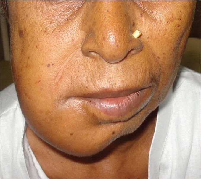
Extraoral examination showed a swelling at body of the right mandible measuring 6 × 6 cm [Figure 1] extending intraorally from right mandibular canine to distal aspect of right mandibular second molar. Swelling was bony hard, ill defined, and caused obliteration of right lower buccal vestibule. There was no regional lymphadenopathy.
Figure 1.

Preoperative photograph of the patient showing swelling of right mandible (or) Extaoral view showing swelling at right side of face
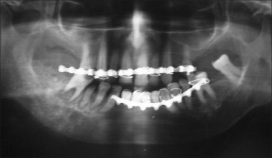
A panoramic radiograph revealed a widened periodontal space and an irregularly-shaped osteoblastic lesion with radiopacity. There were signs of sclerosis and new bone formation in the body of right mandible, which was extending to the angle region. There were signs of patchy bony destruction near the root of molar and premolar teeth [Figure 2].
Figure 2.

Orthopantogram showing signs of sclerosis and new bone formation in the body of right mandible along with signs of patchy bony destruction near the root of molar and premolar teeth (or) OPG showing lesion extending from right side of canine upto ramus of mandible
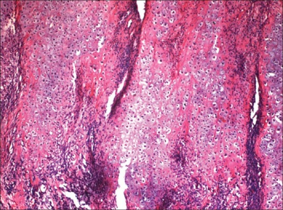
An incisional biopsy was performed which histopathologically revealed areas of hyaline cartilage in lobules with increased cellularity and cellular pleomorphism. Intervening tissue revealed fibrocollagenous stroma. Periphery of the lesion demarcated cellular dedifferentiation into the spindle cells. Mitosis was frequent. Focal calcification and liquefaction were also evident within the cartilage [Figure 3]. The case was hence diagnosed as chondrosarcoma.
Figure 3.

Photomicrograph showing lobular architecture with cellular lobules of chondroid tissue and focal dedifferentiated spindle cell areas (H and E, ×40 digital magnification) (or) Histopahological slide showing areas of hyaline cartilage in lobules with increased cellularity and cellular pleomorphism
A segmental resection under general anesthesia was performed extending from the right lateral incisor and the ramus of the mandible, as the mandibular basal bone appeared to be affected. The surgical margins were free of malignant cells.
DISCUSSION
Chondrosarcoma of the mandible mostly present as a painless swelling or may appear as mass of long duration with pain, paresthesia, trismus, and loosening of the teeth, which points toward the progression of the disease. Radiographically, the appearance of the lesion varies from ill-defined radiolucent to obvious radioopaque shadow. However, these findings are not pathognomonic.[7] Conventional radiograph, Computed tomography scan and magnetic resonance imaging are quite valuable in determining the nature and extent of the lesion,[6] but a definitive diagnosis should be made histologically which states significantly that chondrosarcomas are composed purely of hyaline cartilage and fulfill cytologic malignant criteria.[8]
In the present case, a hard mass was noticed on the body-angle region of right mandible and presented with loosening of right posterior teeth along with periodontal space widening and resorption of the roots of the teeth which was more prominent in relation to the distal root of right first molar. There are several reports of chondrosarcomas of the jaws and facial skeleton that grew rapidly following incisional biopsy,[9,10] and the tumor in the current case acted in the same way.
Radical resection is the most effective primary modality for the treatment of chondrosarcoma, as this is considered to be radioresistant. However, chemoradiation is usually advised for locally recurrent or residual tumor. Although the biopsy in the present case report showed the lesion to be of low-grade nature, but a segmental resection of the mandible was performed as the basal bone was also infiltrated by the growth and the resected specimen showed tumour-free margins. The patient is disease-free after 12 months of resection.
The 5-year survival rate for chondrosarcomas of the jaws and facial bones has been reported to be 67.6%.[3] The prognosis of chondrosarcomas depends on the size, location, grade, and surgical respectability of the tumors as chondrosarcomas show a wide variation in time of recurrence and metastasis.[6]
The present case is unique as a low-grade chondrosarcoma appeared from a rare site, the body of the mandible and also involved the basal bone which was treated with segmental resection. The unusual presentation of chondrosarcoma described in the present case report will facilitate the medical professionals in the management of such mandibular growths. However, a long-term follow-up is required to assess the nature and long-term outcome of chondrosarcomas of mandible.
Footnotes
Source of Support: Nil.
Conflict of Interest: None declared.
REFERENCES
- 1.Rajendran R, Sivapathasundharam B. Shafer's Textbook of Oral Pathology. 6th ed. India: Elsevier; 2009. [Google Scholar]
- 2.Izadi K, Lazow SK, Solomon MP, Berger JR. Chondrosarcoma of the anterior mandible. A case report. N Y State Dent J. 2000;66:32–4. [PubMed] [Google Scholar]
- 3.Saito K, Unni KK, Wollan PC, Lund BA. Chondrosarcoma of the jaws and facial bones. Cancer. 1995;76:1550–8. doi: 10.1002/1097-0142(19951101)76:9<1550::aid-cncr2820760909>3.0.co;2-s. [DOI] [PubMed] [Google Scholar]
- 4.Fronstin MH, James MD, Hutcheson JB, Sanders HL. Chondrosarcoma of the mandibular symphysis. Report of a case. Oral Surg Oral Med Oral Pathol. 1968;25:665–9. doi: 10.1016/0030-4220(68)90031-5. [DOI] [PubMed] [Google Scholar]
- 5.Ruark DS, Schlehaider UK, Shah JP. Chondrosarcomas of the head and neck. World J Surg. 1992;16:1010–6. doi: 10.1007/BF02067021. [DOI] [PubMed] [Google Scholar]
- 6.Murayama S, Suzuki I, Nagase M, Shingaki S, Kawasaki T, Nakajima T, et al. Chondrosarcoma of the mandible. Report of case and a survey of 23 cases in the Japanese literature. J Craniomaxillofac Surg. 1988;16:287–92. doi: 10.1016/s1010-5182(88)80063-5. [DOI] [PubMed] [Google Scholar]
- 7.Cook HE. Chondrosarcoma arising from the zygomatic arch. J Oral Maxillofac Surg. 1986;44:67–9. doi: 10.1016/0278-2391(86)90015-7. [DOI] [PubMed] [Google Scholar]
- 8.Unni KK, Dahlin DC. Grading of bone tumors. Semin Diagn Pathol. 1984;1:165–72. [PubMed] [Google Scholar]
- 9.Shirato T, Onizawa K, Yamagata K, Yusa H, Iljima T, Yoshida H. Chondrosarcoma of the mandibular symphysis. Oral Oncology Extra. 2006;42:247–50. [Google Scholar]
- 10.Sato K, Nukaga H, Horikoshi T. Chondrosarcoma of the jaws and facial skeleton: A review of the Japanese literature. J Oral Surg. 1977;35:892–7. [PubMed] [Google Scholar]


