Abstract
Septic cavernous sinus thrombosis (CST) related to dental infection is a rare clinical event. The septic CST is a disease of high morbidity and mortality. The prompt diagnosis and timely treatment of septic CST is cornerstone of successful outcome. The dental infection should be given due attention, as to prevent CST. In this case report of immunocompetent female, we highlighted the role of dental abscess in producing bilateral CST and facial palsy. The close collaboration between dentist and neurologist and early institution of antibiotics led to complete recovery at follow-up after 3 months. The dental infection should never be neglected as it is the interface of serious intracranial complication like CST.
Keywords: Cavernous sinus thrombosis, facial palsy, ophthalmoplegia
INTRODUCTION
The septic cavernous sinus thrombosis (CST) due to odontogenic infection is uncommon and leads to substantial morbidity and mortality.[1] The peculiar anatomy of cervicofacial planes, dental structures, and its direct communication with cavernous sinus predisposes individual to development of septic CST in background of dental infections. The favorable outcome depends upon prompt diagnosis and early institution of antibiotics.[2] The negligence of dental hygiene and dental infections can lead to disastrous consequences. In this illustration, we intend to present an immunocompetent female having widespread pulmonary and dental infection who developed bilateral CST with facial palsy. The timely referral from dentistry department to neurologist and prompt management led to complete recovery at discharge after a month. In nutshell, dental infection should never be ignored as CST is always a possible complication.
CASE REPORT
A 50-year-old, previously healthy, female patient presented with complaints of fever, pain, and pus discharge from right maxillary molars and swelling of right cheek since 15 days. Three days later she developed puffiness of both eyes.
Patient was drowsy at the time of admission. She was mildly febrile. On examination, there was diffuse inflamed swelling over right maxillary region along with bilateral exophthalmos with chemosis and congestion of conjunctiva. Oral examination revealed right maxillary dental abscess related to molars. Central nervous system examination revealed nuchal rigidity. Bilateral ptosis with external ophthalmoplegia was present along with palsy of lower half of face [Figure 1]. Other neurological examination was normal. Other systems examination did not reveal any abnormality.
Figure 1.
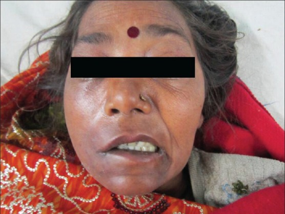
Anterior view photograph of the patient at the time of presentation showing bilateral exophthalmos with chemosis along with palsy of lower half of face on right side
Her baseline investigations revealed the following results: Total leukocyte count: 17000/mm3, Differential leukocyte count: P80L18E1M1, Erythrocyte sedimentation rate: 46 mm/h, serum Na+136 meq/L, serum K+3.4 meq/L, blood urea 20 mg/dL, serum creatinine 0.6 mg/dL, and random blood glucose 106 mg/dL. Her chest X-ray revealed multiple thin-walled abscesses (pneumatoceles) [Figure 2]. HIV Enzyme-linked immunosorbent assay was nonreactive.
Figure 2.
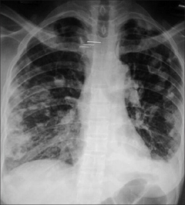
Chest radiogram showing multiple pneumatoceles
The patient was first seen by oral and maxillofacial surgeon. Her orthopantomogram was done which showed opacity at the tips of right second and third maxillary molars [Figure 3]. Drainage of the dental abscess and extraction of the right maxillary molars was done immediately. The pus sent for culture grew Staphylococcus aureus. With suspicion of CST, the patient was transferred to neurology department. Her magnetic resonance imaging (MRI) brain was done which showed bilateral mastoiditis along with bilateral CST with abscess in right cerebellar and right cerebellopontine angle region with infarct in right parietooccipital and right high parietal region [Figures 4 and 5]. Magnetic resonance (MR) venography did not show involvement of any other major sinuses. The patient was started on intravenous fluids and antibiotics (vancomycin 2 g/day, ceftriaxone 2 g/day, and clindamycin 1200 mg/day). Anticoagulant was added in the form of enoxaparin 0.4 mL s.c. twice a day. Oral anticoagulant (warfarin) was added subsequently.
Figure 3.
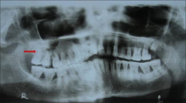
Orthopantomogram showing opacity at the tips of right second and third maxillary molars (arrow)
Figure 4.
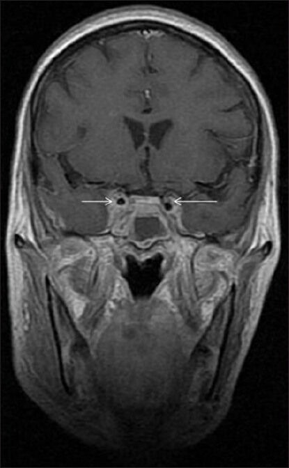
Coronal contrast-enhanced T1WI showing multiple defects in the enhancing bilateral cavernous sinuses (arrows)
Figure 5.
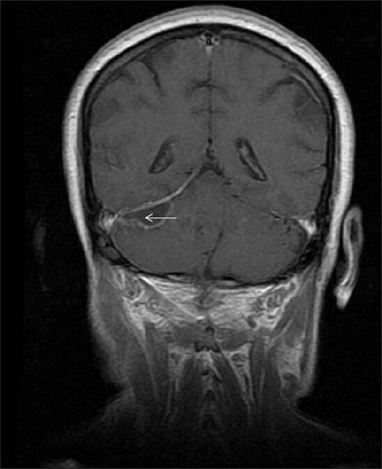
MRI-brain showing right cerebellar abscess (arrow)
The patient improved during the hospital stay of 4 weeks and was discharged satisfactorily. At follow-up after 3 months, she had complete recovery.
DISCUSSION
Septic CST can be defined as thrombophlebitis of the cavernous sinus of infectious origin. Facial infections and paranasal sinusitis are among the most important causes of septic CST. However, less commonly it may be caused by pharyngeal, otogenic, and odontogenic infections.[3] Dental infections constitute less than 10% of the cases.[4] Blood from brain is drained by a complex system of veins and dural sinuses into internal jugular vein. The most important dural sinuses are cavernous sinuses, sagittal sinuses, straight sinus, transverse sinuses, and sigmoid sinus, which are all interconnected. The right and left cavernous sinuses are situated on the lateral aspect of the sella turcica. They extend from the superior orbital fissure to the petrous apex of the temporal bone. They are interconnected by anterior and posterior intercavernous sinuses that encircle the pituitary gland.[3]
Cavernous sinuses receive blood from the ophthalmic veins, the superficial middle and inferior cerebral veins, and the sphenoparietal and sphenoid sinuses. The cavernous sinuses in turn drain into the pterygoid venous plexus via emissary veins, and into the internal jugular vein and the sigmoid sinus via the inferior and the superior petrosal sinuses respectively. Absence of valves in the cavernous sinuses and their connections favor bidirectional spread of infection. This can give rise to extensive thrombi throughout the network of sinuses.[3]
Most commonly, CST results from spread of infection from the sinuses, especially the sphenoid, ethmoid, and frontal sinuses, or from the middle one third of the face. Less commonly infection from teeth, nose, tonsils, soft palate, and ears may constitute primary source of the infection. The dental infection most commonly spreads via the pterygoid venous plexus, where an infected thrombus may extend or disseminate septic emboli.[4]
In 1989, Ogundiya et al.,[2] reported case of a patient with dental infection which got complicated leading to orbital abscess, unilateral blindness, and CST.
In 1991, Yun et al.,[5] reported CST in a 60-year-old diabetic male about 38 days after extraction of an infected upper third molar tooth.
In 2000, Feldman et al.,[6] reported a case of 69-year-old male who was being treated for dental abscess. Computed tomography of brain did not show any intracranial abnormalities at the time of admission, but there was presence of a right parapharyngeal abscess. Later, patient developed bilateral proptosis with ophthalmoplegia. Repeat computed tomography (CT) scan after 48 h revealed a right parapharyngeal abscess and a bilateral CST. The patient was started on intravenous antibiotics and anticoagulation. The parapharyngeal abscess was causing airway compromise. The patient underwent a tracheotomy, incision, and drainage of the parapharyngeal abscess, and extraction of the right third molar. The patient was found to be completely recovered during his follow-up.
In 2007, Embong et al.,[7] described a case of 43-year-old man who presented with a history of right-sided toothache, progressively increasing painful left orbital swelling, and diminution of left eye vision. CT head was suggestive of left orbital cellulitis, right parapharyngeal abscess, tooth abscess over right upper alveolar region with bilateral maxillary, and posterior ethmoidal sinusitis. MRI revealed CST with left superior ophthalmic vein thrombosis.
In 2009, Jones and Arnold[8] reported a case of dental infection complicating with left facial abscess, CST, bilateral internal jugular thrombosis, and multiple lung abscesses. The patient later developed a middle cerebral artery infarct.
The signs of CST result from venous congestion due to impaired venous drainage from the orbit and eye. The onset is acute, usually with unilateral periorbital edema and proptosis associated with headache and photophobia. Examination may reveal ophthalmoplegia. The infection can spread to the contralateral cavernous sinus via intercavernous sinuses, usually within 1 to 2 days of the initial presentation. The diagnosis of CST is usually done on clinical grounds and can be confirmed by appropriate radiographic studies. MRI and MR venography are more sensitive than CT scan for diagnosis. It may show a heterogeneous signal from the abnormal cavernous sinus along with deformity of the cavernous portion of the internal carotid artery, and an obvious hyperintense signal of thrombosed vascular sinuses.[6]
S. aureus is the most frequently cultured organism in these infections (70%), followed by Streptococcus species (20%).[4]
Treatment includes high-dose intravenous antibiotics. Empirically, patients should be started on antibiotics directed at the most common pathogens (Gram-positive, Gram-negative, and anerobes). Antibiotics should be revised as soon as culture and sensitivity results are available. Patients are usually treated for 3 to 4 weeks. The role of anticoagulation therapy is still controversial. Early institution (within 5-7 days)[9] may help in reducing morbidity, but delayed use provides no benefits. No controlled trials have been performed in this regard. Serious complications like septic pulmonary embolism, meningitis, carotid thrombosis, subdural empyema, and brain abscess may occur.[10] With the availability of good broad-spectrum antibiotics, the prognosis of septic CST has improved reducing mortality from near 100% to 20-30%.[11] Residual neurological deficits in the form of squint and numbness and paresthesia in the region of 5th nerve supply have been noted. Recurrence of CST also has been reported as late as 8 months.[9]
The interesting aspect of this case is the association of septic CST with the facial palsy. To the best of our knowledge, 7th nerve palsy has never been reported previously. Only lower half of the face on right side is involved. However, there is no corresponding intracerebral lesion that could explain it. Mechanical compression of lower branches of the facial nerve seems to be more likely possibility causing facial palsy.
Footnotes
Source of Support: Nil.
Conflict of Interest: None declared.
REFERENCES
- 1.Pavlovich P, Looi A, Rootman J. Septic thrombosis of the cavernous sinus: Two different mechanisms. Orbit. 2006;25:39–43. doi: 10.1080/01676830500506077. [DOI] [PubMed] [Google Scholar]
- 2.Ogundiya DA, Keith DA, Mirowski J. Cavernous sinus thrombosis and blindness as complications of an odontogenic infection: Report of a case and review of literature. J Oral Maxillofac Surg. 1989;47:1317–21. doi: 10.1016/0278-2391(89)90733-7. [DOI] [PubMed] [Google Scholar]
- 3.Kiddee W, Preechawai P, Hirunpat S. Bilateral septic cavernous sinus thrombosis following the masticator and parapharyngeal space infection from the odontogenic origin: A case report. J Med Assoc Thai. 2010;93:1107–11. [PubMed] [Google Scholar]
- 4.Abdelwahhab AA, Hussein IA. Cavernous sinus thrombosis as a fatal complication of a dental abscess: A case report. J Royal Med Serv. 2010;17:20–3. [Google Scholar]
- 5.Yun MW, Hwang CF, Lui CC. Cavernous sinus thrombosis following odontogenic and cervico-facial infection. Eur Arch Otorhinolaryngol. 1991;248:422–4. doi: 10.1007/BF01463569. [DOI] [PubMed] [Google Scholar]
- 6.Feldman DP, Picerno NA, Porubsky ES. Cavernous sinus thrombosis complicating odontogenic parapharyngeal space neck abscess: A case report and discussion. Otolaryngol Head Neck Surg. 2000;123:744–5. doi: 10.1067/mhn.2000.110964. [DOI] [PubMed] [Google Scholar]
- 7.Embong Z, Ismail S, Thanaraj A, Hussein A. Dental infection presenting with ipsilateral parapharyngeal abscess and contralateral orbital cellulitis - A case report. Malays J Med Sci. 2007;14:62–6. [PMC free article] [PubMed] [Google Scholar]
- 8.Jones RG, Arnold B. Sudden onset proptosis secondary to cavernous sinus thrombosis from underlying mandibular dental infection. BMJ Case Rep. 2009;2009 doi: 10.1136/bcr.03.2009.1671. [DOI] [PMC free article] [PubMed] [Google Scholar]
- 9.Ebright JR, Pace MT, Niazi AF. Septic thrombosis of the cavernous sinuses. Arch Intern Med. 2001;161:2671–6. doi: 10.1001/archinte.161.22.2671. [DOI] [PubMed] [Google Scholar]
- 10.Kothari R, Shetkar UB, Misra S, Mehta R. Septic cavernous sinus thrombosis - A case report. Pravara Med Rev. 2011;6:37–9. [Google Scholar]
- 11.Yarington CT., Jr Cavernous sinus thrombosis revisited. Proc R Soc Med. 1977;70:456–9. doi: 10.1177/003591577707000703. [DOI] [PMC free article] [PubMed] [Google Scholar]


